C A S E R E P O R T
Open Access
Acute acalculous cholecystitis due to breast
cancer metastasis to the cystic duct
Masakazu Hashimoto
1*, Kei Koide
1, Michinori Arita
1, Koji Kawaguchi
1, Masakazu Tokunaga
1, Yoshihiro Mikuriya
1and Toshiyuki Iwamoto
2Abstract
Background:
Acute acalculous cholecystitis (AAC) is a relatively rare disorder of the gallbladder. Breast cancer
recurrence more than 10 years after curative surgery is also infrequent.
Case presentation:
Here, we report a case of a 59-year-old woman who presented with right flank pain. Her
medical history included a lumpectomy for cancer of the left breast 12 years prior. Laboratory tests showed a
severe inflammatory reaction and mild liver function abnormalities. Ultrasonography and computed tomography
revealed an enlarged gallbladder and inflammation of the surrounding tissues; however, no gallstone was present.
She was diagnosed with AAC. We performed an emergency laparoscopic cholecystectomy, and histopathological
examination revealed a poorly differentiated adenocarcinoma in the cystic duct. Both metastatic and primary tumor
cells were positive for estrogen and progesterone receptors on immunohistochemistry. The final pathological
diagnosis was acute cholecystitis due to breast cancer metastasis to the cystic duct.
Conclusion:
Although AAC secondary to metastatic breast cancer is rare, it should be included in the differential
diagnosis for abdominal pain in patients with a previous history of breast cancer.
Keywords:
Acute cholecystitis, Biliary metastasis, Breast cancer, Late recurrence
Background
Acute acalculous cholecystitis (AAC) is characterized by
gallbladder inflammation without cystic duct obstruction
due to gallstones. It is clinically indistinguishable from
acute calculous cholecystitis (ACC). AAC accounts for
2–12 % of acute cholecystitis cases [1–3]. Most cases of
AAC are related to surgery, total parental nutrition, and
prolonged fasting [4, 5]; AAC caused by metastases to
the gallbladder is relatively infrequent [6].
Breast cancer has a high recurrence rate, and
recur-rences tend to occur within 5 years of surgery.
Recur-rences after more than 10 years of disease-free survival
are rare, although they are still commoner than in other
cancers such as colon and gastric cancer [7–9].
We report a case of AAC secondary to metastatic
breast cancer. This was discovered incidentally after
cholecystectomy in a patient who had 12 years of
disease-free survival.
Case presentation
A 59-year-old woman presented to our hospital
complain-ing of right flank and epigastric pain. An examination of
the abdomen revealed tenderness in the right upper
quad-rant and positive Murphy’s sign. Her laboratory test
re-sults were as follows: white blood cell count, 13,600/mm
3;
hemoglobin, 8.2 g/dL; platelet count, 20.6 × 10
4/mm
3;
as-partate aminotransferase, 45 IU/L; alanine
aminotransfer-ase, 61 IU/L; total bilirubin, 1.0 mg/dL; and C-reactive
protein, 26.2 mg/dL. The levels of carcinoembryonic
anti-gen (CEA) and carbohydrate antianti-gen 19-9 were 6.3 ng/mL
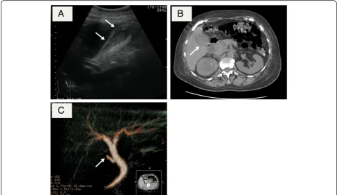
and 224.9 U/mL, respectively. Abdominal ultrasonography
(US) revealed a thickened gallbladder wall and subserosal
edema. Computed tomography (CT) also revealed an
en-larged gallbladder and pericholescystic fluid collection
(Fig. 1b). However, stones were not observed in the
gall-bladder or cystic duct, and the cause of the acute
chole-cystitis
could
not
be
identified.
Drip-infusion
cholangiography-CT (DIC-CT) confirmed the lack of
pa-tency of the cystic duct and showed no gallstones in the
common bile duct (Fig. 1c).
* Correspondence:oita521@yahoo.co.jp
1Department of Surgery, Chuden Hospital, 3-4-27 Otemachi, Naka-Ku,
Hiroshima 730-8562, Japan
Full list of author information is available at the end of the article
The patient’s medical history included a lumpectomy
of invasive ductal carcinoma of the left breast and
nega-tive sentinel lymph node (pT1c/pN0) 12 years before.
She had been followed up with US, CT, and
fluorine-18-fluorodeoxyglucose
positron-emission
tomography
(FDG-PET) imaging for 10 years, and no recurrence had
been observed as of her last follow-up.
The patient was diagnosed with AAC and underwent a
laparoscopic cholecystectomy without any complications.
Macroscopically, the gallbladder mucosa appeared
nec-rotic. Histopathological examination revealed a poorly
differentiated adenocarcinoma in the cystic duct and
gall-bladder neck. On immunohistochemical examination, the
tumor cells were positive for estrogen and progesterone
receptors (ER and PR). The tumor cells were also positive
for cytokeratin-7 and epithelial membrane antigen and
were negative for human epidermal growth factor receptor
2 (HER2), gross cystic disease fluid protein-15, and
cytokeratin-20. These results were similar to the
immuno-histochemical findings from the primary breast cancer
(Fig. 2f). Accordingly, the pathological diagnosis of
meta-static breast cancer was made. One month after
cholecyst-ectomy, an FDG-PET scan revealed abdominal para-aortic
lymph node metastases and a lumbar vertebra metastasis.
The patient was treated with chemotherapy and hormone
therapy, and she died 5 years later (17 years later after
breast surgery).
Discussion
AAC is associated with a higher mortality rate and has a
worse prognosis than ACC [4, 10, 11]. Most cases of
AAC occurs in critically ill patients and are related to
surgery, burns, severe trauma, bacterial sepsis, shock,
congestive heart failure, total parenteral nutrition, and
prolonged fasting [4, 5]. AAC is also associated with
gallbladder cancer and bile duct cancer [12, 13]. Ida et
al. reported a 6.9 % incidence of AAC in gallbladder
can-cer [14]. In contrast, cystic duct cancan-cer is extremely rare
and is not usually diagnosed prior to cholecystectomy
[15]. Thickening of the gallbladder neck and cystic duct
walls in the context of AAC can suggest the presence of
cancer. Endoscopic ultrasonography and cytodiagnosis
from endoscopic naso-gallbladder drainage may be
use-ful in identifying this thickening [16–18].
Metastasis to the gallbladder is very rare. Metastases
ori-ginating from malignant melanomas, and lung, renal,
pan-creatic, and colorectal cancers have been reported [6, 19].
Breast cancer metastasis to the gallbladder is rare and our
literature search revealed only 25 cases. Only one case of
cystic duct metastasis had been reported [20]. In this case,
cystic duct metastasis occurred after metastases to both
lobes of the liver, and right-supraclavicular node, which
was found 3 years after mastectomy. Acalculous
cholecyst-itis was indicated based on the clinical finding of
obstruc-tion of the cystic duct by liver metastasis. Laparotomy
Fig. 1aUS showed thickening of the gallbladder wall and subserosal edema.bCT showed the enlarged gallbladder and the thickened gallbladder wall.cDIC-CT showed no gallstones in the common bile duct and an interruption of cystic duct
revealed a solitary metastatic deposit surrounding the
proximal cystic duct. In our case, prior to the
cholecystec-tomy, the suspicion of breast cancer metastasis to the
cystic duct was low because the patient had remained
cancer-free for over 10 years.
Breast cancer is the commonest form of malignancy in
females. Postoperative recurrence occurs in
approxi-mately 30 % of cases [21]. The commonest sites of
re-currence are the bone, lung, and liver [7–9]. Owing to
advances in chemotherapy and endocrine therapy, the
prognosis for breast cancer has improved over the years.
Despite this, many patients continue to experience
dis-ease recurrence. Recurrences tend to occur within the
first 5 years after surgery; late recurrences after more
than 10 years are very uncommon [7, 9, 22]. Late
recur-rences have been found to affect the bone and lung in
33.3 % of patients, and the recurrence patterns of late
and early recurrences were not found to be significantly
different [22]. Lymph node metastases [23], ER-positive
status [9], and HER2-negative status [24] are reported to
be risk factors for late recurrence in breast cancer
pa-tients. In our case, the patient’s tumor cells were positive
for ER and PR and negative for HER2 on
immunohisto-chemical examination. Moreover, it was recently
re-ported that extension of hormonal treatment to 10 years
was useful for preventing recurrences in such patients
[25]. On the other hand, post-relapse survival was
sig-nificantly longer in patients with late recurrences than in
patients with early recurrences [26, 27]. In the present
case, after multiple metastases were diagnosed, the
pa-tient was treated with aromatase inhibitor therapy,
bisphosphonate therapy, and chemotherapy such as
pac-litaxel and epirubicin, and she survived for 5 years.
In this patient, even if her breast cancer metastases
were diagnosed before cholecystectomy, this knowledge
might not have been useful in guiding the clinical
decision-making process because multiple metastases
were detected on FDG-PET only 2 weeks after the
sur-gery. However, most cases of late breast cancer
recur-rence involve solitary tumors, which can be radically
treated to improve patient survival [28]. As such, it is
important to be able to identify AAC due to metastases
from breast cancer recurrence prior to surgery.
Conclusions
In conclusion, we have reported a case of AAC secondary
to cystic duct metastasis from recurrent breast cancer. It
is necessary to consider metastatic breast cancer as a
cause of AAC in patients with a history of breast cancer.
Consent
Written informed consent was obtained from the patient
for publication of this case report and any accompanying
images.
Abbreviations
AAC:Acute acalculous cholecystitis; ACC: Acute calculous cholecystitis;
CEA: Carcinoembryonic antigen; CT: Computed tomography; DIC-CT: Drip-infusion cholangiography computed tomography; ER: Estrogen receptor;
FDG-PET: Fluorine-18-fluorodeoxyglucose positron-emission tomography; HER2: Human epidermal growth factor receptor 2; PR: Progesterone receptor;
US: Ultrasonography
Acknowledgements None.
Funding None.
Authors’contributions
MH, KK, MA, KK, MT, YM, and TI made substantial contributions to the conception and design and acquisition, analysis, and interpretation of the data. MH was involved in drafting the manuscript or revising it critically for important intellectual content. KK gave the final approval of the version to be published. All authors read and approved the final manuscript.
Authors’information
M. Hashimoto, K. Koide, M. Arita, K. Kawaguchi, M. Tokunaga, and Y. Mikuriya are clinicians of the Department of Surgery, Chuden Hospital. T. Iwamoto is a pathologist of the Department of Pathology, Chuden Hospital.
Competing interests
The authors declare that they have no competing interests.
Author details
1Department of Surgery, Chuden Hospital, 3-4-27 Otemachi, Naka-Ku,
Hiroshima 730-8562, Japan.2Department of Pathology, Chuden Hospital,
3-4-27 Otemachi, Naka-Ku, Hiroshima 730-8562, Japan.
Received: 8 September 2016 Accepted: 5 October 2016
References
1. Savoca PE, Longo WE, Zucker KA, McMillen MM, Modlin IM. The increasing prevalence of acalculous cholecystitis in outpatients. Results of a 7-year study. Ann Surg. 1990;211(4):433–7.
2. Howard RJ. Acute acalculous cholecystitis. Am J Surg. 1981;141(2):194–8. 3. Warren BL, Carstens CA, Falck VG. Acute acalculous cholecystitis—a
clinical-pathological disease spectrum. S Afr J Surg. 1999;37(4):99–104.
4. Kalliafas S, Ziegler DW, Flancbaum L, Choban PS. Acute acalculous cholecystitis: incidence, risk factors, diagnosis, and outcome. Am Surg. 1998;64(5):471–5. 5. Barie PS, Eachempati SR. Acute acalculous cholecystitis. Curr Gastroenterol
Rep. 2003;5(4):302–9.
6. Satoh H, Iyama A, Hidaka K, Nakashiro H, Harada S, Hisatsugu T. Metastatic carcinoma of the gallbladder from renal cancer presenting as intraluminal polypoid mass. Dig Dis Sci. 1991;36(4):520–3.
7. Quiet CA, Ferguson DJ, Weichselbaum RR, Hellman S. Natural history of node-negative breast cancer: a study of 826 patients with long-term follow-up. J Clin Oncol. 1995;13(5):1144–51.
8. Takeuchi H, Baba H, Kano T, Maehara Y. The time-related changes of the importance of prognostic factors in breast cancer. A sequential multivariate analysis of 1423 Japanese patients. Breast Cancer Res Treat. 2005;94(3):273– 8. doi:10.1007/s10549-005-9014-x.
9. Takeuchi H, Tsuji K, Ueo H. Prediction of early and late recurrence in patients with breast carcinoma. Breast Cancer (Dove Med Press). 2005;12(3):161–5.
10. Laurila J, Syrjala H, Laurila PA, Saarnio J, Ala-Kokko TI. Acute acalculous cholecystitis in critically ill patients. Acta Anaesthesiol Scand. 2004;48(8):986–91. doi:10.1111/j.0001-5172.2004.00426.x.
11. Gu MG, Kim TN, Song J, Nam YJ, Lee JY, Park JS. Risk factors and therapeutic outcomes of acute acalculous cholecystitis. Digestion. 2014;90(2):75–80. doi:10.1159/000362444.
12. Barnett KT, Malafa MP. Complications of hepatic artery infusion: a review of 4580 reported cases. Int J Gastrointest Cancer. 2001;30(3):147–60. doi:10.1385/IJGC:30:3:147.
13. Yasuda H, Takada T, Kawarada Y, Nimura Y, Hirata K, Kimura Y, et al. Unusual cases of acute cholecystitis and cholangitis: Tokyo Guidelines. J Hepatobiliary Pancreat Surg. 2007;14(1):98–113. doi:10.1007/s00534-006-1162-9.
14. Ida T, Morimoto T, Tarumi T, Yamato T, Hisano S, Nakagawa M, et al. Current status of benign biliary disorders in Japan and accuracy rates of
preoperative diagnoses. Collective review of 14,654 patients. Am J Surg. 1983;146(2):269–73.
15. Takahisa HJN, Eiji O, Kenichi N, Hisaharu O, Tetsuo N, Takahiro M, Shingo K, Shyoji H. A case of primary cystic duct carcinoma based on Farrar’s criteria necessitating additional resection because of difficult preoperative diagnosis. Jpn J Gastroenterol Surg. 2009;42(11):1687–92.
16. Kimura K, Fujita N, Noda Y, Kobayashi G, Ito K. Differential diagnosis of large-sized pedunculated polypoid lesions of the gallbladder by endoscopic ultrasonography: a prospective study. J Gastroenterol. 2001;36(9):619–22. 17. Sadamoto Y, Oda S, Tanaka M, Harada N, Kubo H, Eguchi T, et al. A useful
approach to the differential diagnosis of small polypoid lesions of the gallbladder, utilizing an endoscopic ultrasound scoring system. Endoscopy. 2002;34(12):959–65. doi:10.1055/s-2002-35859.
18. Itoi T, Sofuni A, Itokawa F, Kurihara T, Tsuchiya T, Moriyasu F, et al. Preoperative diagnosis and management of thick-walled gallbladder based on bile cytology obtained by endoscopic transpapillary gallbladder drainage tube. Gastrointest Endosc. 2006;64(4):512–9. doi:10.1016/j.gie.2006.01.024. 19. Kawahara T, Ohshiro H, Sekiguchi Z, Furuya M, Namura K, Itoh H, et al.
Gallbladder metastasis from renal cell carcinoma. Case Rep Oncol. 2010;3(1): 30–4. doi:10.1159/000279308.
20. Beaver BL, Denning DA, Minton JP. Metastatic breast carcinoma of the gallbladder. J Surg Oncol. 1986;31(4):240–2.
21. Fisher B, Anderson S, Bryant J, Margolese RG, Deutsch M, Fisher ER, et al. Twenty-year follow-up of a randomized trial comparing total mastectomy, lumpectomy, and lumpectomy plus irradiation for the treatment of invasive breast cancer. N Engl J Med. 2002;347(16):1233–41. doi:10.1056/NEJMoa022152. 22. Takeuchi H, Muto Y, Tashiro H. Clinicopathological characteristics of
recurrence more than 10 years after surgery in patients with breast carcinoma. Anticancer Res. 2009;29(8):3445–8.
23. Nishimura R, Osako T, Nishiyama Y, Tashima R, Nakano M, Fujisue M, et al. Evaluation of factors related to late recurrence—later than 10 years after the initial treatment—in primary breast cancer. Oncology. 2013;85(2):100–10. doi:10.1159/000353099.
24. Oven Ustaalioglu BB, Balvan O, Bilici A, Develi A, Aliustaoglu M, Vardar FA, et al. The differences of clinicopathological factors for breast cancer in respect to time of recurrence and effect on recurrence-free survival. Clin Transl Oncol. 2015;17(11):895–902. doi:10.1007/s12094-015-1323-x. 25. Goss PE, Ingle JN, Pritchard KI, Robert NJ, Muss H, Gralow J, et al. Extending
aromatase-inhibitor adjuvant therapy to 10 years. N Engl J Med. 2016;375(3): 209–19. doi:10.1056/NEJMoa1604700.
26. Courdi A, Largillier R, Ferrero JM, Lallement M, Raoust I, Ettore F, et al. Early versus late local recurrences after conservative treatment of breast carcinoma: differences in primary tumor characteristics and patient outcome. Oncology. 2006;71(5-6):361–8. doi:10.1159/000107771. 27. Ogiya A, Yamazaki K, Horii R, Shien T, Horimoto Y, Masuda N et al.
Post-relapse survival in patients with the early and late distant recurrence in estrogen receptor-positive HER2-negative breast cancer. Breast Cancer (Dove Med Press). 2016. doi:10.1007/s12282-016-0730-3.
28. Zagouri F, Sergentanis TN, Koulocheri D, Nonni A, Bousiotou A, Domeyer P, et al. Bilateral synchronous breast carcinomas followed by a metastasis to the gallbladder: a case report. World J Surg Oncol. 2007;5:101. doi:10.1186/ 1477-7819-5-101.
Submit your manuscript to a
journal and benefi t from:
7Convenient online submission
7Rigorous peer review
7Immediate publication on acceptance
7Open access: articles freely available online
7High visibility within the fi eld
7Retaining the copyright to your article