REVIEW
Cell membrane protein functionalization
of nanoparticles as a new tumor-targeting
strategy
Anna Pasto
1,3, Federica Giordano
2,3, Michael Evangelopoulos
3, Alberto Amadori
1,4and Ennio Tasciotti
3,5*Abstract
Nanoparticles have seen considerable popularity as effective tools for drug delivery. However, non-specific target-ing continues to remain a challenge. Recently, biomimetic nanoparticles have emerged as an innovative solution that exploits biologically-derived components to improve therapeutic potential. Specifically, cell membrane proteins extracted from various cells (i.e., leukocytes, erythrocytes, platelets, mesenchymal stem cells, cancer) have shown considerable promise in bestowing nanoparticles with increased circulation and targeting efficacy. Traditional nano-particles can be detected and removed by the immune system which significantly hinders their clinical success. Bio-mimicry has been proposed as a promising approach to overcome these limitations. In this review, we highlight the current trends in biomimetic nanoparticles and describe how they are being used to increase their chemotherapeutic effect in cancer treatment.
Keywords: Bioinspired, Biomimicry, Drug delivery, Nanoparticles, Cancer
© The Author(s) 2019. This article is distributed under the terms of the Creative Commons Attribution 4.0 International License (http://creat iveco mmons .org/licen ses/by/4.0/), which permits unrestricted use, distribution, and reproduction in any medium, provided you give appropriate credit to the original author(s) and the source, provide a link to the Creative Commons license, and indicate if changes were made.
Background
Cancer is the second leading cause of death in the world with over 8 million deaths worldwide. Current treatment option range from surgery and radiation to chemo and hormone therapy [1]. Although technologies to combat cancer have made significant advances in eradicating the tumor mass and reducing metastasis, chemotherapy continues to remain the mainstay of tumor treatment. Unfortunately, chemotherapeutic administration often results in unwanted side effects and the development of drug resistance that can lead to cancer recurrence and metastatic dissemination. To mitigate the side effects, nanotechnology has been showcased as a versatile tool to increase drug potency and localize treatment [2–6]. Indeed, nanoparticles (NPs) are endowed with: (i) opti-mal drug loading properties [7], (ii) increased payload stability [8], prolonged circulation, (iii) enhanced per-meability and retention (EPR), and (iv) biocompatibility
via tunable chemical compositions [9, 10]. Despite these advantages, NP efficacy is often limited by the presence of biological barriers such as mononuclear phagocyte system (MPS), hemorheological forces [11] and endothe-lial vessel wall [12]. In an effort to overcome these hur-dles, we and others have developed several classes of NPs characterized by different types of functions, responsive triggers, and surface modifications that contribute to increased their efficacy [13].
Initially, our group engineered multi-stage porous silicon NPs to mimic red blood cells and serve as a car-rier to shuttle loaded nanoparticulates [14]. Strategically designed using fine-tuned size and shape parameters, these biocompatible [15] and biodegradable [10] multi-stage NPs leverage unique design properties [9] to enhance circulation and margination within vascular endothelia, thereby protecting the payload from various biological hurdles [16]. Nevertheless, targeted and local-ized delivery still require the use of various moieties (i.e., antibodies [17, 18], photosensitizers [19]) functionalized onto the NP surface, as well as the use of ‘smart’ materials [20] and lipid NPs [21]. Although substantial increases in targeting and efficacy were observed, MPS sequestration
Open Access
*Correspondence: [email protected]
3 Present Address: Center for Biomimetic Medicine, Houston Methodist
resulted in high accumulation in the liver and spleen, thereby hindering ideal localization at the tumor site. In addition, due to a number of biological barriers found within the body, it is paramount to strategically design a system that has the potential to circumvent these barri-ers sequentially while still maintaining the payload. For a thorough review in the biological barriers a nanoparticle is exposed to, we direct the reader to the following paper: [22].
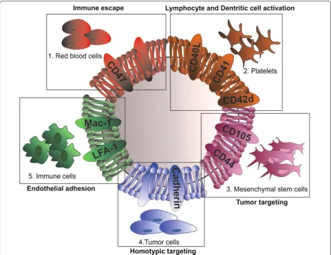
In an effort to mitigate immunorecognition and sequestration by filtering organs, our group modified multi-stage NPs with a leukocyte-derived cell membrane shell as a strategy to bypass critical biological barriers [23]. Other groups have developed similar strategies con-sisting in using surface coatings derived from various cel-lular sources (i.e., mesenchymal stem cells [24], platelets
[25], etc.), which transfer intrinsic cellular properties to a synthetic nanomaterial. Designed to leverage activated endothelia as a targeting method [26], this strategy has been shown to improve tumor targeting, provide pro-longed circulation, and reduce immunoreactivity (Fig. 1).
The aim of this review is to highlight the various strate-gies in which biomimetic NPs are being used in cancer treatment. In addition, this review will cover the various cell sources employed for NP design and the intrinsic effects these cells provide in tumor targeting.
Main text
Source of cells for biomimetic nanoparticles 1. Red blood cells
Due to unique biological properties such as prolonged blood circulation time, lack of organelles (i.e., nucleus),
Fig. 1 Schematic illustration of an empty biomimetic nanoparticle (NP) developed from the membrane sourced of different cells: platelets, red blood, mesenchymal, tumor and immune cells. Thanks to the cells of origin, NPs are endowed with specific features (in bold) that mediate their ability to escape the immune system, extravasate blood vessels and target tumor site. LFA-1 lymphocyte function-associated antigen 1, Mac-1
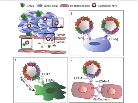
and abundance in the body, red blood cells (RBCs) rep-resent the most convenient cell membrane protein source to produce cell-based NPs. In addition, thanks to the expression of specific inhibitory proteins such as CD47, also known as the “do not eat me” signal, RBCs can easily escape immune system recognition, inhibit-ing macrophage-mediated phagocytosis [27]. Zhang and coworkers were pioneers in the use of RBC membranes to develop biomimetic NPs. Specifically, they combined PLGA NPs with RBC membranes purified from fresh RBCs. The resulting RBC-NPs were validated for their protein content and long-term stability features, demon-strating successful translocation of the associated RBC membrane proteins to the NP surface. Thanks to the presence of immunosuppressive proteins on the RBC membrane (i.e., CD47), RBC-NPs showcased higher cir-culation half-life with significant retention in the blood and decreased macrophage uptake compared to conven-tional polyethylene glycol (PEG)-funcconven-tionalized lipid-pol-ymer hybrid nanoparticles (PEG-NPs). Overall, RBC-NPs resulted in higher structural rigidity, increased stability, and superior cargo encapsulation and delivery compared to uncoated NPs [28]. Further assessment of this technol-ogy in a lymphoma tumor murine model demonstrated the efficient delivery of doxorubicin (DOX) to tumor sites, leading to significant tumor growth inhibition while demonstrating positive immunocompatibility and safety relative to free drug [29].
Similarly, Su et al. formulated paclitaxel-loaded NPs using a polymeric core and a hydrophilic RBC vesicle shell (called RVPNs) that were co-administrated with the tumor-penetrating peptide, iRGD, to enhance antitumor therapy [30]. The authors demonstrated the advantages of the prolonged circulation of RVPNs and the tumor-penetration properties of iRGD in a murine breast cancer model. This strategy displayed remarkably higher reten-tion of paclitaxel in the blood compared to convenreten-tional paclitaxel-loaded NPs. Specifically, RVPNs and iRGD achieved 90% tumor growth inhibition. In addition, this strategy showed positive results in the treatment of metastasis, exhibiting a 95% reduction of lung metastasis and substantially lower hematological toxicity compared to uncoated NPs, NPs/iRGD, or RVPNs alone [30].
2. Platelets
Recently, platelets have also garnered significant atten-tion as a source for biomimetic NPs. Derived from the bone marrow, these enucleated cells are involved in hemostasis, clotting, inflammation, as well as tissue repair [31]. Several studies have also demonstrated that platelets play a crucial role in carcinogenesis [32, 33]. Indeed, inflammation occurring during neoplastic pro-gression recalls platelets to the tumor site, stimulating
tumor angiogenesis. In addition, platelets sustain tumor cell extravasation and the survival of circulating tumor cells in the bloodstream [33], thus favoring metastatic spreading.
Taking advantage of the interactions between plate-lets and tumor cells, and thanks to their physical and biochemical properties such as discoidal shape and flex-ibility, biomimetic platelet-like NPs have been exploited for targeted drug delivery [34]. Li et al. produced silica (Si) NPs coated with membranes isolated from activated platelets (PMDV-coated Si particles) and functional-ized with tumor necrosis factor (TNF)-related apopto-sis inducing ligand (TRAIL) [35]. PMVD-coated Si-NPs were shown to express most of the platelet surface pro-teins (i.e., CD41, CD42b and CD61) and glycans relevant for targeting circulating tumor cells (CTCs) and escaping phagocytosis. Indeed, evaluation of a variety of cancer-bearing murine models (i.e., human breast cancer, colon cancer, and a syngeneic metastatic colon cancer and melanoma mouse model) demonstrated that TRAIL-con-jugated PMDV-Si particles were able to efficiently target CTCs in lung vasculature and to dramatically decrease lung metastases compared to untreated mice, empty PMDV-coated Si particles, and soluble TRAIL. In addi-tion, despite TRAIL is associated with an increase in liver toxicity, this strategy exhibited no substantial effect on hepatic apoptosis following a 24 h treatment.
A similar approach was used by Hu et al. that developed platelet membrane (PM)—coated core–shell nanovesicles (called PM-NVs) loaded with two anticancer compo-nents: TRAIL and DOX. The administration of PM-NVs in a breast cancer mouse model demonstrated NP accu-mulation at the tumor site and efficient delivery of TRAIL toward cancer cell membrane, resulting in the activation of the extrinsic apoptosis signaling pathway. Moreover, thanks to their acid-responsive encapsulation matrix, the PM-NVs were better digested after endocytosis, thus enhancing DOX intracellular accumulation. This resulted in the inhibition of tumor growth and a reduction in lung metastasis [36]. The same group, recently, exploited a combined strategy of two nanocarriers to enhance antitu-mor activity [37]. The first one was a signal transmission nanocarrier (NCA) functionalized with an RGD peptide
to specifically target integrins (i.e., ανβ3) within tumor blood vessels. The second was a biomimetic nanocarrier (NCB) made of integrated platelet membranes and loaded
the specific affinity between P-selectin, transferred onto the NP surface, and CD44, overexpressed on the tumor cells, the authors demonstrated that their platelet mem-brane-coated biomimetic nanocarriers (PM-NPs) were endowed with the capability to target bone microenvi-ronment and myeloma cells. PM-NPs showed high inter-nalization, high tumor targeting, enhanced intracellular drug release, and decrease in off-target effects. Overall, compared to traditional active targeting, platelet-derived coating strategies increased selective bone targeting, as well as cytotoxicity against myeloma cells and the effi-cient thrombus dissolution in an in vivo multiple mye-loma mouse model [38].
3. Mesenchymal stem cells
It has been largely accepted that most current anti-tumor therapies fail as a result of a small cell popula-tion (referred as cancer stem cells, CSCs) responsible for tumor progression, tumor maintenance and metastases formation. CSCs have been identified and described in almost all types of cancer (i.e., prostate [39, 40], lung [41], colon [42], pancreatic [43], gastric [44], breast [45], gli-oma [46] and ovarian cancer [47, 48]). To date, no treat-ment has been successful in destroying or reducing CSCs due to their intrinsic properties (i.e., drug-resistance and ability to enter a quiescence state) and the lack of a uni-versal marker for their identification.
Mesenchymal stem cells (MSCs) are multipotent stem cells endowed with the ability to differentiate into osteo-blasts, adipocytes, chondroosteo-blasts, fibroblasts and peri-cytes. Stimulated by microenvironmental factors such as hypoxia, ligands of Toll-like receptors or cytokine gra-dients within the extracellular matrix, MSCs migrate to injured and inflamed sites. With cancer being considered as a disease of chronic inflammation, it’s appropriate that the last decade has seen a considerable rise in MSCs used as a therapeutic tool in tumor treatment [19, 49].
In a biomimetic strategy employing MSCs, Timaner et al. developed nanoghosts (NGs) derived from the cyto-plasmic membranes of MSCs. MSC-NGs retained the ability of MSCs to target inflamed endothelium, allow-ing safe and effective targeted delivery of their payload both in vitro and in vivo using a prostatic mouse cancer model. In addition, the authors demonstrated that MSC-NGs accumulated in proximity of CSCs within the tumor mass. The membrane proteins expressed on the NG sur-face mediated cell-to-cell interaction with CSCs [50], thus suggesting their potential role in eradicating this CSC subset.
A similar approach was used by Machluf and cow-orkers that resulted in a significantly higher accu-mulation of MSC-derived NGs within the tumor as compared to smooth muscle cell-derived NGs (used as a
non-mesenchymal control). MSC-NGs that retained on their surface specific MSC integrins were shown to bind and fuse to the tumor cell surface and disrupt the plasma membrane, leading to a cytotoxic effect and tumor growth inhibition. The authors suggested that the anti-tumor effect observed using empty MSC-NGs was likely due to the interaction of NG with the different compo-nents of the tumor microenvironment [24]. Conversely, treatment with empty synthetic liposomes did not result in any interaction with the tumor microenvironment. Thus, it is fair to speculate that MSC-derived integrins may mediate MSC-NG interaction with infiltrat-ing immune cells, blood vessel endothelium, and tumor-associated fibroblasts.
On the other hand, Gao et al. demonstrated that MSC-biomimetic NPs were endowed MSC-derived hypoim-munogenicity due to different molecular recognition moieties. Indeed, bone marrow derived MSC membrane-coated gelatin nanogels (SCMGs) showed reduced uptake by the MPS, reduced clearance by the MPS and reduced internalization by filtration organs (such as spleen, kid-ney, liver and lung) [51]. As a result, SCMGs displayed longer blood circulation time, higher in vitro targeting and increased in vivo accumulation within the tumor site, compared to uncoated nanogels. All together, these results suggest the use of MSC-derived biomimetic NPs as a promising tool for anti-cancer therapies by exploit-ing the unique tropism of MSCs to sites of inflammation.
4. Tumor cells
facilitate tumor-specific immune response through the increase of antigen presentation and the interaction with lymphocytes, that leads to increase IFNγ secretion.
Chen et al. developed biomimetic NPs by fusing a cell membrane isolated from MCF-7 human breast cancer cells to an indocyanine green (ICG)/PLGA core [53]. Dubbed ICNPs, this strategy resulted in reduced liver and kidney uptake, as well as higher tumor accumulation due to cell affinity. In addition, ICNPs showed both effi-cient real-time dual-modal tumor imaging and enhanced photothermal therapy properties. Specifically, this strat-egy resulted in complete ablation of the tumor using a single dose of ICNPs.
Similarly, Sun et al. developed a strategy to exploit cancer cell membrane-coated NPs for targeted chemo-therapy of homotypic tumors [54]. The authors devel-oped paclitaxel-loaded polymeric nanoparticles (PPNs) using polycaprolactone (PCL) and pluronic copolymer F68 coated with 4T1 mammary breast cancer cell mem-branes (called CPPNs). In vitro studies demonstrated that CPPNs are endowed with high targeting specificity to the homotypic tumor cells and reduced macrophage uptake. These properties were confirmed in vivo in an orthotopic breast cancer and in a metastatic tumor mouse model. Indeed, in both murine models, administration of CPPNs was associated with an increase in NP accumulation both at the primary tumor site and at metastatic lesions. Interestingly, this strategy exhibited superior inhibition of tumor and lung metastasis growth when compared to paclitaxel alone.
5. Immune cells
In recent years, immune cells have also been proven to be a promising source of coating for biomimetic NPs. Specifically, our group was the first to demonstrate the beneficial properties endowed to NPs when a leukocyte-derived cell source was used. This proof-of-concept was demonstrated with the functionalization of nanoporous silicon particles with cellular membranes isolated from leukocyte cells [23]. Dubbed leukolike vectors (LLVs), we successfully grafted over 300 proteins on the NP sur-face using isolated cell membrane patches, with over 50% being associated with cellular membrane [55]. Among them, we identified proteins that promote reduced MPS uptake (i.e., clusterin [56]), immune tolerance (i.e., CD47 and CD45 [57, 58]), and endothelium/tumor targeting (i.e., lymphocyte function-associated antigen 1 [LFA-1] and macrophage-1 antigen [Mac-1] [12, 23]). In addi-tion, beyond simply targeting activated endothelium, we further dissected the specific role LLVs attain following functionalization. Specifically, we exhibited, following the transfer of membrane onto NPs, proteins grafted onto
the NPs surface retained biological activity, resulting in molecular interplay between particles: cell that fostered increased drug targeting. To this end, LLVs were able to preferentially accumulate at sites of activated endothe-lium, resulting in the downstream phosphorylation of vascular endothelium (VE)-cadherin proteins [12]. This increase in phosphorylation led to a reduction of endothelial tight junctions, thereby facilitating the trans-port of a payload into the tumor microenvironment.
To further evaluate the role cell source may have in the process of transport and targeting, we next compared syngeneic- and xenogeneic-derived cell membrane with uncoated NPs [59]. This study revealed when a syngeneic cell source was employed as a biomimetic coating, mac-rophage avoidance was higher than xenogeneic-coated and uncoated NPs. This was further demonstrated with a delay in liver sequestration following in vivo adminis-tration, with an increase in blood circulation also being observed. Taken together, these results indicate that the presence of various proteins on the nanoparticle surface contributed to an increase in targeting, vascular perme-ability, and payload delivery.
Similar observations of anti-inflammatory effects were observed when biomimetic NPs were further functional-ized with the α4β7 integrin commonly overexpressed in a T lymphocyte-subset that specializes in targeting gastro-intestinal inflammation [61]. This strategy demonstrated that doping biomimetic NPs with this integrin resulted in increased association with the inflamed vessel area while bare NPs tended to diffuse from blood vasculature into the interstitial space, indicating poor adhesion with inflamed vasculature. Gene expression following treat-ment with doped NPs also indicated a significant reduc-tion in anti- (i.e., MRC-1) and pro-inflammatory (i.e., IL6 and TNFα) markers compared to bare and non-doped biomimetic NPs.
Similar strategies have also been employed in the use of peripheral blood mononuclear cells (PBMCs) as a prom-ising source of cell membrane for NP coating. Compared to conventional NPs, PBMC-derived biomimetic nano-particles are endowed with leukocyte properties such as immune evasion, longer blood circulation, and tumor recognition and targeting. As such, several attempts have been made to develop immune cell-derived NPs. For example, Cao et al. cloaked drug-carrying liposomes with cell membranes isolated from macrophages for target-ing metastatic lungs sites in a 4T1 breast cancer mouse model. These membrane-decorated emtansine liposomes (MELs) were endowed with enhanced uptake in meta-static cells resulting in inhibitory effects on cell viability. Furthermore, MELs exhibited specific metastatic-tar-geting potential in vivo with anti-metastatic activity observed in a metastatic breast cancer mouse model [62]. Based on a similar strategy, Kang et al. demonstrated the efficacy of neutrophil-mimetic nanoparticles loaded with proteasome inhibitor, carfilzomib, in selectively targeting CTCs both in blood and pre-metastatic niche [63]. Referred to as NM-NP-CFZs, these biomimetic NPs exhibited an enhanced in vitro binding ability to 4T1 breast cancer cells along with higher CTC associa-tion in vivo and improved homing to the pre-metastatic niche compared to uncoated NPs, thereby resulting in a decrease in the formation of tumor nodules.
Conclusions
The tumor microenvironment is composed of tumor cells surrounded by a variety of additional cells (i.e., fibroblast, immune and stromal cells) that participate in the cell-signaling network to create a neoplastic niche while sup-porting tumor growth and metastasis [64, 65]. As such, disruption of the cross-talk occurring in this microenvi-ronment could potentially minimize tumor cell prolifera-tion, leading to a reduction in aggressiveness that favors increased therapeutic potency [66]. Taking inspiration
from the natural trafficking that occurs within the body, biomimetic NPs have been developed as a novel tool capable of interacting with the various cellular compo-nents within the tumor microenvironment [12], with the potential to interfere with cellular cross-talk. Indeed, beyond serving as simple drug carriers, biomimetic NPs are endowed with specific properties inherited from the donor cell source such as the ability to: (i) avoid mac-rophage phagocytosis; (ii) adhere to activated endothe-lia and (iii) preferentially accumulate at the tumor site (Fig. 2).
Altogether, the surface modification with mem-brane proteins provides biomimetic NPs with the abil-ity to decrease opsonization and prolong circulation within the body, thus facilitating NP targeting. More recently, researchers have begun to explore combining proteins extracted from multiple cell sources as a strat-egy to develop biomimetic NPs with multiple functions and overall greater therapeutic efficacy (Fig. 1). Dehaini et al. pioneered this approach coating NPs with both RBC and platelet membranes. This chimeric formulation presented a higher systemic circulation and enhanced tumor cell targeting [67]. A potential risk that could arise from prolonged circulation time and avoidance of filter-ing organs (e.g., liver, spleen) is the non-specific target-ing of NPs in tissues and organs where they could elicit potential toxicity. In general, we expect biomimetic NPs to be less toxic compared to inorganic and polymeric NPs due to their cell-derived elements. Indeed, the ance of biomimetic NPs follows the physiological clear-ance of protein and lipid organic components. To date, our work has demonstrated that biomimetic NPs present decreased liver sequestration as well as decreased lung and spleen accumulation [59] with reduction in systemic toxicity [56].
Another important player in biomimetic NP function is the protein corona (PC): a layer of biomolecules that binds on the surface of NPs when dispersed in biologi-cal fluids. Contrarily to uncoated NPs, the presence of proteins on the biomimetic NP’s surface could favor the adsorption of specific proteins over others. For example, leukocyte membrane proteins in the liposome bilayer affect the adsorption of blood soluble proteins in the corona. We studied in vivo the evolution of the PC over time and revealed that the integration of leukocyte mem-brane proteins into the leukosome bilayer influenced the number, amount, orientation and type of plasma proteins adsorbed by the NPs, thus affecting NP biodistribution and interaction with cells [56].
into a clinically-viable tool. To minimize immunoreac-tivity, biomimetic NPs could be ideally fabricated from the cells of the patient to be treated. However, due to the limited number of some cell subpopulations within the body, an ex vivo amplification step would be nec-essary upon isolation of the right cell types. Conse-quently, there is the need to create standard protocols to guarantee reproducibility, test batch-to-batch prop-erties and achieve consistent systemic effects in vivo. While more studies need to be performed en route to clinical translation, experimental evidence suggests that biomimetic NPs could provide a solution to over-come major limitations and drawbacks of previous gen-erations of NPs and could represent a promising tool in the treatment of cancer.
Abbreviations
NPs: nanoparticles; NGs: nanoghosts; RBCs: red blood cells; PEG: polyethylene glycol; DOX: doxorubicin; TRAIL: tumor necrosis factor (TNF)-related apoptosis inducing ligand; CTCs: circulating tumor cells; CSCs: cancer stem cells; MSCs: mesenchymal stem cells; MPS: mononuclear phagocyte system.
Authors’ contributions
AP, FG and ME collected the information and wrote the manuscript. AP designed the figures. AA and ET revised the manuscript. All authors read and approved the final manuscript.
Author details
1 Veneto Institute of Oncology-IRCCS, Padua, Italy. 2 School of Medicine
and Surgery, University of Milano-Bicocca, Monza, Italy. 3 Present Address:
Center for Biomimetic Medicine, Houston Methodist Research Institute, 6670 Bertner Ave, Houston, TX 77030, USA. 4 Department of Surgery, Oncology
and Gastroenterology, University of Padova, Padua, Italy. 5 Houston Methodist
Orthopedics and Sports Medicine, Houston Methodist Hospital, Houston, TX, USA.
Acknowledgements
Not applicable.
Competing interests
The authors declare that they have no competing interests.
Availability of data and materials
Not applicable.
Consent for publication
Not applicable.
Ethics approval and consent to participate
Not applicable.
Funding
This work was financially supported by: NCI and the Office of Research on Women’s Health (1R56CA213859), Cancer Prevention & Research Institute of Texas (RP170466 and RP180394), William Randolph Hearst Foundation, Robert J. Kleberg, Jr. and Helen C. Kleberg Foundation and IOV 5 per mille.
Publisher’s Note
Springer Nature remains neutral with regard to jurisdictional claims in pub-lished maps and institutional affiliations.
Received: 19 November 2018 Accepted: 8 March 2019
References
1. Zhai Y, Su J, Ran W, Zhang P, Yin Q, Zhang Z et al (2017) Preparation and application of cell membrane-camouflaged nanoparticles for cancer therapy. Theranostics. 7(10):2575–2592
2. Tasciotti E, Cabrera FJ, Evangelopoulos M, Martinez JO, Thekkedath UR, Kloc M et al (2016) The emerging role of nanotechnology in cell and organ transplantation. Transplantation 100(8):1629–1638
3. Fernandez-Moure JS, Evangelopoulos M, Colvill K, Van Eps JL, Tasciotti E (2017) Nanoantibiotics: a new paradigm for the treatment of surgical infection. Nanomedicine (Lond). 12(11):1319–1334
4. Weiwei G, Liangfang Z (2015) Engineering red-blood-cell-membrane— coated nanoparticles for broad biomedical applications. AIChE J 61(3):738–746
5. Arrighetti N, Corbo C, Evangelopoulos M, Pasto A, Zuco V, Tasciotti E (2018) Exosome-like nanovectors for drug delivery in cancer. Curr Med Chem. https ://doi.org/10.2174/09298 67325 66618 08311 50259 6. Tsao C, Pandolfi L, Wang X, Minardi S, Lupo C, Evangelopoulos M et al
(2018) Electrospun patch functionalized with nanoparticles allows for spatiotemporal release of VEGF and PDGF-BB promoting in vivo neovas-cularization. ACS Appl Mater Interfaces 10(51):44344–44353. https ://doi. org/10.1021/acsam i.8b199 75
7. Martinez JO, Evangelopoulos M, Bhavane R, Acciardo S, Salvatore F, Liu X et al (2015) Multistage nanovectors enhance the delivery of free and encapsulated drugs. Curr Drug Targets 16(14):1582–1590
8. De Rosa E, Chiappini C, Fan D, Liu X, Ferrari M, Tasciotti E (2011) Agarose surface coating influences intracellular accumulation and enhances payload stability of a nano-delivery system. Pharm Res 28(7):1520–1530 9. Yazdi IK, Ziemys A, Evangelopoulos M, Martinez JO, Kojic M, Tasciotti
E (2015) Physicochemical properties affect the synthesis, controlled delivery, degradation and pharmacokinetics of inorganic nanoporous materials. Nanomedicine (Lond). 10(19):3057–3075
10. Martinez JO, Evangelopoulos M, Chiappini C, Liu X, Ferrari M, Tasciotti E (2014) Degradation and biocompatibility of multistage nanovectors in physiological systems. J Biomed Mater Res A. 102(10):3540–3549 11. D’Apolito R, Tomaiuolo G, Taraballi F, Minardi S, Kirui D, Liu X et al (2015)
Red blood cells affect the margination of microparticles in synthetic microcapillaries and intravital microcirculation as a function of their size and shape. J Control Release 217:263–272
12. Palomba R, Parodi A, Evangelopoulos M, Acciardo S, Corbo C, de Rosa E et al (2016) Biomimetic carriers mimicking leukocyte plasma membrane to increase tumor vasculature permeability. Sci Rep. 6:34422
13. Parodi A, Molinaro R, Sushnitha M, Evangelopoulos M, Martinez JO, Arrighetti N et al (2017) Bio-inspired engineering of cell- and virus-like nanoparticles for drug delivery. Biomaterials 147:155–168
14. Tasciotti E, Liu X, Bhavane R, Plant K, Leonard AD, Price BK et al (2008) Mesoporous silicon particles as a multistage delivery system for imaging and therapeutic applications. Nat Nanotechnol 3(3):151–157
15. Martinez JO, Boada C, Yazdi IK, Evangelopoulos M, Brown BS, Liu X et al (2013) Short and long term, in vitro and in vivo correlations of cellular and tissue responses to mesoporous silicon nanovectors. Small 9(9–10):1722–1733
16. Martinez JO, Brown BS, Quattrocchi N, Evangelopoulos M, Ferrari M, Tasciotti E (2012) Multifunctional to multistage delivery systems: the evolution of nanoparticles for biomedical applications. Chin Sci Bull 57(31):3961–3971
17. Scavo MP, Gentile E, Wolfram J, Gu J, Barone M, Evangelopoulos M et al (2015) Multistage vector delivery of sulindac and silymarin for prevention of colon cancer. Colloids Surf B Biointerfaces. 136:694–703
18. Martinez JO, Evangelopoulos M, Karun V, Shegog E, Wang JA, Boada C et al (2014) The effect of multistage nanovector targeting of VEGFR2 posi-tive tumor endothelia on cell adhesion and local payload accumulation. Biomaterials 35(37):9824–9832
19. Nakki S, Martinez JO, Evangelopoulos M, Xu W, Lehto VP, Tasciotti E (2017) Chlorin e6 functionalized theranostic multistage nanovectors trans-ported by stem cells for effective photodynamic therapy. ACS Appl Mater Interfaces 9(28):23441–23449
20. Khaled SZ, Cevenini A, Yazdi IK, Parodi A, Evangelopoulos M, Corbo C et al (2016) One-pot synthesis of pH-responsive hybrid nanogel particles for the intracellular delivery of small interfering RNA. Biomaterials 87:57–68 21. Balasubramanian K, Evangelopoulos M, Brown BS, Parodi A, Celia C, Yazdi
IK et al (2017) Ghee butter as a therapeutic delivery system. J Nanosci Nanotechnol 17(2):977–982
22. Blanco E, Shen H, Ferrari M (2015) Principles of nanoparticle design for overcoming biological barriers to drug delivery. Nat Biotechnol 33(9):941–951
23. Parodi A, Quattrocchi N, van de Ven AL, Chiappini C, Evangelopoulos M, Martinez JO et al (2013) Synthetic nanoparticles functionalized with biomimetic leukocyte membranes possess cell-like functions. Nat Nano-technol 8(1):61–68
24. Toledano Furman NE, Lupu-Haber Y, Bronshtein T, Kaneti L, Letko N, Wein-stein E et al (2013) Reconstructed stem cell nanoghosts: a natural tumor targeting platform. Nano Lett 13(7):3248–3255
25. Hu CM, Fang RH, Wang KC, Luk BT, Thamphiwatana S, Dehaini D et al (2015) Nanoparticle biointerfacing by platelet membrane cloaking. Nature 526(7571):118–121
26. Molinaro R, Corbo C, Livingston M, Evangelopoulos M, Parodi A, Boada C et al (2018) Inflammation and cancer: in medio stat nano. Curr Med Chem 25(34):4208–4223. https ://doi.org/10.2174/09298 67324 66617 09201 60030
27. Oldenborg PA (2004) Role of CD47 in erythroid cells and in autoimmun-ity. Leuk Lymphoma 45(7):1319–1327
28. Hu CM, Zhang L, Aryal S, Cheung C, Fang RH, Zhang L (2011) Erythrocyte membrane-camouflaged polymeric nanoparticles as a biomimetic deliv-ery platform. Proc Natl Acad Sci USA 108(27):10980–10985
29. Luk BT, Fang RH, Hu CM, Copp JA, Thamphiwatana S, Dehaini D et al (2016) Safe and immunocompatible nanocarriers cloaked in RBC membranes for drug delivery to treat solid tumors. Theranostics. 6(7):1004–1011
30. Jinghan S, Huiping S, Qingshuo M, Qi Y, Shan T, Pengcheng Z et al (2016) Long circulation red-blood-cell-mimetic nanoparticles with peptide-enhanced tumor penetration for simultaneously inhibiting growth and lung metastasis of breast cancer. Adv Func Mater 26(8):1243–1252 31. Gentry PA (1992) The mammalian blood platelet: its role in haemostasis,
inflammation and tissue repair. J Comp Pathol 107(3):243–270 32. Schumacher D, Strilic B, Sivaraj KK, Wettschureck N, Offermanns S (2013)
33. Gay LJ, Felding-Habermann B (2011) Contribution of platelets to tumour metastasis. Nat Rev Cancer 11(2):123–134
34. Anselmo AC, Modery-Pawlowski CL, Menegatti S, Kumar S, Vogus DR, Tian LL et al (2014) Platelet-like nanoparticles: mimicking shape, flexibility, and surface biology of platelets to target vascular injuries. ACS Nano 8(11):11243–11253
35. Li J, Ai Y, Wang L, Bu P, Sharkey CC, Wu Q et al (2016) Targeted drug delivery to circulating tumor cells via platelet membrane-functionalized particles. Biomaterials 76:52–65
36. Hu Q, Sun W, Qian C, Wang C, Bomba HN, Gu Z (2015) Anticancer platelet-mimicking nanovehicles. Adv Mater 27(44):7043–7050 37. Hu Q, Sun W, Qian C, Bomba HN, Xin H, Gu Z (2017) Relay drug delivery
for amplifying targeting signal and enhancing anticancer efficacy. Adv Mater. 29(13):1605803
38. Hu Q, Qian C, Sun W, Wang J, Chen Z, Bomba HN et al (2016) engineered nanoplatelets for enhanced treatment of multiple myeloma and throm-bus. Adv Mater 28(43):9573–9580
39. Lang SH, Frame FM, Collins AT (2009) Prostate cancer stem cells. J Pathol. 217(2):299–306
40. Collins AT, Berry PA, Hyde C, Stower MJ, Maitland NJ (2005) Prospective identification of tumorigenic prostate cancer stem cells. Cancer Res 65(23):10946–10951
41. Eramo A, Lotti F, Sette G, Pilozzi E, Biffoni M, Di Virgilio A et al (2008) Identification and expansion of the tumorigenic lung cancer stem cell population. Cell Death Differ 15(3):504–514
42. O’Brien CA, Pollett A, Gallinger S, Dick JE (2007) A human colon cancer cell capable of initiating tumour growth in immunodeficient mice. Nature 445(7123):106–110
43. Hermann PC, Huber SL, Herrler T, Aicher A, Ellwart JW, Guba M et al (2007) Distinct populations of cancer stem cells determine tumor growth and metastatic activity in human pancreatic cancer. Cell Stem Cell 1(3):313–323
44. Fukuda K, Saikawa Y, Ohashi M, Kumagai K, Kitajima M, Okano H et al (2009) Tumor initiating potential of side population cells in human gastric cancer. Int J Oncol 34(5):1201–1207
45. Dontu G, Al-Hajj M, Abdallah WM, Clarke MF, Wicha MS (2003) Stem cells in normal breast development and breast cancer. Cell Prolif 36(Suppl 1):59–72
46. Galli R, Binda E, Orfanelli U, Cipelletti B, Gritti A, De Vitis S et al (2004) Isolation and characterization of tumorigenic, stem-like neural precursors from human glioblastoma. Cancer Res 64(19):7011–7021
47. Pasto A, Pagotto A, Pilotto G, De Paoli A, De Salvo GL, Baldoni A et al (2017) Resistance to glucose starvation as metabolic trait of platinum-resistant human epithelial ovarian cancer cells. Oncotarget. 8(4):6433–6445
48. Pasto A, Bellio C, Pilotto G, Ciminale V, Silic-Benussi M, Guzzo G et al (2014) Cancer stem cells from epithelial ovarian cancer patients privilege oxidative phosphorylation, and resist glucose deprivation. Oncotarget. 5(12):4305–4319
49. Corradetti B, Taraballi F, Martinez JO, Minardi S, Basu N, Bauza G et al (2017) Hyaluronic acid coatings as a simple and efficient approach to improve MSC homing toward the site of inflammation. Sci Rep. 7(1):7991 50. Timaner M, Letko-Khait N, Kotsofruk R, Benguigui M, Beyar-Katz O,
Rachman-Tzemah C et al (2018) Therapy-educated mesenchymal stem cells enrich for tumor-initiating cells. Cancer Res 78(5):1253–1265 51. Gao C, Lin Z, Jurado-Sanchez B, Lin X, Wu Z, He Q (2016) Stem cell
membrane-coated nanogels for highly efficient in vivo tumor targeted drug delivery. Small 12(30):4056–4062
52. Fang RH, Hu CM, Luk BT, Gao W, Copp JA, Tai Y et al (2014) Cancer cell membrane-coated nanoparticles for anticancer vaccination and drug delivery. Nano Lett 14(4):2181–2188
53. Chen Z, Zhao P, Luo Z, Zheng M, Tian H, Gong P et al (2016) Can-cer cell membrane-biomimetic nanoparticles for homologous-targeting dual-modal imaging and photothermal therapy. ACS Nano 10(11):10049–10057
54. Sun H, Su J, Meng Q, Yin Q, Chen L, Gu W et al (2016) Cancer-cell-bio-mimetic nanoparticles for targeted therapy of homotypic tumors. Adv Mater 28(43):9581–9588
55. Corbo C, Parodi A, Evangelopoulos M, Engler DA, Matsunami RK, Engler AC et al (2015) Proteomic profiling of a biomimetic drug delivery plat-form. Curr Drug Targets 16(13):1540–1547
56. Corbo C, Molinaro R, Taraballi F, Toledano Furman NE, Hartman KA, Sher-man MB et al (2017) Unveiling the in vivo protein corona of circulating leukocyte-like carriers. ACS Nano 11(3):3262–3273
57. Molinaro R, Corbo C, Martinez JO, Taraballi F, Evangelopoulos M, Minardi S et al (2016) Biomimetic proteolipid vesicles for targeting inflamed tissues. Nat Mater 15(9):1037–1046
58. Martinez JO, Molinaro R, Hartman KA, Boada C, Sukhovershin R, De Rosa E et al (2018) Biomimetic nanoparticles with enhanced affinity towards activated endothelium as versatile tools for theranostic drug delivery. Theranostics. 8(4):1131–1145
59. Evangelopoulos M, Parodi A, Martinez JO, Yazdi IK, Cevenini A, van de Ven AL et al (2016) Cell source determines the immunological impact of biomimetic nanoparticles. Biomaterials 82:168–177
60. Evangelopoulos M, Tasciotti E (2017) Bioinspired approaches for cancer nanotheranostics. Nanomedicine (Lond). 12(1):5–7
61. Corbo C, Cromer WE, Molinaro R, Toledano Furman NE, Hartman KA, De Rosa E et al (2017) Engineered biomimetic nanovesicles show intrinsic anti-inflammatory properties for the treatment of inflammatory bowel diseases. Nanoscale. 9(38):14581–14591
62. Cao H, Dan Z, He X, Zhang Z, Yu H, Yin Q et al (2016) Liposomes coated with isolated macrophage membrane can target lung metastasis of breast cancer. ACS Nano 10(8):7738–7748
63. Kang T, Zhu Q, Wei D, Feng J, Yao J, Jiang T et al (2017) Nanoparticles coated with neutrophil membranes can effectively treat cancer metasta-sis. ACS Nano 11(2):1397–1411
64. Stakheyeva M, Riabov V, Mitrofanova I, Litviakov N, Choynzonov E, Cherdyntseva N et al (2017) Role of the immune component of tumor microenvironment in the efficiency of cancer treatment: perspectives for the personalized therapy. Curr Pharm Des 23(32):4807–4826
65. Roy A, Li SD (2016) Modifying the tumor microenvironment using nano-particle therapeutics. Wiley Interdiscip Rev Nanomed Nanobiotechnol. 8(6):891–908
66. Melamed JR, Riley RS, Valcourt DM, Day ES (2016) Using gold nanoparti-cles to disrupt the tumor microenvironment: an emerging therapeutic strategy. ACS Nano 10(12):10631–10635