Pregnancy Prognosis Associated With an Isolated
Single Umbilical Artery in Twin Pregnancy
Thomas J. Cade,1Fabricio Da Silva Costa,1,2,3,4Karen Reidy,3,4Lex W. Doyle,3,4Sarah E. Mitchell,1
Ricardo Palma-Dias,1,2,3,4and Mark P. Umstad1,4
1Division of Maternity Services, The Royal Women’s Hospital, Melbourne, Victoria, Australia 2Pauline Gandel Imaging Centre, The Royal Women’s Hospital, Melbourne, Victoria, Australia 3Pregnancy Research Centre, The Royal Women’s Hospital, Melbourne, Victoria, Australia
4Department of Obstetrics and Gynaecology, University of Melbourne, Melbourne, Victoria, Australia
To determine the prognosis of an isolated single umbilical artery (SUA) in a twin pregnancy, we selected twin pregnancies with a second trimester ultrasound diagnosing a SUA in at least one fetus at our tertiary hospital. This was confirmed by placental histopathology or by expert review of ultrasound images. Cases were identified by searching the hospital ultrasound database over a period of 7.5 years. Higher order multiples or coexistent aneuploidy or major anomalies were excluded. Each case of an isolated SUA was assigned three consecutive twin pregnancy controls paired for chorionicity and maternal age. Primary out-comes were preterm birth<34 weeks, small for gestational age (SGA) or perinatal death. Other outcomes included antenatal growth restriction, mode of delivery, and admission to neonatal intensive care or special care nursery. Nine pregnancies (18 fetuses) were identified for analysis as cases. Isolated SUA was asso-ciated with preterm birth<34 weeks (odds ratio=12.2; 95% CI=2.0–75.2;p=.005) but not for SGA. There was also no difference in SGA between the affected twin and its normal co-twin. Perinatal death was increased but after controlling for gestational age and clustering this finding was no longer significant. We conclude that isolated SUA in twins adds a degree of risk to an already high-risk pregnancy but does not increase the need for surveillance for growth restriction.
Keywords:two-vessel cord, diseases in twins, umbilical arteries, preterm birth, small for gestational age
The incidence of a single umbilical artery (SUA) approxi-mates 0.5% in singleton pregnancies (Granese et al.,2007; Hua et al.,2010). Such a finding may be associated with fetal aneuploidy (Dagklis et al.,2010) or malformations (most commonly cardiac or renal; Hua et al.,2010; Thummala et al.,1998), but an isolated finding can present clinicians with a pregnancy challenge. While some studies have re-ported an increased incidence of fetal growth restriction and adverse outcome (Burchstein et al.,2011; Hua et al., 2010), other studies have reported no difference in fetal growth restriction when compared with a normal three-vessel cord (Bombrys et al.,2008; Wiegand et al.,2008).
An isolated SUA in twin pregnancies can present an even greater challenge than management in singleton pregnan-cies. Discordance with the co-twin, issues of chorionicity, and the inherent risks of any twin pregnancy all require consideration. An early review of autopsy cases found that a SUA was more prevalent in twin pregnancies than single-tons and that most are discordant (Heifetz,1984).
More recent studies have found significantly lower birth weights in a twin with a SUA compared to a co-twin with
a normal three-vessel cord (Byers et al.,2013; Klatt et al., 2012). When compared with a cohort of twins both with three-vessel cords, the outcomes of a twin pregnancy with a SUA include increased risks of growth discordance (Klatt et al., 2012), small for gestational age (SGA; Klatt et al., 2012; Stout et al., 2013), and preterm delivery before 28 weeks (Stout et al.,2013).
As no current study has compared cases of isolated SUA in twin pregnancies with individually matched consecutive controls, and given the apparent controversy in the man-agement of an isolated SUA in singleton pregnancies, we have aimed to investigate the outcomes further.
RECEIVED27 April 2014;ACCEPTED11 June 2014. First published
online 5 August 2014.
ADDRESS FOR CORRESPONDENCE: Dr Thomas J. Cade,
Divi-sion of Maternity Services, The Royal Women’s Hospital, 20 Flemington Road, Parkville, Victoria 3053, Australia. E-mail:
Patients and Methods
All patients who had a twin pregnancy and a second trimester morphology ultrasound at the Royal Women’s Hospital, Melbourne, over a period of 7.5 years were con-sidered for inclusion. Patients were identified through the hospital’s Picture Archiving System (PACS; Viewpoint, GE Healthcare, Buckinghamshire, United Kingdom), with the start point of the study defined as the introduction of the computerized PACS in our hospital.
All patients were scanned at a tertiary obstetric hospi-tal by consultant obstetricians with particular expertise in obstetric ultrasound. Color Doppler was used to visual-ize the umbilical arteries, both adjacent to the fetal blad-der and in a section of a free loop of cord. Twin preg-nancies diagnosed with a SUA in one or both twins were identified.
Placental histopathology was performed by experienced perinatal pathologists, and those cases in which a three-vessel cord was identified were excluded. If placental histopathology was not performed, the ultrasound images were reviewed by one of the authors (FDSC).
For those cases in which the finding was isolated, three controls were selected (cases with a major coexistent anomaly or aneuploidy were excluded from analysis). These were defined as the next three consecutive twin pregnancies undergoing a second trimester ultrasound at our hospi-tal, with neither twin being diagnosed with any congenital anomaly. Controls were matched for chorionicity and ma-ternal age (within 5 years). Background and outcome data were collected for the mother and both twins for cases and controls.
Primary outcomes were preterm delivery in less than 34 weeks, SGA (birth weight< -2SDrelative to the British Growth (Cole et al., 1998)), and perinatal death. Other outcomes were mode of delivery, twin-twin transfusion syndrome (for monochorionic twins), oligohydramnios, growth restriction or abnormal umbilical Doppler studies, and neonatal intensive care (NICU) or special care nursery (SCN) admission.
Data were analyzed using Stata version 13.1 (StataCorp, 2013). Differences between groups for continuous vari-ables were assessed using Student’sttest, both paired and unpaired, where appropriate, and mean differences with 95% CIs were calculated. Dichotomous variables were an-alyzed with either chi-square or Fisher’s exact test if sam-ple sizes were small, and odds ratios (ORs) and 95% CIs were calculated. To adjust for confounding variables, con-tinuous variables were analyzed by linear regression and dichotomous variables by logistic regression. Models were fitted using generalizing estimating equations to account for clustering of twins for fetal/infant outcomes, where necessary.
This study was approved as an audit by the hospital’s Human Research Ethics Committee.
Results
Initially, 34 multiple pregnancies were identified as a ‘SUA’ or ‘two-vessel cord’ on the hospital software over the speci-fied period (7.5 years). Four cases were excluded when pla-cental histopathology revealed a three-vessel cord. In seven cases, no placental histopathology was performed and ul-trasound images were not available for review: these cases were also excluded.
Of the remaining 23 cases, three were triplet pregnancies, one was a conjoined twin and nine had co-existing anoma-lies and were excluded. One case was a fetal death in utero in the early second trimester and was also excluded.
Of the 10 cases with coexistent anomalies, three had cardiac anomalies (two coarctations of the aorta and one right-sided aortic arch with enlarged pulmonary trunk), two were cases of trisomy 18, two had multiple serious anomalies, one had an absent lung, and one had severe ventriculomegaly.
This left a total of nine cases of isolated SUA out of 1,243 twin pregnancies over the same period (0.72%). All were discordant for SUA. The exclusions are summarized inFigure 1.
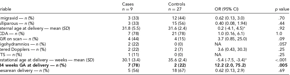
There were no differences between cases and controls in background or antenatal variables (Table 1). There was an increased chance of delivery before 34 weeks but no differ-ence in the mode of delivery. There was also no statistically significant increase in growth restriction on antenatal ul-trasound.
Although there was a significantly lower mean birth weight there was not a significantly lower incidence of SGA (Table 2). Within the cases, there was no difference in birth weightSD score between the fetus with and without the SUA (mean (SD): with SUA -0.7 (1.5); without SUA -1.4 (1.0); mean difference=0.6; 95% CI=-0.2, 1.4;p=.11). In fact, the twin with the SUA tended to be the larger one.
There was a statistically significant increase in any peri-natal death in the cases (OR=10.6, 95% CI=1.03–109, p=.046) but not after adjusting for clustering of multi-ples (Table 2). Any perinatal death was strongly related to gestational age: odds of mortality fell by 0.66 (95% CI= 0.48–0.91) for each week’s increase in gestational age. Of the three cases of perinatal death, two were in an unaffected infant and one was in an infant with SUA. When adjusted for clustering of multiples and for gestational age, the odds of perinatal death became non-significant (OR=1.26; 95% CI=0.27–5.8;p=.77).
34 multiple pregnancies with single umbilical artery on ultrasound
Eleven excluded Placental histopathology
conirms two-vessel cord or ultrasound images available for
review
Non-conjoined twin
pregnancies only Four excluded (3
triplets, 1 conjoined twin)
No co-existing anomaly or
aneuploidy (n = 9) Ten excluded Y
N
N = 23
N
N = 19
N
Total inclusions N = 9
Y
Y
FIGURE 1.
Inclusions and exclusions.
however, it was not possible to assess the effect of adjusting for gestational age on the odds of being admitted to SCN or NICU.
Discussion
The literature on isolated SUA in twin pregnancies is scarce, with concerns generally focusing on preterm birth, in-trauterine growth restriction, or SGA. In the largest and most recent series, Stout et al. (2013) examined 40 twin pregnancies and reported an incidence of 1.7% and a slight increase in preterm birth rates and moderate increase in
SGA. Our series of nine patients represents a lower rate of 0.72%, possibly because we mandated a histopathologi-cal diagnosis of SUA in the placenta postnatally or review of ultrasound images by a single experienced operator. We cannot calculate an accurate incidence because not all twin pregnancies have had the same scrutiny of ultrasound and histopathology as the cases with SUA.
TABLE 1
Maternal Data
Cases Controls
Variable n=9 n=27 OR (95% CI) p value
Primigravid —n (%) 3 (33) 12 (44) 0.62 (0.13, 3.0) .70
Nulliparous —n (%) 3 (33) 15 (56) 0.40 (0.08, 1.94) .44
Maternal age at delivery — mean (SD) 31.8 (5.5) 31.6 (2.4) 0.2 (-4.1, 4.5)∗ .92
DCDA —n (%) 7 (78) 21 (78) 1.0 (0.16, 6.1) 1.0
IUGR on scan —n (%) 4 (44) 4 (15) 3.7 (0.85, 25.0) .09
Oligohydramnios —n (%) 2 (22) 0 (0) NA .057
Altered Dopplers —n (%) 2 (22) 2 (7) 3.6 (0.43, 30.3) .25
TTTS —n (%) 1 (11) 0 (0) NA .25
Gestational age at delivery — weeks — mean (SD) 30.1 (3.4) 35.6 (2.4) -5.4 (-7.5, -3.4)∗ <.001
<34 weeks GA at delivery —n (%) 7 (78) 2 (22) 12.2 (2.0, 75.2) .005
Caesarean delivery —n (%) 5 (56) 18 (67) 0.62 (0.13, 2.9) .69
Note: Primary outcomes in bold type.
DCDA=dichorionic, diamniotic; IUGR=intrauterine growth restriction; TTTS=twin-twin transfusion syndrome; GA=gestational age; OR=odds ratio; CI=confidence interval.
∗mean difference (95% CI).
TABLE 2
Fetal Data
Cases Controls
Variable n=18 n=54 OR (95% CI)∗ p value
Male —n (%) 7 (39) 21 (39) 1.0 (0.24, 4.16) 1.0
BW — mean (SD) 1,290 (638) 2,334 (476) -1,044 (-1,494, -594)† <.001
BWSD score — mean (SD) -1.03 (1.27) -0.51 (-0.86) -0.52 (-1.32, -0.27)† .19
BWSD score<-2SD — n (%) 3 (17) 2 (4) 5.2 (0.62, 43.4) .10
Admitted to NICU —n (%) 11 (61) 5 (9) 15.4 (2.6, 91.7) .003
Admitted to SCN or NICU —n (%) 18 (100) 27 (51) NA <.001
FDIU —n (%) 0 (0) 1 (2) NA 1.0
Neonatal death —n (%) 3 (17) 0/53 (0) NA .014‡
Any perinatal (FDIU or neonatal) death —n (%) 3 (17) 1 (2) 10.6 (0.83, 135) .069
Note: Primary outcomes in bold type.
BW=birthweight; NICU=neonatal intensive care nursery; SCN=special care nursery; FDIU=fetal death in utero; OR=odds ratio; CI=confidence interval; NA=not available.
∗Adjusted for clustering of multiples;†mean difference (95% CI, adjusted for clustering of multiples);‡Fisher’s Exact Test.
most preterm deliveries were spontaneous at the onset and not iatrogenic. The only iatrogenic case was due to severe pre-eclampsia related to pre-existing maternal renal disease. We also showed a statistically significant increase in the risk of perinatal death before adjustment for confounders. Interestingly, of the three cases of perinatal death in preg-nancies with a SUA, one was in the infant with the SUA and two were in the unaffected co-twin. This raises the question whether the presence of SUA is an adverse predictor for the twin pregnancy as a whole, or if the increase in perinatal death was due to the earlier gestation at delivery. As control-ling for gestational age reduced the odds of perinatal death, the latter is more likely.
The case-control methodology with strict inclusion cri-teria for individually matched, consecutive controls is a strength of our study. Another is the requirement for all patients to have been scanned at our tertiary referral cen-ter and histopathological confirmation of the diagnosis or expert review of the antenatal images. While these factors contribute to a more robust methodology, we are also lim-ited by the small sample size. The relatively low rate of
isolated SUA, even in a large tertiary referral center such as The Royal Women’s Hospital, makes it difficult to undertake properly designed prospective studies without multicenter collaboration.
Our series thus has quite different findings to others that have commented on an increase in SGA for isolated SUA in twin pregnancies (Byers et al., 2013; Stout et al., 2013). Klatt et al. in their series in 2012 commented on the trend to increased SGA, but this did not reach statistical significance. It is unclear why our findings are different; however, the strict methodology we have employed for both case and control identification and inclusion (as detailed above) may be one factor. The significantly increased risk of preterm birth in the cases in our study may also suggest that growth restriction occurs at a later gestation in these pregnancies; however, more data is required to definitively support such a hypothesis.
Acknowledgment
Funding from the Centre of Research Excellence Grant in Neonatal Medicine (National Health and Medical Research Council of Australia).
References
Bombrys, A. E., Neiger, R., Hawkins, S., Sonek, J., Croom, C., McKenna, D., . . . Sibai, B. (2008). Pregnancy outcome in isolated single umbilical artery.American Journal of Perina-tology,25, 239–242.
Burchstein, S., Levy, A., Holcberg, G., Zlotnik, A., & Sheiner, E. (2011). Is single umbilical artery an independent risk factor for perinatal mortality?Archives of Gynecology and Obstetrics,283, 191–194.
Byers, B. D., Saade, G. R., & Harirah, H. M. (2013). Twin preg-nancies discordant for single umbilical artery.Journal of Reproductive Medicine,58, 241–245.
Cole, T. J., Freeman, J. V., & Preece, M. A. (1998). British 1990 growth reference centiles for weight, height, body mass in-dex and head circumference fitted by maximum penalized likelihood.Statistics in Medicine,17, 407–429.
Dagklis, T., Defigueiredo, D., Staboulidou, I., Casagrandi, D., & Nicolaides, K. H. (2010). Isolated single umbilical artery and fetal karyotype.Ultrasound in Obstetrics and Gynecol-ogy,36, 291–295.
Granese, R., Coco, C., & Jeanty, P. (2007). The value of single umbilical artery in prediction of fetal aneuploidy:
Find-ings in 12,672 pregnant women.Ultrasound Q, 23,117– 121.
Heifetz, S. A. (1984). Single umbilical artery. A statisti-cal analysis of 237 autopsy cases and review of the literature. Perspectives in Pediatric Pathology, 8, 345– 378.
Hua, M., Odibo, A. O., Macones, G. A., Roehl, K. A., Crane, J. P., & Cahill, A. G. (2010). Single umbilical artery and its associated findings.Obstetrics and Gynecology, 115, 930– 934.
Klatt, J., Kuhn, A., Baumann, M., & Raio, L. (2012). Single um-bilical artery in twin pregnancies.Ultrasound in Obstetrics and Gynecology,39, 505–509.
StataCorp (2013). Stata statistical software, release 13.1. Col-lege Station, TX.
Stout, M. J., Obido, A. O., Longman, R., Shanks, A. L., & Cahill, A. G. (2013). The incidence of isolated single umbilical artery in twins and adverse pregnancy outcomes. Prena-tal Diagnosis,33, 269–272.
Thummala, M. R., Raju, T. N., & Langenberg, P. (1998). Iso-lated single umbilical artery anomaly and the risk for con-genital malformations: A meta-analysis.Journal of Pediatric Surgery,33, 580–585.