Research Article
CODEN: IJPNL6
Diagnostic criteria of different species of leishmaniasis in Balochistan, Pakistan
Darshna Kumari, Muhammad Nadeem*, Ayaz Ali Khan
and Javeid Iqbal
Department of Pharmacology, Faculty of Pharmacy, Hamdard University, Karachi-Pakistan
*Corresponding author e-mail: nadeem_d30@yahoo.co.in
ABSTRACT
Leishmaniasis is a parasitic disease that results into severe lesion/lesions after bitten by the sandfly. The proper and immediate diagnosis is necessary for this lethal disease until it become worse in condition. The purpose of this research study is to present diagnostic criteria of leishmaniasis of different species which is helpful in early diagnosis. Diagnosis criteria include location and number of lesions, type and size of lesions located on the body of the patients. This study was conducted at public and private hospitals and clinics of Karachi Pakistan. Total 200 patients were included in this study. All patients were included in the study after fulfilling the inclusion criteria. Data was collected from the February, 2011 – December, 2012 in the form of well organized questionnaire which had following information as; age, sex, location of patients, site, number, and type of lesion. The mean age of patient was 35±5 years. Out of all selected patients, 40 % were male and rest of 60 % was female. The highest ratio of patients was natives of Baluchistan Pakistan due to its climate which is favorable for sandfly. Majority of patients have single lesion>multiple lesion>double lesion>binary lesion. The common type of lesion seen was the wet ulcerative type open and reddish lesion and the dry nodules. The size of the lesion was observed ranging from 1cm - 3cm respectively. It is observable from the lesion types that the cutaneous leishmaniasis is more common in Pakistan especially in Balochistan than other species of leishmaniasis. In the light of this study, it is concluded that the leishmaniasis is still highly dominated in the Pakistan especially in Balochistan province which is due to its favorable climate for the causative agent. Beside this, there is no proper treatment available and due to poor socioeconomic condition of Pakistan, delayed in diagnosis and treatment of such disease is common. For the purpose of treatment, derivative of pentavalent antimony have been applied that is also more poisonous. There is need of extensive study about parasite of leishmaniasis and their proper treatment and awareness should be provided in the dominated area of Pakistan.
Key words: leishmaniasis, Diagnostic criteria, lesions and management of leishmaniasis
INTRODUCTION
Leishmaniasis is a parasitic disease caused by various species of leishmanial parasite as well as by the piercing skin of human body by infected sandfly [1]. Leishmaniasis is called by various different names according to its location such as; chiclero ulcer, bush yaws, Utah, oriental sore, Aleppo boil, Baghdad sore, saldana. When sandfly bites the host, after several weeks or month the patients notice a rashly bump on any of our body part. This parasite is found in many areas of world especially; entire western border, northern area of Baluchistan and Sindh are the characteristic area of this limited geographic range of
Pakistan. Earlier, it was confined to India, China, Middle East, and other countries. Now it is recognized in all continents of the world except Australia and Antarctica [2]. There are about 21 different species of leishmaniasis. According to disease condition, it may be cutaneous, muco-cutaneous, and visceral leishmaniasis; while on the basis of the parasite multiplication in different body’s organs like; macrophages, skin, nasal, oral mucosa and internal organ. The lesion may heal from time to time and may be spontaneously leaving a scar to eradicate the amastigote and to reduce the size of lesion for rapid healing and leaving no scar is the main treatment of cutaneous leishmaniasis [3].
International Journal of Pharmacy
Therapeutic treatment involves pentavalent antimony compound, sodium sibogluconate and meglumine antimonite. Other treatment includes lipid formulation of intravenous amphotericinb b, oral ketoconazole, topical paromomycin, local heat, or freezing with liquid nitrogen, but still no one give satisfaction results. From 50 years, scientists are going to use pentavalent antimony compound which is highly resistant and administered intravenously which shows various side effects such as; anorexia, myalgias, arthralgia, cardiac abnormalities, intellectual level aminotransferase level and pancreatitis. Another treatment for leishmaniasis is found effective; is photodynamic therapy by which we can eradicate amastigote from the lesion.
Treatment should be repeated at weekly interval so that amastigote were no longer found in direct smear from the lesion. Leishmaniasis is worldwide increasing health problem [4]. The patient with single ulcer can treat simply by topical treatment only. The leishmania braziliensis which involve in the mucocutaneous form causing bone marrow cessation, liver problem, chemical disturb pancreas require systemic antimony [5]. Millions cases of cutaneous leishmaniasis and many cases of visceral leishmaniasis are reported every year. In 21stcentury cutaneous leishmaniasis becomes major health hazardous [6]. A skin scraping with microscopic analysis is the authentic technique when there is possible leishmanial skin lesion seen [7]. Other possible technique which is available are biopsies with tissue-impression smears[8]], aspiration by needle of tissue fluid from the outer of a lesion can yield fluid for culture to isolate the organism and identify the species, Immunologic tests are being tested recently including a highly degree of sensitive polymerase chain reaction test.
MATERIAL AND METHOD
This investigational and survey based study was conducted in different public and private hospitals, and clinics of Karachi, Pakistan. Total 325 patient’s data was collected but 200 patients were included in the study on the basis of presence of leishmaniasis parasite. The majority of patients belong to endemic areas and coastal areas of Baluchistan. There are some patients who are having lesions but they were excluded from study due to absence of parasites in their microscopic sample slides. The cases other than the leishmaniasis like some bacterial infection and some with fibrosis were excluded from the study. There are some patients who were not agreed to
Study Design: According to our study, the patients of leishmaniasis were divided into following categories. They were distributed into different groups on the basis of age and sex. Diagnosis was done by making the glass slides (which contain sample of lesion) under compound microscope. Diagnosis was confirmed pathologically, when the parasite of leishmaniasis was seen. In some patients parasites seen inside the macrophages where as in other cases parasites scattered out of macrophages. There were some exceptional cases where the granulomatous infiltrate are seen, that also indicating leishmaniasis.
Study protocols
Protocol no: 1.The patients having ulcerative lesion more than 3 month old.
Protocol no: 2.The patients having ulcerative lesion more than 8 month old.
Protocol no: 3.The patients having ulcerative lesion more than one year old.
Types of clinical finding: These clinical impressions were noted on the basis of presence of wet ulcerative open lesion, lesion as a dry nodule and typical type of multiple lesions.
Data collection and Data analysis: Patient data was collected from November, 2011 to October, 2012. Data was collected by using a questionnaire which contained the information as; age, sex, family history, number of lesions, type and location of parasitic lesion. Leishmaniasis test was conducted and on the basis of this test, patients were included in study. The basic diagnostic tests performed were, scraping of lesion which is the simplest and most common test, but it is only 70 to 75 % sensitive. Slides were fixed by using methanol, stained with Giemsa, and examined under oil immersion lens of compound microscope. Amastigotes were seen in monocytes or extracellular. It is important to see the nucleus and the rod-shaped kinetoplast, a mitochondrial structure containing extra nuclear DNA, to diagnose leishmaniasis. After collection of data, it was put in IBM-SPSS (Version-19) and generated different parameters required.
RESULT AND DISCUSSION
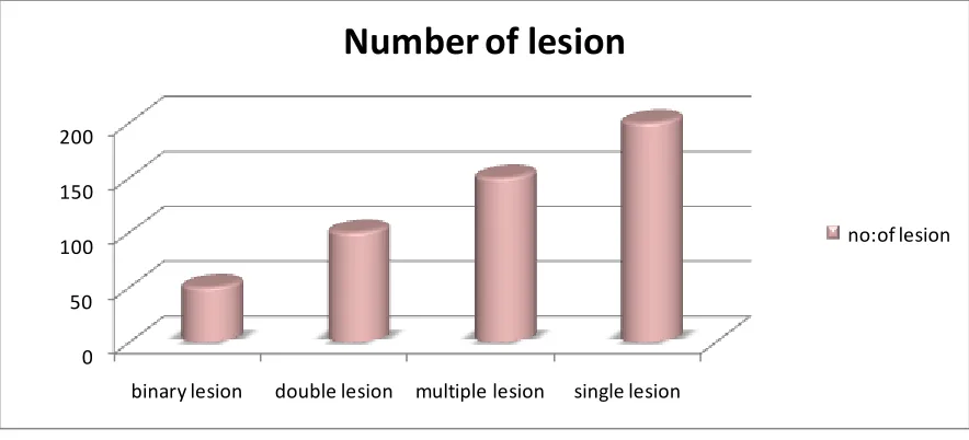
such as hands, nose, and feet. Classically it affects the exposed parts of the body and presents with a skin ulcer at the site of a sand fly bite as shown in Figure 2. The majority of patients, selected in this study, were natives of Baluchistan. It is because in Baluchistan, there is the good flow of sand flies is found at the working premises during the dawn and dusk time. The atmosphere of Baluchistan is favorable for the occurrence of sand flies. In other areas of Pakistan minimum ratio of leishmaniasis patients was seen, as shown in figure 3. Patients of different ages were seen with leishmaniasis. The median age was 30-50 years. Except some patients who got multiple lesions, double lesion, binary lesion, the majority of patients have got single lesion as shown in Figure 4.
In Pakistan, leishmaniasis is either caused by Leishmania major (zoonotic) or Leishmania
tropica (anthroponotic). It generally heals
spontaneously within a year but the clinic - pathological picture of leishmaniasis is variable depending upon several host parasite related factors. The incubation period is usually measured in months, but may range from a few days to over a year. The initial lesion appears as a red furuncle-like papule. The papule gradually enlarges in size over a period of several weeks and becomes more dusky violaceous hue. Eventually, the lesion becomes crusted with an underlying shallow ulcer, often having risen and somewhat indurate borders. The healing is usually with a scar that is typically atrophic, hyper pigmented and irregular (cribriform). In addition, the classical types may often show a clustering of lesions, skin crease orientation, volcanic nodules, satellite papules, subcutaneous nodules, and iceberg nodules. Some other uncommon morphological features of localized leishmaniasis, in addition to the above-mentioned classical picture, are described in following sections. According to type of lesion we can conclude that the cutaneous leishmaniasis is more common type of leishmaniasis seen in areas of Pakistan. Cutaneous leishmaniasis is endemic in Balochistan province of Pakistan. Upto 4000 new cases are registered yearly at Bolan Medical Complex, a tertiary care hospital in Quetta while a lot more attend other public and private hospitals. The wet/rural type of CL which is a zoonotic disorder caused by L. major is more prevalent in the region [9] It is commonly known as Kaaldana (kaal = year, dana = lesion) by local pushtoon population and Saldana by other people of region. Typical lesions of cutaneous leishmaniasis do not make any diagnostic problem, while some cases
presenting with an atypical morphological picture make diagnosis a difficult task. Different clinical presentation is determined by the host parasite interaction.6 Scalp leishmaniasis, CL of the eyelids and cheilitis-like CL have been reported rarely in the literature [10] Leishmaniasis on the scalp in our patients is probably because of the tradition of shaving the scalp, thus providing an easy approach for the sand fly to bite at this unusual site. DLE-like lesions and leishmaniasis with an eczematous appearance are being reported for the first time. Zosteriform pattern of leishmaniasis is only rarely mentioned previously.8 Raja et al. have described a number of patients with chancriform CL in the non-immune military recruits who were posted temporarily in the endemic region [11]. Lesions of this kind were not seen in our series in the native population of Balochistan, although annular pattern of CL were observed in both studies [12]. Keeping in mind all these unique, uncommon presentations of leishmaniasis, dermatologists should always consider CL as a possibility in different cases.
CONCLUSION
First of all, we confirm the pathology of leishmaniasis parasites by the presence of leishmaniasis bodies. The presence of granulomatous seen so this can also indication of leishmaniasis. This diagnostic criterion of leishmaniasis of different species was seen by collecting all above data and history. The confirmation of parasite was made by pathologically by making glass slides which are the simplest and less time consuming choice. Other techniques of diagnosing are very time consuming. Skin biopsy technique is mostly used in secondarily infected patients. Conclusion of our research study is that the leishmaniasis is mostly seen in area of Baluchistan Pakistan than other provinces. Mostly the working males are affected than females. Severity of lesion was seen which need a proper attention, awareness and suitable treatment. On the basis of lesion it is confirmed that the leishmaniasis is a cutaneous type of leishmaniasis.
ACKNOWLEDGEMENT
Figure 1 Ratio of infected individual according to sex.
Figure 2 The more prominent site of lesion
0 0.2 0.4 0.6 0.8 1 1.2 1.4 1.6
Baluchistan punjab sindh kpk
0 50 100 150 200
binary lesion double lesion multiple lesion single lesion
Number of lesion
no:of lesion
Figure 4 Number of lesion in leishmaniasis patients.
REFERNCES
1. S, A., et al., Mucocutaneous leishmaniasis: an imported infection among travellers to central and South America. BMJ, 2004. 329(7470): p. 842–844.
2. J, B., et al., Treatment of cutaneous leishmaniasis among travellers. J Antimicrob Chemother, 2004 Feb. 53(2): p. 158–166.
3. CR, D., et al., Leishmaniasis: new approaches to disease control BMJ 2003. 326 ( ): p. 377–382 4. P, D., The increase in risk factors for leishmaniasis worldwide. Tropical Medicine Hygiene, 2001. 95:
p. 239–243.
5. PD, R., L. R, and S. JJ, Prevalence of “enzootic rodent leishmaniasis” (Leishmania mexicana amazonensis), and apparent absence of “pian bois” (Le. braziliensis guyanensis), in plantations of introduced tree species and in other nonclimax forests in eastern Amazonia. Trans Royal Society Tropical Medicine Hygiene, 1983 77 ( ): p. 775–785
6. RW, A., D. P, and d.R. P, Estimation of population at risk of infection and number of cases of leishmaniasis. Parasitol Today 1992. 8: p. 104-105.
7. (CDC), C.f.D.C.a.P., Cutaneous leishmaniasis in U.S. military personnel. Southwest/Central Asia, oct: 24, 2003. 52(42): p. 1009–1012.
8. (CDC), C.f.D.C.a.P., Cutaneous leishmaniasis in U.S. military personnel—Southwest/Central Asia. MMWR Morb Mortal Wkly Rep, 2003. 52: p. 1009–1012.
9. Bryceson ADM. Clinical variations associated with taxa of leishmania. In: Col Inl CNRS/Interm 1948 Montpelier: IMEEE 1986; 221-228.
10. Momeni AZ, Amin Javaheri M, Clinical picture of cutaneous leishmaniasis in Isphahan Iran. Int J Dermatol 1994; 33: 260-265.
11. Bienzle U, Ebert F, Dietrich M. Cutaneous leishmaniasis in Eastern Saudi Arabia. Epidemiology and clinical features in a non-immune population living in the area. Trop Med Parasitol 1978; 29: 188-93. 12. Raja KM, Khan AA, Hameed A. Rehman SB. Unusual clinical variants of cutaneous leishmaniasis in