TUMOUR INFILTRATING LYMPHOCYTES AS A PROGNOSTIC AND PREDICTIVE
FACTOR FOR NEOADJUVANT CHEMOTHERAPY IN TRIPLE NEGATIVE BREAST
CANCER
*Rana Saeed El-Sayed Ata1, Lamiss Mohamed Abd El-Aziz2, Ayman Mohamed El-Saqa MD3 and Asmaa Mohamed El-Kady4
1
M.B.B.Ch, Department of Clinical Oncology and Nuclear Medicine, Faculty of Medicine, Tanta University.
2Assisstant Professor, Clinical Oncology and Nuclear Medicine Department, Assisstant Professor. 3Assisstant Professor, Department of Pathology, Faculty of Medicine, Tanta University.
4
Lecturer, Clinical Oncology and Nuclear Medicine Department, Assisstant Professor.
Article Received on 17/10/2018 Article Revised on 07/11/2018 Article Accepted on 28/11/2018
BACKGROUND
Triple Negative Breast Cancer (TNBC) is a subtype of breast cancer that shows minimal or no immunohistochemical expression of estrogens receptor, progesterone receptor and human epidermal growth factor receptor. Various TNBC molecular subtypes have been further identified. For example, six specific subtypes, namely, basal-like1, basal-like2, mesenchymal, mesenchymal stem like, immune response, and luminal androgen receptor positive were identified.[1]
Triple negative breast cancer is known for its aggressive behavior and onset at a younger age, high mean tumour
size, higher grade tumours and sometimes, a higher rate of node positivity.[2]
An early peak of recurrence between the first and third year after diagnosis, and more aggressive metastases which are more likely to occur in viscera particularly in the lungs and brain.[3,4]
About 34.2% of patients with a TNBC tumor carried BRCA1 and 4.8% BRCA2 alterations.[5]
There is association between response to chemotherapy and the extent of the local immune reaction within the TNBC indicating tumor-infiltrating lymphocytes and
ISSN 2455-3301
WJPMR
AND MEDICAL RESEARCH
www.wjpmr.com*Corresponding Author: Rana Saeed El-Sayed Ata1
M.B.B.Ch, Department of Clinical Oncology and Nuclear Medicine, Faculty of Medicine, Tanta University.
ABSTRACT
Background: Triple negative breast cancer is a subtype of breast cancer that shows minimal or no immunohistochemical expression of estrogens receptor, progesterone receptor and human epidermal growth factor receptor 2 (HER2), & accounts for ~12% of invasive breast cancers. Patients with TNBC who received carboplatin had a significant three-fold increased probability of pathological complete response if the breast cancer was lymphocytic predominant. The presence of CD8+ cells in the tumor infiltrate prior to the onset of neoadjuvant chemotherapy (NAC) predicted pCR in several studies. Aim: Comparison between paclitaxel and doxorubicin versus paclitaxel and carboplatin as neoadjuvant chemotherapy in triple negative breast cancer as regard treatment response, survival rates and toxicity analysis of different prognostic factors with special emphasis on tumour infiltrating lymphocytes (TILs) and their immunophenotyping profile as a prognostic and predictive marker. Patients and methods: This retrospective study included 40 patients with TNBC stage II, III treated with NAC at Clinical Oncology and Nuclear Medicine Department Tanta University Hospitals and Oncology Department of Health Insurance hospitals throughout the period from January 2011 to December 2015. Twenty patients were treated with paclitaxel and doxorubicin (Group A) while 20 patients were treated with paclitaxel and carboplatin (Group B). Results: Paclitaxel and carboplatin showed higher overall response (OAR) and pCR (90%, 40%) respectively vs (75%, 5%) for paclitaxel and doxorubicin. Five-year OS, DFS were higher in carboplatin, paclitaxel group. High TILs tumors showed higher pCR, 5-year OS and DFS versus low, moderate TILs tumors. Conclusion: Platinum-based chemotherapy drugs are more effective in TNBC as neoadjuvant chemotherapy. Increasing numbers of stromal TILs were associated with improved pCR, OS, DFS in TNBC. Increased stromal expressions of CD3 and CD8 were all significantly associated with pCR.
level of tumor cell apoptosis as predictive markers for response to neoadjuvant chemotherapy.[6]
Tumour infiltrating lymphocytes (TILs) are associated with increased pathological complete response (pCR), longer disease-free survival, and improved overall survival.[7]
In recent study, patients with TNBC who received carboplatin had a significant three-fold increased probability of pCR if the breast cancer was lymphocytic predominant breast cancer (LPBC).[8,9]
Aim
Comparison between paclitaxel and doxorubicin versus paclitaxel and carboplatin as neoadjuvant chemotherapy in triple negative breast cancer as regard treatment response, survival rates and toxicity analysis of different prognostic factors with special emphasis on tumour infiltrating lymphocytes (TILs) and their immunophenotyping profile as a prognostic and predictive marker.
PATIENTS AND METHODS
This retrospective study included 40 patients with stage II, III TNBC treated with neoadjuvant chemotherapy at Clinical Oncology and Nuclear Medicine Department Tanta University Hospitals and Oncology Department of Health Insurance hospitals throughout the period from January 2011 to December 2015.
Twenty patients were treated with paclitaxel and doxorubicin (Group A) while 20 patients were treated with paclitaxel and carboplatin (Group B).
Inclusion criteria: Female patients aged between 18 and ≤ 72 years old of ECOG performance status 0-1 with stage II, III TNBC treated with 3 cycles of paclitaxel and doxorubicin or 3 cycles of paclitaxel and carboplatin NAC then underwent either breast conservative surgery or mastectomy with an available surgical specimen to evaluate histopathological response, adequate bone marrow reserve, renal function.
Exclusion criteria: Performance status more than 2, any uncontrolled medical illness, second malignancy disease, pregnant or lactating mothers.
Methods
Data of all patients were collected from patients' medical records including: complete history, clinical examination, assessment of performance status, initial clinical T and N staging according to AJCC eighth edition 2017, pathological T and N staging after NAC, evaluation of post-chemotherapy specimen for: tumor size, nodal status, histology, in situ component, histological grade, vascular invasion, complete blood picture, renal function tests, liver function tests, serum alkaline phosphatase and CA15.3, diagnostic mammography and ultrasound, MRI breast,
echocardiography, plain chest x-ray, abdominal ultrasound, CT of chest and abdomen ,bone scan.
Stored Hematoxylin & Eosin (H&E) samples of pre-chemotherapy biopsy were retrieved for TILs assessment including routine H&E staining for TILs and is classified into: low sTILs: <10 % stromal TILs moderate sTILs: 0-40 % stromal TILs, high sTILs: >0-40 % stromal TILs. CD3 and CD8 immunophenotyping was done for detected TILs.
Immunohistochemical staining was done by deparaffinization and rehydration of sections, blocking endogenous peroxidase activity, microwave antigen retrieval, blocking nonspecific staining. The anti-CD8, anti-CD3 antibody reaction was performed using Novocastra and DAKO, antibodies were detected using the DABMap kit (Ventana).
HE samples were reviewed by the pathologist who was blinded to the patient profiles. The evaluation of TILs was done by visual assessment on HE sections and the TILs were quantitated in deciles. TIL score is the proportion of the area infiltrated by lymphocytes within the tumor itself plus the adjacent stroma, and classified the scores as low (<10 %), intermediate (10–40 %), and high (>40 %). Two cut-off points (10 and 40%) were chosen. The number of intratumoural TILs infiltrated into tumour cell nests had generally lower concentrations. So, this study focused on stromal TILs. In this study the lymphocyte-predominant breast cancer (LPBC) involved > 40% stromal lymphocytic infiltration. CD3+ and CD8+ TILs were counted in five randomly selected high-power fields at 40X magnification, the counts were averaged. Initially TIL count was recorded as: Mild infiltration (1-25 cells), Moderate (26-50 cells), and high infiltration (≥51 cells) in the stroma.
Treatment
All patients were treated with NAC as follows:Group A: 20 patients treated with paclitaxel 80 mg/m²over on day 1,8,15 intravenous and doxorubicin 60 mg/m² on day 1 intravenous one cycle every 3 weeks for three cycles followed by primary surgery, Group B: 20 patients treated with paclitaxel 80 mg/m²over 2 hours on day1,8,15 intravenous and carboplatin area under curve (AUC) 2 on day 1,8,15 intravenous one cycle every 3 weeks for three cycles followed by primary surgery.
the neoadjuvant setting. Post operative radiation therapy was given to all patients (40) within 4 weeks after the last cycle of chemotherapy. Thirty-two patients were treated by conventional fractionation and 8 patients were treated by hypofractionated schedules.
Patient assessment
Assessment of tumour response
It was done after 3 cycles according to Response Evaluation Criteria in Solid Tumors, version 1.1 criteria (RECIST 1.1). Pathological complete response (pCR) is defined as the absence of residual invasive and in situ cancer on hematoxylin and eosin evaluation of the complete resected breast specimen and all sampled regional lymph nodes after the completion of neoadjuvant systemic therapy.
Assessment of toxicity
Any adverse events were recorded. Chemotherapy toxicity grading was based on the common terminology criteria for adverse event (NCI-CTC version 5.0). Radiotherapy toxicity grading was based on acute and late toxicity criteria of the Radiation Therapy Oncology Group (RTOG) and the European organization for research and treatment of cancer (EORTC).
Follow-up
It was recorded as follows: monthly self-examination, history and physical examination every 3 months for the
first two years then every 6 months for 5 years, and then annually. Bilateral breast mammography was performed annually. Laboratory and imaging studies were done if there was evidence of recurrent or metastatic disease. Monitoring and management of lymphoedma were performed if needed.
Statistical analysis
The collected data was organized, tabulated and statistically analyzed using SPSS software statistical computer package version 21. Patient characteristics were compared using Chi- square test. Two tailed P values 0.05 were considered significant. Borderline statistical significance was defined as P values between 0.05 and 0.10. Survival plots and cumulative survival probabilities were estimated using the Kaplan-Meier method. Disease free survival (DFS): was calculated from the date of diagnosis to the date of disease recurrence & or distant metastasis. Overall survival (OS) was calculated from the date of diagnosis to the date of death or last follow up.
Results
Age ranged from 25 to 72 years with mean age was 49 years with SD ± 10.62. Patients’ and tumor characteristics are shown in table (1).
Table (1): Correlation between baseline patients' and tumor characteristics and treatment groups A, B. Group A N (%) Group B N (%) Total N (%) P-value
Age <50 50 or more
9 (45%) 11 (55%)
11 (55%) 9 (45%)
20 (50%) 20 (50%)
0.527
Performance 0
1
16 (80%) 4 (20%)
15 (75%) 5 (25%)
31 (77.5%)
9 (22.5%) 0.705 Tumor Size
T1 T2 T3 T4
0 (0%) 14 (70%)
3 (15%) 3 (15%)
1 (5%) 14 (70%)
3 (15%) 2 (10%)
1 (2.5%) 28 (70%) 6 (15%) 5 (12.5%)
0.753
Lymph Nodes N0
N1 N2 N3
5 (25%) 10 (50) 3 (15%) 2 (10%)
9 (45%) 5 (25%) 3 (15%) 3 (15%)
14 (35%) 15 (37.5%)
6 (15%) 5 (12.5%)
0.390
Stage IIa IIb IIIa IIIb IIIc
3 (15%) 10 (50%)
2 (10%) 3 (15%) 2 (10 %)
6 (30%) 6 (30%) 3 (15%) 2 (10%) 3 (15%)
9 (22.5%) 16 (40%) 5 (12.5%) 5 (12.5%) 5 (12.5%)
0.627
Grade 2 3
16 (80%) 4 (20%)
11 (55%) 9 (45%)
27 (67.5%)
13 (32.5%) 0.091 LVI
positive 11 (55%) 14 (70%) 25 (62.5%) Multifocal No Yes 16 (80%) 4 (20%) 19 (95%) 1 (5%) 35 (87.5%)
5 (12.5%) 0.151 KI67
<20 20 or more
10 (50%) 10 (50%) 6 (30%) 14 (70%) 16 (40%) 24 (60%) 0.197 Surgery MRM BCS 14 (70%) 6 (30%) 12 (60%) 8 (40%) 26 (65%)
14 (35%) 0.507
Ten patients (25%) were low TILs versus 19 patients (47.5%) were moderate TILs while 11 patients (27.5%) were high TILs including 5 patients in group A and 6 patients in group B. Most patients had moderate lymphocytic infiltration by IHC, CD3 and CD8 in group
A, while in group B most patients had low lymphocytic infiltration. Significant difference between the two groups was found as regard TILs by IHC (P=0.044) and CD3 (P=0.014) but not CD8 (P=0.064) as shown in table (2).
Table (2): Correlation between sTILs IHC, CD3, CD8 and treatment groups A, B.
Immunohistochemistry, Immunophenotyping
Group A Group B Total Chi-square
N % N % N % X2 P-value
sTILs IHC Low (<10%) Moderate (10-40%) High (>40%) 2 13 5 10.0 65.0 25.0 8 6 6 40.0 30.0 30.0 10 19 11 25.0 47.5 27.5
6.270 0.044*
CD3 Low Moderate High 3 13 4 15.0 65.0 20.0 9 4 7 45.0 20.0 35.0 12 17 11 30.0 42.5 27.5
8.583 0.014*
CD8 Low Moderate High 3 12 5 15.0 60.0 25.0 8 5 7 40.0 25.0 35.0 11 17 12 27.5 42.5 30.0
5.488 0.064
There were no significant differences between clinicopathological characteristics of patients and sTILs IHC groups. Except for; High TILs tumors had higher histological grade 3 (63.6%) vs 10.5% for moderate TILs and 40% for low TILs, (P=0.010).
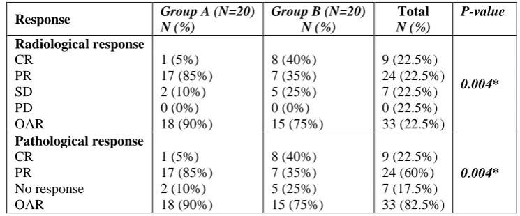
Pathological complete response
Overall response (OAR) was 90%, 75% in group A, B respectively. Of total 40 patients, 9 patients (22.5%)
achieved pCR including 8 (40% of group B) patients in group B and 1 patient (5% of group A) in group A which was statistically significant (P=0.004). Nine patients achieved pathological CR and were distributed as follows: 10%, 10.5%, 54.4% in low, moderate, high sTILs IHC groups respectively which was statistically significant (P<0.001) as shown in table (3), (4).
Table (3): Correlation between radiological, pathological response and treatment groups A, B.
Response Group A (N=20)
N (%)
Table (4): Correlation between pathological response and patients’ clinicopathological characteristics. Patients' clinicopathological characteristics CR (N=9) N (%) PR (N=24) N (%) No response (N=7) N (%)
P-value
Treatment groups Group A Group B 1 (11.1%) 8 (88.9%) 17 (70.8%) 7 (29.2%) 2 (28.6%)
5 (71.4%) 0.004*
Age <50 50
or more 5 (55.6%) 4 (44.4%) 12 (50%) 12 (50%) 3 (42.9%) 4 (57.1%) 0.881
Performance 0
1 7 (77.8%) 2 (22.2%) 20 (83.3%) 4 (16.7%) 4 (57.1%) 3 (42.9%) 0.344 Tumor Size T1 T2 T3 T4 1 (11.1%) 7 (77.8%) 0 (0%) 1 (11.1%) 0 (0%) 17 (70.8%) 5 (20.8%) 2 (8.3%) 0 (0%) 4 (57.1%) 1 (14.3%) 2 (28.6%) 0.287 Lymph Nodes N0 N1 N2 N3 4 (44.4%) 2 (22.2%) 1 (11.1%) 2 (22.2%) 9 (37.5%) 11 (45.8%) 2 (8.3%) 2 (8.3%) 1 (14.3%) 2 (28.6%) 3 (42.9%) 1 (14.3%) 0.258 Stage IIa IIb IIIa IIIb IIIc 4 (44.4%) 2 (22.2%) 1 (11.1%) 0 (0%) 2 (22.2%) 5 (20.8%) 12 (50%) 3 (12.5%) 2 (8.3%) 2 (8.3%) 0 (0%) 2 (28.6%) 1 (14.3%) 3 (42.9%) 1 (14.3%) 0.121
Grade 2
3 4 (44.4%) 5 (55.6%) 17 (70.8%) 7 (29.2%) 6 (85.7%) 1 (14.3%) 0.186
LVI negative
positive 3 (33.3%) 6 (66.7%) 10 (41.7%) 14 (58.3%) 2 (28.6%) 5 (71.4%) 0.786
Multifocal No
Yes 9 (100%) 0 (0%) 20 (83.3%) 4 (16.7%) 6 (85.7%)
1 (14.3%) 0.430
KI67 <20 20
or more 3 (33.3%) 6 (66.7%) 12 (50%) 12 (50%) 1 (14.3%) 6 (85.7%) 0.213
Surgery MRM
BCS 4 (44.4%) 5 (55.6%) 16 (66.7%) 8 (33.3%) 6 (85.7%) 1 (14.3%) 0.221
Table (5): Correlation between pathological response and sTILs IHC, CD3, CD8.
Immunophenotyping CR (N=9)
N (%)
PR (N=24)
N (%) No response (N=7) N (%) P-value
sTILs IHC
Low 1 (11.1%) 3 (12.5%) 6 (85.7%)
<0.001** Moderate 2 (22.2%) 16 (66.7%) 1 (14.3%)
High 6 (66.7%) 5 (20.8%) 0 (0%)
sTILs CD3
Low 1 (11.1%) 5 (20.8%) 6 (85.7%)
<0.001** Moderate 2 (22.2%) 14 (58.3%) 1 (14.3%)
High 6 (66.7%) 5 (20.8%) 0 (0%)
sTILs CD8
Low 1 (11.1%) 4 (16.7%) 6 (85.7%)
<0.001** Moderate 1 (11.1%) 15 (62.5%) 1 (14.3%)
High 7 (77.8%) 5 (20.8%) 0 (0%)
Treatment groups Group A Group B 1 (11.1%) 8 (88.9%) 17 (70.8%) 7 (29.2%) 2 (28.6%)
5 (71.4%) 0.004*
Five-year overall survival
The 5-year OS was 92.5% with median follow up of 62 months. Five-year OS was 90% in group A versus 95% in group B which was statistically insignificant (P=0.564). Five-year OS was 80%, 94.7%,100% in low, moderate and high sTILs by IHC respectively, but it was not statistically significant (P= 0.160). About 83.3% in low sTILs CD3, 94.1% in moderate, 100% in high groups achieved 5-year OS but it was not statistically
Figure (1): Overall survival for all patients.
Figure (2): Correlation between 5-year OS with treatment groups A, B
Figure (4): Correlation between 5-year OS and pathological response
Five-year Disease-free survival
The 5-year DFS was 55% with median follow up of 62 months. Five-year DFS was 35% in group A versus 74.7% in group B which was statistically significant (P=0.029). Five-year DFS was 50%, 45.1%, 72.7% in low, moderate and high sTILs by IHC respectively, and it was statistically insignificant (P= 0.395). About 58.3% in low sTILs CD3, 35.3% in moderate, 81.8% in high groups achieved 5-year DFS but it was not statistically significant (P= 0.118). Five-year DFS was 45.5%,
50.4%, 66.7% in low, moderate and high sTILs by CD8 respectively, and it was statistically insignificant (P= 0.383). Five-year DFS was significantly better with node negative, stage IIa, absence of multifocality and negative LVI with (P<0.001), (P=0.015), (P=0.002), (P=0.029) respectively, but no significant differences were found as regard performance, tumor size, age, grade, surgery and KI67. Five-year DFS was 77.8%, 52.8%, 28.6%, in patients who achieved pCR, PR, no response respectively with (P=0.023).
Figure (6): Correlation between 5-year DFS with treatment groups A, B.
Figure (7): Correlation between 5-year DFS and pathological response.
Toxicity
None of the toxicity differences between the 2 groups were statistically Significant as regard neutropenia, vomiting, anemia, peripheral neuropathy, cardiotoxicity or hepatotoxicity. Pregabalin was used as prophylaxis to avoid and to treat peripheral neuropathy. As regard radiation therapy side effects; none of the toxicity differences between the 2 groups were statistically Significant as regard skin changes and dermatitis, respiratory complications in the form of cough or dyspnea or cardiac adverse effects such as asymptomatic ECG or echo changes.
DISCUSSION
Neoadjuvant chemotherapy is an effective alternative to adjuvant chemotherapy, particularly in patients with locally advanced breast cancer who are not operable or not candidates for breast conservation at diagnosis.[10]
subtypes, including TNBC and HER2-positive breast cancer.[11,12]
Platinum-based chemotherapy drugs are evaluated in TNBC because they have defective DNA repair pathways and may be more sensitive to platinum-based DNA cross-linking agents such as carboplatin than other breast cancer subtypes.[13] Addition of carboplatin to anthracycline/taxane NAC has shown to improve pCR in TNBC as in GeparSixto, CALGB 40603 studies, In GeparSixto, Denkert et al,2014 showed increased pCR to 74% in LPBC vs 43% for LPBC treated without carboplatin (P=0.005).[14] It was found that NAC of TNBC with addition of carboplatin to paclitaxel was superior to doxorubicin plus paclitaxel as regard pCR, DFS and OS. This may be due to more deficiencies in BRCA associated DNA repairing mechanism in TNBC as it was reported in many studies.[15,16] Tumour infiltrating lymphocytes (TILs) IHC score is the proportion of the area infiltrated by lymphocytes within the tumor itself plus the adjacent stroma, and classified the scores as low (<10 %), intermediate (10–40 %), and high (>40 %). The number of intratumoural TILs that directly infiltrated into the tumour cell nests had generally lower concentrations. So, this study is focused on stromal TILs.[17] The presence of CD8+ cells in the tumor infiltrate prior to the onset of NAC predicted pCR in several studies.[18]
The present study enrolled 40 patients with stage II, III TNBC treated with NAC including 20 patients treated with paclitaxel and doxorubicin (Group A) and 20 patients treated with paclitaxel and carboplatin (Group B).
In the current study TNBC 25%, 47.5%, 27.5% of patients were with low, intermediate and high sTILs respectively compared to Denkert et al,2015 who reported that 24.5% of cases were LPBC.[19] Also, Herrero-Vicent et al, 2017 demonstrated that LPBC with TILs >40% represented 35.5% of cases and 64.6% for non-LPBC.[20] In the present study, no significant differences in patients’ characteristics were found between groups of sTILs IHC as regard age, performance status, tumor size, lymph node, stage, LVI, multifocality, and surgery. With positive significant relation for grade in high group that had more grade 3 tumors (P=0.010) similar to Herrero-Vicent et al, 2017 who reported that high TILs tumors were significantly associated with grade 3 (64% vs 34%, P=0.005).[20]
As regard Pathological complete response, it was achieved in 5% of patients in group A, compared to 40% of patients in group B with total 22.5% who achieved pCR. Overall response (OAR) was 90% in group A, 75% in group B (P=0.004) in harmony with Zhang et al,2016 who reported that pCR in PC arm was 38% vs 14% for EP arm with P=0.014).[16] There was a significant relation between pathological and radiological response and sTILs IHC, CD3 and CD8; 66.7%, 66.7%, 77.8%
who achieved pCR were high TILs by IHC, high CD3, high CD8 respectively (P<0.001 for each) in agreement with West et al,2011 who reported that pCR was better in high TILs group than moderate, low groups (74.2%, 29.6% and 34.6% respectively, P<0.0001).[21] Six patients (54.4%) in high TILs group, 2 (10.5%) in moderate group and 1 patient (10%) in low TILs group achieved pCR with (P<0.001) compared to Herrero-Vicent et al,2017 with 87% in LPBC that included both moderate and high sTILs, 9.4% in non-LPBC achieved pCR in anthracycline, taxane-based NAC, P=0.001.[20]
Overall survival and prognostic factors
In the present study, 92.5% achieved 5-year OS with median follow up period of 62 months. The 5-year OS was 90% in group A versus 95% in group B which was statistically insignificant (P=0.564) compared to Zhang et al,2016 who reported that 5-year OS in PC arm was 83.3% vs 70.7% for EP arm with (P=0.350).[16] There was no significant relation between 5-year OS and sTILs IHC, CD3, CD8. Of total 40 patients, 80%, 94.7%,100% in low, moderate and high sTILs by IHC respectively achieved 5-year OS but it was not statistically significant (P= 0.160). About 83.3% in low sTILs CD3, 94.1% in moderate, 100% in high groups achieved 5-year OS but it was not statistically significant (P= 0.271). Of total 40 patients, 81.8%, 94.1%,100% in low, moderate and high sTILs by CD8 respectively achieved 5-year OS but it was not statistically significant (P= 0.210) compared to West et al,2011 that reported improved survival in high CD3 group (P= 0.0056) and in high CD8 group (P=0.0390).[21]
Five-year OS was 100%, 95.8%, 68.6%, in patients who achieved pCR, PR, no response respectively with (P=0.057) which was not significant compared to Zhang et al,2016 who reported that 5-year OS was 100% for pCR patients vs 67.2% for residual disease with (P=0.004).[16]
Disease-free survival
52.8%, 28.6%, in patients who achieved pCR, PR, no response respectively with (P=0.023) compared to Tian et al,2015 who reported that 5-year DFS rate was significantly higher in patients with TNBC who achieved a pCR than those who did not achieve a pCR (OR, 7.42; 95% CI, 4.09–13.48) and Namini et al,2017 who reported that Patients with a pCR had significantly higher 3-year DFS than non-pCR (97% vs 55%, p<0.001).[22,23]
Toxicity
None of the toxicity differences between the 2 groups were statistically significant. The most common adverse events were neutropenia, vomiting and anemia which were almost the same in both groups A, B which were not statistically significant. Peripheral neuropathy was more in group B (70% vs 45%), cardiotoxicity was more in group A (45% vs 35%) but they were statistically insignificant similar to Zhang et al,2016 who reported that neutropenia and grade 3/4 neutropenia, vomiting and peripheral neuropathy was more in PC group, cardiotoxicity was more with EP group (25% versus 19.2%) but they were not statistically significant (P=0.344, P=0.567, P=1.000 respectively).[16] Both groups were similar as regard hepatotoxicity in the form of elevated liver enzymes. As regard radiation therapy side effects; radiation dermatitis was more in group A (60%) (P=0.525), but both groups had the same grade 3/4 skin toxicity (13%). Both groups were similar as regard respiratory complications. Only 15%, 5% in group A, B respectively developed cardiac adverse effects (P=0.292), with no grade 3/4 respiratory or cardiac toxicity in both groups.
CONCLUSION
● Tumor infiltrating lymphocytes represent an important predictive and prognostic biomarker in patients with breast cancer.
● Increasing numbers and expressions of CD3 and CD8 of stromal TILs were associated with improved pCR, overall survival and disease-free survival in TNBC.
● Platinum-based chemotherapy drugs are more effective in TNBC as neoadjuvant chemotherapy such as carboplatin than other breast cancer subtypes resulting in higher pCR.
RECOMMENDATION
More studies in larger set of TNBC patients my possibly help in confirming the current study results and focused how to increase the survivors in TN population.
CONFLICT OF INTEREST
The authors declare no conflict of interest.
REFERENCES
1. Tseng LM, Chiu JH, Liu CY, Tsai YF, Wang YL, Yang CW and Shyr YM. A comparison of the
molecular subtypes of triple-negative breast cancer among non-Asian and Taiwanese women. Breast Cancer Res Treat, 2017 Jun; 163(2): 241-54. 2. Bareche Y, Venet D, Ignatiadis M , Aftimos P,
Piccart M, Rothe F and Sotiriou C. Unravelling triple-negative breast cancer molecular heterogeneity using an integrative multiomic analysis. Ann Oncol., 2018 Apr 1; 29(4): 895-902. 3. Brosnan EM and Anders CK. Understanding
patterns of brain metastasis in breast cancer and designing rational therapeutic strategies. Ann Transl Med, 2018 May; 6(9): 163.
4. Joyce DP, Murphy D, Lowery AJ, Curran C, Barry K, Malone C, McLaughlin R et al. Prospective comparison of outcome after treatment for triple-negative and non-triple-triple-negative breast cancer. Surgeon, 2017 Oct; 15(5): 272-7.
5. Hahnen E, Hauke J, Engel C, Neidhardt G, Rhiem K, and Schmutzlera RK. Germline mutations in triple-negative breast cancer. Breast Care (Basel), 2017 Mar; 12(1): 15–9.
6. Galvez M, Castaneda C A, Sanchez J, Castillo M, Rebaza LP, Calderon G, De La Cruz M et al. Clinicopathological predictors of long-term benefit in breast cancer treated with neoadjuvant chemotherapy. World J Clin Oncol. Apr 10, 2018; 9(2): 33-41.
7. Asano Y, Kashiwagi S, Goto W, Takada K, Takahashi K, Hatano T, Takashima T et al. Prediction of treatment response to neoadjuvant chemotherapy in breast cancer by subtype using tumor-infiltrating lymphocytes. Anticancer Res., 2018 Apr; 38(4): 2311-21.
8. Herrero-Vicent C, Guerrero A, Gavilá J Gozalbo F, Hernández A, Sandiego S, Algarra MA et al. Predictive and prognostic impact of tumour-infiltrating lymphocytes in triple-negative breast cancer treated with neoadjuvant chemotherapy. Ecancermedicalscience., 2017 Aug 15; 11: 759. 9. Echavarria I, López-Tarruella S, Picornell A,
García-Saenz JÁ, Jerez Y, Hoadley K, Gómez HL et al. Pathological response in a triple-negative breast cancer cohort treated with neoadjuvant carboplatin and docetaxel according to Lehmann's refined classification. Clin Cancer Res., 2018 Apr 15; 24(8): 1845-52.
10. Biswas T, Efird JT, Prasad S, Jindal C and Walker PR. The survival benefit of neoadjuvant chemotherapy and pCR among patients with advanced stage triple negative breast cancer. Oncotarget. 2017 Dec 22; 8(68): 112712–9.
11. Cortazar P, Zhang L, Untch M, Mehta K, Costantino JP, Wolmark N, Bonnefoi H et al. Pathological complete response and long-term clinical benefit in breast cancer: the CTNeoBC pooled analysis. Lancet. 2014 Jul 12; 384(9938): 164-72.
residual cancer burden and breast cancer subtype. J Clin Oncol., 2017 Apr 1; 35(10): 1049-60.
13. Jin J, Zhang W, Ji W, Yang F1 and Guan X. Predictive biomarkers for triple negative breast cancer treated with platinum-based chemotherapy. Cancer Biol Ther, 2017 Jun 3; 18(6): 369-78. 14. Denkert C, Von Minckwitz G, Schneeweiss A, Loibl
S, Salat C, Rezai M, Blohmer JU et al. Neoadjuvant carboplatin in patients with triple-negative and HER2-positive early breast cancer (GeparSixto; GBG 66): a randomised phase 2 trial. Lancet Oncol, 2014 Jun; 15(7): 747-56.
15. Telli ML, Jensen KC, Vinayak S, Kurian AW, Lipson JA, Flaherty PJ, Timms K et al. Phase II study of gemcitabine, carboplatin, and iniparib as neoadjuvant therapy for triple-negative and BRCA1/2 mutation-associated breast cancer with assessment of a tumor-based measure of genomic instability: PrECOG 0105. J Clin Oncol, 2015 Jun 10; 33(17): 1895-901.
16. Zhang P, Yin Y, Mo H, Zhang B, Wang X, Li Q, Yuan P et al. Better pathologic complete response and relapse-free survival after carboplatin plus paclitaxel compared with epirubicin plus paclitaxel as neoadjuvant chemotherapy for locally advanced triple-negative breast cancer: a randomized phase 2 trial. Oncotarget, 2016 Sep 13; 7(37): 60647–56. 17. Ogiya R, Niikura N, Kumaki N, Bianchini G, Kitano
S, Iwamoto T, Hayashi N et al. Comparison of tumor‐ infiltrating lymphocytes between primary and metastatic tumors in breast cancer patients. Cancer Sci., 2016 Dec; 107(12): 1730–5.
18. Barnes TA and Amir E. HYPE or HOPE: the prognostic value of infiltrating immune cells in cancer. Br J Cancer, 2017 Aug 8; 117(4): 451–60. 19. Denkert C, von Minckwitz G, Brase JC, Sinn BV,
Gade S, Kronenwett R, Pfitzner BM et al. Tumor-infiltrating lymphocytes and response to neoadjuvant chemotherapy with or without carboplatin in human epidermal growth factor receptor 2-positiveand triple-negative primary breast cancers. J Clin Oncol, 2015 Mar 20; 33(9): 983-91. 20. Herrero-Vicent C, Guerrero A, Gavilá J Gozalbo F,
Hernández A, Sandiego S, Algarra MA et al. Predictive and prognostic impact of tumour-infiltrating lymphocytes in triple-negative breast cancer treated with neoadjuvant chemotherapy. Ecancermedicalscience, 2017 Aug 15; 11: 759. 21. West NR, Milne K, Truong PT, Macpherson N,
Nelson BH and Watson PH. Tumor-infiltrating lymphocytes predict response to anthracycline-based chemotherapy in estrogen receptor-negative breast cancer. Breast Cancer Res. 2011; 13(6): R126. 22. Tian M, Zhong Y, Zhou F, Xie C, Zhou Y and Liao
Z. Effect of neoadjuvant chemotherapy in patients with triple-negative breast cancer: A meta-analysis. Oncol Lett. 2015 Jun; 9(6): 2825–32.
23. Namini SN, Swain M, Al-Rashdan A, McCready DR, Fleming R, Miller N, Magnati M. Predictors of outcome and patterns of failure for high risk triple