Payel et al. World Journal of Pharmaceutical and Medical Research
PRODUCTION, OPTIMIZATION AND APPLICATION OF TYROSINASE FROM
BACILLUS SP
CGR6 IN DYE DEGRADATION
Pranita Pradhan and Payel Sarkar*
Department of Microbiology, University of North Bengal, Raja Rammohunpur, Siliguri-734013, West Bengal, India.
Article Received on 04/07/2017 Article Revised on 25/07/2017 Article Accepted on 15/08/2017
INTRODUCTION
Tyrosinase is also known as polyphenol oxidase, DOPA oxidase, phenolase and catechol oxidase. Tyrosinases (E.C. 1.14.18.1) are copper-containing enzymes which are nearly ubiquitously distributed in all domains of life.
This property of tyrosinase can be successfully utilised for the biological treatment of wastewater containing phenol. (Mita et al., 2007). Virtually all phenolic derivatives are toxic and are considered hazardous pollutants. As little as 0.005 mgJ/l phenol imparts an objectionable taste and odour to drinking water when combined with chlorine during routine water treatment (Lanouett et al., 1977). About 100,000 commercially available dyes are known and nearly 1 million tons of dyes are produced throughout the year, whereas, out of the total usage, 10% of dyes are released in environment as dye waste (Selvam et al., 2003) and most of the dyes are phenol derivatives. Further, their discharge into surface water leads to aesthetic problems and obstructs light penetration and oxygen transfer into water bodies, hence affecting aquatic life.
MATERIALS AND METHODS
Isolation of dye degrading bacteria
Soil sample was collected from dye effluent release region in Siliguri (West Bengal, India). 1g of soil sample was serially diluted into sterile peptone saline water in
order to reduce the initial number of microorganisms. The dilutions were then spread plated on minimal agar supplemented with 0.01% Congo red and incubated at 37⁰C for 48 hours. The colonies obtained were purified by repeatedly streaking them so as to obtain discrete isolated colonies.
Selection and biochemical characterization of dye degrading bacteria
All isolated strains were single streaked on minimal agar supplemented with 0.01% Congo red. The best strain was selected on the basis of the biggest zone of dye decolourization. Morphological and biochemical characterizations were performed in order to identify the strain.
Measurment of growth
For bacterial growth, the inoculum was prepared by inoculating a loopful of culture into nutrient broth followed by incubation at 37ºC and 180 rpm.1ml of overnight grown culture was used to inoculate 100ml of nutrient broth supplemented with 0.01% Congo red. The incubation was carried out at 37ºC with constant shaking at 180 rpm in an orbital shaker. The bacterial culture growing in absence of Congo red under similar conditions served as control. Growth was followed by recording absorbance at 660 nm at a constant interval of 6h till 96h.
wjpmr, 2017,3(8), 212-218 SJIF Impact Factor: 4.103
Research Article ISSN 2455-3301
WJPMR
WORLD JOURNAL OF PHARMACEUTICAL
AND MEDICAL RESEARCH
www.wjpmr.com
*Corresponding Author: Payel Sarkar
Department of Microbiology, University of North Bengal, Raja Rammohunpur, Siliguri-734013, West Bengal, India.
ABSTRACT
The aim of the present study is to isolate a bacterial strain from dye contaminated soil. Bacterial strain CgR6 was selected as the best dye degrading isolate. Biochemical characterization concluded that CgR6 strain belongs to the genus Bacillus sp. After optimising the conditions in minimal media it was found that tyrosinase productionwas maximum in presence of glucose (1.09 EU/ml) and sodium nitrate (0.92 EU/ml). Addition of NaCl enhanced tyrosinase production (0.59 EU/ml. Sodium azide was found to be a potent inhibitor of tyrosinase enzyme causing reduction in tyrosinase production. Maximum tyrosinase activity was seen at dye concentration of 0.01% (1.2 EU/ml). the optimum temperature for tyrosinase was 50 ºC at pH 10. The enzyme was stable at a wide range of temperature (10ºC - 80 ºC) and a pH range of 8-11. Tyrosinase produced from strain CgR6 also had the ability to decolourization a wide range of dyes such as Congo red, Methyl red, Malachite green and Crystal violet. Highest percentage of decolourization was seen in Malachite green (91.9%). Thus, the tyrosinase produced by Bacillus
CgR6 strain has immense potential in removal of dye wastes.
Payel et al. World Journal of Pharmaceutical and Medical Research
Production media and enzyme preparation
A 1% (v/v) bacterial suspension was transferred from an overnight nutrient broth culture to the basal production medium (TPM) composed of (g/ l): peptone, beef extract , sodium chloride , and Congo red at pH 7. Bacterial cells were grown at 37 °C under shaking condition at 180 rpm for 96 h. After every 24 h cells were removed by centrifugation and the supernatant was desalted and used for measurement of tyrosinase activity.
Tyrosinase assay
Activity of tyrosianse was determined spectrophotometrically at room temperature in case of control and test organism. Tyrosinase activity was assayed using L- tyrosine as substrate. 5ml of culture was centrifuged at 10000g at 4ºC for 10min. The reaction mixture consisted of 2ml treated dye supernatant, 1.5ml of 0.1M potassium phosphate buffer at pH 7 and 0.5ml L-tyrosine as substrate. The increase in absorbance due to oxidation of L-tyrosine to dihydroxyphenylalanine (DOPA) was observed spectrophotometrically at 475nm. One unit of tyrosinase activity was determined by the following formula:
1EU/ml= (OD of sample-OD of blank) X vol. of assay mixture Vol. of enzyme
Optimization of tyrosinase production by OFAT method
For optimization of tyrosinase production, the effect of various process variables such as nitrogen source, carbon source, substrate concentration, salt concentration were studied using OFAT method. Each parameter optimized was incorporated further in the subsequent experiments.
For these, the bacterial cells were grown for 96 h at 37 °C under shaking condition. The cells were removed by centrifugation at 10,000 rpm for 10 min and the supernatants were used for measurement of tyrosinase activity at each step.
Effect of nitrogen source: To determine the effect of nitrogen sources, different organic and inorganic nitrogen sources such as peptone, yeast extract, sodium nitrate, malt extract and urea at a concentration of 1% (w/v) each, were used in the production medium.
Effect of carbon source: The production media was replaced with glucose, fructose, sucrose, lactose, and maltose (1%w/v) to determine the effect of various carbon sources on tyrosinase production.
Effect of salt: Sodium chloride in the TPM media was replaced with potassium chloride, calcium chloride and ammonium chloride.
Effect of substrate concentration: Different concentration of Congo red (0.01%, 0.05%, 0.1%, 0.2%) was supplemented in the TPM media and inoculated with 1ml of 24h old culture. It was incubated at 37ºC with constant shaking at 180 rpm.
Characterization of enzyme
Determination optimum temperature: The effect of temperature on tyrosinase was determined by incubating the reaction mixture at a temperature range of 30o - 60o C under standard assay conditions.
Determination of optimum pH: The pH optimum was determined by measuring the enzyme activity at pH 4-9 under standard condition. Buffer systems (50 mM) used were 50mM acetate buffer (pH 3-5) and 50mM phosphate buffer (pH 6-8) and 50mM tris HCl buffer (pH 9).
Effect of chemicals on tyrosinase stability: In order to determine the effect of chemicals on the stability of tyrosinase, different chemicals (0.1mM) such as EDTA, SDS, Sodium azide, and benzaldehyde were used. The enzyme was pre-incubated with either of these chemicals for 1 h at 40⁰C. The tyrosinase activity in absence of chemicals served as control (100% activity).
Application of tyrosinase in decolorization of different dyes
Decolorization experiment was performed by growing the culture in 25ml of sterile optimized media with 0.01% of Congo red and incubated for 96h incubation. 10ml of sample was centrifuged at 10000 rpm for 10min. After that the supernatant was inoculated in optimized media with different dye (0.1%) such as crystal violet, Congo red, malachite green, methyl red. The supernatant along with the dyes were then incubated at 37º C with constant shaking at 180 rpm in an orbital shaker. Sample was withdrawn after 24h intervals and centrifuged at 10000 rpm at 4ºC for 10 min. Decolourization was assessed by measuring absorbance of the supernatant at wavelength maxima (λm) of respective dye. Decolorization assay was determined in the terms of percentage decolorization using UV spectrophotometer. The percentage decolourization was calculated from the following formula,
Payel et al. World Journal of Pharmaceutical and Medical Research
Isolation of dye degrading bacteria
15 bacterial colonies were seen on the minimal agar plate supplemented with 0.01% Congo red of which 6 showed a zone of hydrolysis.
Strain Gram nature Morphology Shape Elevation colour Surface Decolourization CgR1 + Cocci Circular Raised Cream Smooth - CgR2 + Rod Regular Raised White Smooth + CgR3 + Cocci Circular Raised White Rough - CgR4 + Rod irregular Raised White Rough + CgR5 + Cocci Circular Raised White Rough - CgR6 + Rod Circular Raised White Smooth +++ CgR7 + Long rod irregular Raised White Smooth - CgR8 - Rod irregular Raised Yellow Smooth - CgR9 + Rod irregular Raised White Rough + CgR10 + Small rod Circular Raised Yellow Smooth - CgR11 + Cocci Circular Raised White Smooth - CgR12 + Rod Circular Raised White Rough + CgR13 + Long rod Circular Raised White Smooth - CgR14 + Cocci Circular Raised Yellow Smooth ++ CgR15 - Cocci regular Raised Cream Smooth -
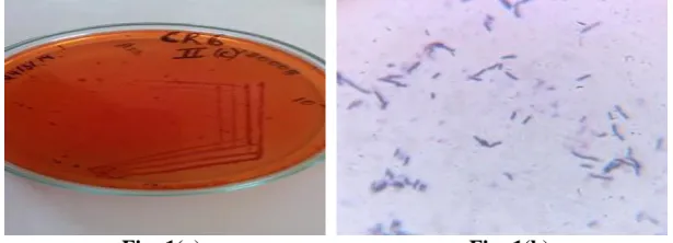
Of the 15 strains isolated, CgR6 was found to show maximum zone of hydrolysis. The strain was found to be rod shaped and Gram positive in nature. Circular white raised colonies were obtained in minimal agar plate.
Selection and characterization of best dye degrading bacteria
It was found that the selected strain CgR6 was a rod shaped, aerobic and Gram positive bacteria. The physiological and biochemical characteristics are shown in fig 1 and table 2 respectively. Altogether, the characteristics indicated that the strain belongs to genus
Bacillus.
Fig: 1(a) Fig: 1(b)
Fig 1: CgR6 growing on the minimal agar media supplemented with 0.01% Congo red (a) and the microscopic view of Bacillus sp CgR6 (b).
Payel et al. World Journal of Pharmaceutical and Medical Research
Table 2.Biochemical characterization of Bacillus sp CgR6.
Biochemical test Observation
Catalase
++
Oxidase
_
Starch hydrolysis
_
Caseinase
++
Gelatinase
_
Lipase
+
Nitrate reduction
+++
Indole
_
Methyl Red
_
VP Test
++
Citrate utilization
_
Tyrosinase Assay
The formation of dihydroxyphenylalanine (DOPA) was used to measure the tyrosinase activity. The production as estimated by the tyrosinase assay is shown in fig 3. It is evident from the figure that the highest tyrosinase activity was seen after 72 hours of inoculation (0.89 EU/ml) and decreased gradually with minimum at 96 hours (0.16 EU/ml).
Fig 3: Tyrosinase assay.
Optimization of tyrosinase production media Effect of nitrogen
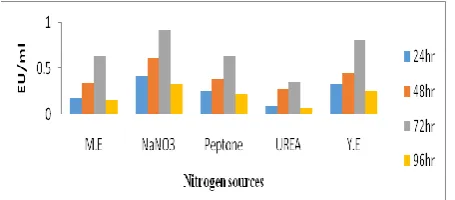
Sodium nitrate showed maximum enzyme activity (0.92 EU/ml) followed by yeast extract (0.81 EU/ml). Less tyrosinase synthesis was seen in the presence of Peptone, malt extract and urea as shown in fig 4.
Fig 4: Effect of nitrogen source on tyrosinase activity.
Effect of Carbon source
Least tyrosinase production was seen when lactose was used as the carbon source. Glucose was found to be the
best inducer for tyrosinase production as it is the simplest sugar and can be easily utilised by the microorganisms.
Fig 5: Effect of various carbon sources on tyrosinase activity.
Effect of Salt
Maximum tyrosinase activity was observed in media containing NaCl (0.59 EU/ml). Gradual fall in the enzyme activity was seen when other salts like potassium chloride, Ammonium chloride and Calcium chloride were used as illustrated by fig 6. According to the experiment NaCl was found to be the best suitable for tyrosinase production.
Fig 6: Effect of salt on tyrosinase production.
Effect of substrate concentration
The ability of strain to produce tyrosinase in different dye concentrations was tested by inoculating the culture media with 0.01%, 0.05%, 0.1% and 0.2% Congo red. Interestingly, maximum tyrosinase activity was seen at dye concentration of 0.01% (1.2 EU/ml) and gradual decrease in the enzyme synthesis was observed with almost no activity at 0.2% dye concentration.
Fig 7: Effect of dye concentration on tyrosinase activity.
Characterization of Tyrosinase
Effect of pH and temperature on activity and stability of tyrosinase
Payel et al. World Journal of Pharmaceutical and Medical Research
sharp decline in enzymatic activity was noted with in temperature above the optimum value. For determination of thermal stability, the crude enzyme was pre-incubated at 10o to 80o C for 2h and then assayed for enzymatic activity. The results in figure 8 indicates that tyrosinase was moderately thermostable as it retained 100% of activity at 60oC while the thermal stability profile at the other higher investigated temperatures showed that the enzyme is not stable above 60ºC.as shown in fig 8.
Fig 8:Determination of optimum temperature ( ) and temperature stability ( ) of tyrosinase.
Effect of pH on tyrosinase production
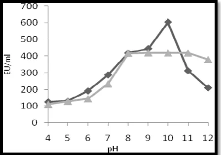
The crude enzyme was active in the pH range 4-12 with pH optimum at 10. The effect of pH on the stability of tyrosinase was tested by incubating the enzymes over a range of pH values. The enzyme was alkalostable showing stability over a pH from 8 to 11 (Fig 9).The enzyme could not retain its activity in acidic pH. shown in figure 9.
Fig 9: Determination of optimum pH ( ) and pH stability ( ) of tyrosinase.
3.6.3 Effect of inhibitors on enzyme stability
The bacterial strain CgR6 was incubated in optimised media containing inhibitors like SDS, EDTA, Benzaldehyde and Sodium azide to check their effect on enzyme stability. Sodium azide acted as a strong inhibitor of tyrosinase with minimum activity of (0.12 EU/ml) at 72 h followed by benzaldehyde (0.29 EU/ml.
Fig 10:Effect of inhibitors on tyrosinase activity.
Dye decolourization assay
To test the ability of the strain CgR6 to decolourize other dyes, it was inoculated in an optimised media containing (0.01%) methyl red, Congo red, Malachite green and Crystal violet. 5ml sample was withdrawn at an interval of about 6 hr, centrifuged at 10,000rpm for about 10mins, after which the optical density was measured at 620nm. The decolourization percentage was calculated using previously mentioned formula.
Fig 11: Decolourization of dyes by stain CgR6.
Payel et al. World Journal of Pharmaceutical and Medical Research
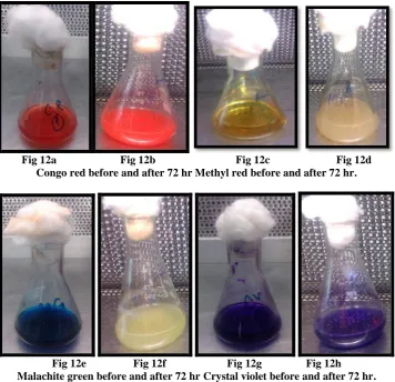
Fig 12a Fig 12b Fig 12c Fig 12d Congo red before and after 72 hr Methyl red before and after 72 hr.
Fig 12e Fig 12f Fig 12g Fig 12h Malachite green before and after 72 hr Crystal violet before and after 72 hr.
Fig 12: Decolorisation of various dyes by strain CgR6.
CONCLUSION
In this study, the isolated strain CgR6 was capable of showing tyrosinase activity. After optimising the conditions in minimal media it was found that glucose showed maximum tyrosinase production (1.09 EU/ml) as shown in (fig: 5). Among the nitrogen sources sodium nitrate enhanced tyrosinase production (0.92 EU/ml) (fig: 4). pH is a vital criteria for the growth of any microorganisms. Maximum enzyme activity was seen in neutral or alkaline pH with decrease in its activity at acidic pH (0.61 EU/ml) (fig: 9). Addition of NaCl enhanced tyrosinase production (0.59 EU/ml) (fig: 6). Maximum yield was seen at a temperature of 40⁰C (0.89 EU/ml) (fig: 9). Sodium azide was found to be a potent inhibitor of tyrosinase enzyme causing reduction in tyrosinase production. (0.12 EU/ml) (fig:10). Optimum temperature for tyrosinase was found to be 40˚C (EU/ml) (fig: 8) .At higher temperature the enzyme gets denatured. Increasing concentrations of dye may have a toxic effect on bacteria. Maximum tyrosinase activity was seen at dye concentration of 0.01% (1.2 EU/ml) (fig: 7). The strain CgR6 also had the ability to bring about the decolourization of other dyes like Methyl red, Malachite green and Crystal violet. Highest percentage of decolourization was seen in Malachite green (91.9%) as depicted in (fig: 12).
REFERENCE
1. Claus, H., Decker, H., ‘Bacterial tyrosinases’.
System Appl Microbiol, 2006; 29: 3-14.
2. Chen, K.C., Wu, J.Y., Liou, D.J., Hwang, S.J., ‘Decolorization of textile dyes by newly isolated bacterial strains.’ Journal of Biotechnology, 2003; 101: 57–68.
3. Claus, H., Filip, Z., ‘Enzymatic oxidation of some substituted phenols and aromatic amines, and the behaviour of some phenoloxidases in the presence of soil related adsorbents’. Water Sci. Technol, 1990; 22: 69–77.
4. Gasowska, B., Kafarski, P., Wojtasek, H., ’Interaction of mushroom tyrosinase with aromatic amines, o-diamines and o-aminophenols’. Biochim. Biophys. Acta, 2004; 1673: 170–177.
5. Kalyani DC, Patil PS, Jadhav JP, Govindwar SP,’ Biodegradation of reactive textile dye Red BLI by an isolated bacterium Pseudomonas sp. SUK1.’
Bioresoure Technol, 2008; 99: 4635–4641.
6. K Selvam K Swaminatham, keong sang chae, ‘Decolourization of azo dyes and a dye industry effluent by a white rot fungus Thelephora sp.’ Elseiver, 2003; 88(2): 115-119.
7. Lanouette, K.H.,’ Treatment of Phenolic wastes.’ Chem. Eng, 1977; 84: 99-106.
Payel et al. World Journal of Pharmaceutical and Medical Research
decolourization and degradation of textile dyes.’
Applied Microbiology and Biotechnology, 2001; 56:
81-87.
9. Mita, D.G., Attanasio, A., Arduini, F., Diano, N., Grano, V., Bencivenga, U., Rossi, S., Amine, A. Moscone D,’ Enzymatic determination of BPA by means of tyrosinase immobilized on different carbon carriers.’ Biosens Bioelectron, 2007; 23: 60ñ65. 10. Paraskevi, N., Polmenakou, C., and Euripides, G.S.,
‘Effect of temperature and additional carbon sources on phenol degradation by an indigenous soil Pseudomonas,’ Biodegradation, 2005; 16: 403-413. 11. R Muthezilan, N Yoganath et al, ‘Dye Degrading
Microflora from Industrial Effluents’, Research
journal of Microbiology, 2008; 3(3): ISSN
1816-4935.
12. Selinheimo,E., NiEidhin, D., Steffensen, C., Nielsen, J., Lomascolo, A., Halaouli, S., Record, E., O’Beirne, D., Buchert, J., Kruus, K., ‘Comparison of the characteristics of fungal and plant tyrosinases.’