Pharmacophore
ISSN-2229-5402
Journal home page: http://www.pharmacophorejournal.com
Corresponding Author:Azman Abdullah, Department of Pharmacology, Faculty of Medicine, Universiti Kebangsaan Malaysia Medical Centre (UKMMC), Jalan Yaacob Latif, Bandar Tun Razak, 56000 Cheras, Kuala Lumpur, Malaysia E-mail manlah1969@yahoo.com
THE EFFECT OF ADMINISTRATION OF AN EQUAL DOSE OF
DIFFERENT CLASSES OF DIETARY CHEMICALS ON NQO1
EXPRESSIONAL LEVEL IN MICE LIVER
Nadia Salem Alrawaiq
1, Ahmed Atia
1,2, Azman Abdullah
1*
1. Department of Pharmacology, Faculty of Medicine, Universiti Kebangsaan Malaysia Medical Centre (UKMMC), Jalan Yaacob Latif, Bandar Tun Razak, 56000 Cheras, Kuala Lumpur, Malaysia.
2. Department of Anesthesia and Intensive Care, Faculty of Medical Technology, Tripoli University, Tripoli, Libya.
A R T I C L E I N F O A B S T R A C T
Received: 10th Dec 2016
Received in revised form: 03th Jan 2017
Accepted: 24th Aug 2017 Available online:
04th Sep 2017
Keywords: NQO1, Gene/Protein Expression, Sulforaphane, Quercetin, Curcumin, Butylated Hydroxyanisole, Indole-3-Carbinol
Objective: NAD(P)H: quinone oxidoreductase 1 (NQO1) is important in xenobiotic and carcinogenic detoxifications. NQO1-mediated detoxification of quinones is thought to be an important strategy for cancer chemoprevention. The objective of this study was to determine the expressional levels of liver NQO1 that could be observed by administration of an equal dose (50 mg/kg) of five different dietary chemicals (sulforaphane, quercetin, curcumin, butylated hydroxyanisole, indole-3-carbinol) to mice.
Methods: Adult male ICR white mice were divided into 8 groups (n=6 per group) i.e. normal control, sulforaphane, quercetin, curcumin, butylated hydroxyanisole, indole-3-carbinol, vehicle 1 control and vehicle 2 control groups. The chemicals were administered intraperitoneally for 14 days at a dose of 50 mg/kg body weight. At day 15, mice were sacrificed and their livers harvested. Total RNA was extracted, reverse transcribed and subjected to quantitative real-time PCR to detect NQO1 gene expression. Agarose gel electrophoresis was performed to verify the specificity of amplification. Western blots were performed to detect NQO1 protein expression.
Results: There was 3.1-, 1.5-, 2.2-, 2.5- and 2.5-fold increase in mice liver NQO1 gene expression after treatment with 50 mg/kg sulforaphane, curcumin, quercetin, indole 3 carbinol and butylated hydroxyanisole respectively (P<0.05). The results also showed that NQO1 protein expression in the livers of mice treated with 50 mg/kg sulforaphane, curcumin, quercetin, indole-3-carbinol and butylated hydroxyanisole was increased by 2.3-, 1.7-, 1.8-, 1.9- and 1.9-fold respectively (P<0.05). Conclusions: At the dose of 50 mg/kg, sulforaphane exhibited the highest level of liver NQO1 expression, followed by indole-3-carbinol and butylated hydroxyanisole (equivalent expression levels), quercetin and curcumin.
Copyright © 2013 - All Rights Reserved - Pharmacophore To Cite This Article: Nadia Salem Alrawaiq, Ahmed Atia, Azman Abdullah, (2017), “The effect of administration of an equal dose of different classes of dietary chemicals on NQO1 expressional level in mice liver”, Pharmacophore, 8(5), 1-9.
Introduction
NAD(P)H: quinone oxidoreductase-1 (NQO1) is a widespread flavoprotein that acts as an antioxidant enzyme [3]. The enzyme increases the chemical reaction rate of the two-electron reduction of quinones to hydroquinones, thus averting a one-electron reduction and the related redox cycling which produces a reactive oxygen species (ROS) [4]. Some of the functions of NQO1 include xenobiotic detoxification, superoxide scavenging and preservation of endogenous antioxidant vitamins [3]. The antioxidant role of NQO1 was particularly important in situations where the disruption of the NQO1 gene or genetic polymorphism raised the threat of chemical-induced toxicity and cancers [5,6,7].
Previous studies had suggested that cancer chemoprevention was due to consistent intake of dietary phytochemicals which were able to induce phase II enzymes, including NQO1 [2]. The dietary chemicals used in this study include curcumin (CUR), indole-3-carbinol (I3C), sulforaphane (SUL), butylated hydroxyanisole (BHA) and quercetin (QRC) (Table 1). In this study, we examined the expression of NQO1 gene and protein in the livers of mice treated with equal dose (50 mg/kg body weight) of sulforaphane, curcumin, quercetin, butylated hydroxyanisole and indole-3-carbinol for 14 days using quantitative real-PCR and Western blotting. Therefore, the objective of this study is to determine which of these chemicals could induce NQO1 expression level at the highest if given at equal doses.
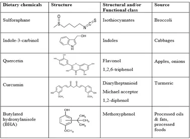
Table 1. Structures of dietary chemicals that were studied
Materials and Methods
Chemicals and reagents
Primers were purchased from Vivantis Technologies (Oceanside, CA, USA). TRIzol Reagent was purchased from Life Technologies (Carlsbad, California, USA). iScriptTM cDNA Synthesis kit and iQTM SYBR Green Supermix (2X) were
purchased from Bio-Rad (Hercules, California, USA). Sulforaphane was purchased from Santa Cruz Biotechnology (Paso Robles, California, USA). Gel Red Nucleic Acid Gel Stain (10,000X in water) was purchased from Biotium (Hayward,
California, USA). NQO1 rabbit polyclonal primary antibody and β-actin rabbit polyclonal primary antibody were purchased
Animals treatment
48 adult male ICR white mice (25–30 g) were divided into 8 groups: normal control group (n=6), sulforaphane treated group (n=6), quercetin treated group (n=6), curcumin treated group (n=6), BHA treated group (n=6), I3C treated group (n=6), vehicle 1 control group (n=6) and vehicle 2 control group (n=6). All chemicals were administered intraperitoneally at a dose of 50 mg/kg body weight for 14 days. Vehicle 1 (DMSO, Tween 20 and normal saline at a ratio of 0.05:0.1:0.85) was used to dissolve sulforaphane, quercetin and curcumin. Vehicle 2 (corn oil) was used to dissolve BHA and I3C. At day 15, the animals were sacrificed and their livers isolated. The animal study protocol was approved by the University Kebangsaan Malaysia Animal Ethics Committee (UKMAEC), and the approval code is: FP/FAR/2012/AZMAN/23-MAY/442-JUNE-2012-JUNE-2015.
RNA Extraction
Total RNA from frozen liver tissues was isolated using TRIzol reagent, according to the manufacturer’s instructions. Isopropyl alcohol (Sigma, USA) was added in each extraction step to precipitate the total RNA. Extracted total RNA pellet was then washed with 75% ethanol and dried before being dissolved in RNase free water. Total RNA was stored at -800C immediately
after extraction. Concentration and purity of the extracted RNA were determined by NanoDrop spectrophotometer 2000c (Thermo Scientific, USA) at a wavelength of 260 nm (OD260). RNA with RNA integrity number (RIN) ranging from 7 to 10 and absorbance ratio of A260 to A280 ranging from 1.5 to 2.0 was used for cDNA synthesis.
Reverse transcription
Generation of cDNA from RNA was done using iScript cDNA synthesis kit (Bio-Rad, USA) according to the manufacturer's
instructions. Briefly, a volume of total RNA (containing 1 μg) from each sample was added to a mixture of 4 µl of 5X iScript
reaction mix, 1 µl of iScript reverse transcriptase, and a volume of nuclease-free water in a total volume of 20 μl. The final reaction mix was kept at 25°C for 5 min, 42°C for 30 min, and heated to 85°C for 5 min in a thermocycler (TC-412, Techne, Barloworld Scientific, UK). The cDNA was then used as a template for amplification by PCR.
Quantification of NQO1 gene expression by quantitative real-time PCR
Quantitative real-time PCR was performed on the MiniOpticon cycler (Bio-Rad, USA). The total reaction volume used was 20μl, consisting of 1μl of 10μM forward primerand 1μl of 10μM reverse primer (500 nM final concentration of each primer),
10.0 μl of iQTM SYBRGreen Supermix (2X) (Bio-Rad, USA), 6.0 μl of nuclease-free water and 2.0 μl of cDNA. Both forward and reverse primers for the genes of interest in this study were designed according to previous studies and synthesized by Vivantis Technologies (Oceanside, CA, USA). The primer sequences for our gene of interest are shown in Table 2.The thermocycling conditions were initiated at 95°C for 30 sec, followed by 40 PCR cycles of denaturation at 95°C for 15 sec and annealing/extension at 60°C for 30 sec. At the end of each cycle, a melting curve (dissociation stage) was performed in order to determine the specificity of the primers and the purity of the final PCR product. All the measurements were performed in triplicate and no-template controls (NTC) were incorporated onto the same set of PCR tubes to test for the contamination by any assay reagents. Threshold cycles were determined for each gene and quantification of templates was performed according to the relative standard curve method. The relative gene expression (∆∆Ct) technique, as defined in the Applied Biosystems User Bulletin No. 2 [8] was used to analyse the real-time PCR data. In short, the expression level of each target gene was given as relative amount normalized against GAPDH standard controls. Subsequently, agarose gel electrophoresis was performed to determine the reliability of the melting curve analysis and to confirm the size of the PCR product. Briefly, electrophoresis was performed using 1% agarose gel in order to separate the real-time PCR products. GelRedTM nucleic acid gel stain (Biotium, USA) was used to stain the gels for 30 min and the gels were subsequently de-stained in distilled water for 30 min. Bands were
then visualized under ultraviolet light using a gel documentation system (FluorChem FC2, Alpha Innotech, USA).
Table 2. Primer sequence for GAPDH and NQO1
Gene
description Accession No. Primer sequence
Reference
GADPH NM_008084 F: 5’-GTGGAGTCTACTGGTGTCTTCA-3’
R: 5’-TTGCTGACAATCTTGAGTGAGT-3’
Kong et al., 2007 [54]
NQO1 NM_008706 F: 5’-GCATTGGCCACAATCCACCAG-3’ R: 5’-ATGGCCCACAGAGAGGCCAAA-3’
Western blotting
Frozen tissue samples (100 mg) were homogenized in 0.5 ml of RIPA buffer containing 10 μl PMSF solution, 10 μl sodium
orthovanadate solution and 10 μl protease inhibitor cocktail solution per 1 ml of 1X RIPA Lysis buffer. Briefly, 0.1 g of frozen
mouse liver was washed with phosphate buffer, and then lysed by using 0.5 ml of RIPA lysis buffer [RIPA buffer and protease inhibitor cocktail (Santa Cruz Biotechnology, USA)] to extract the protein from cytosol. After centrifugation at 13,000 × g for 30 min at 40C, supernatants were collected and soluble protein concentrations were determined by the Lowry method using
bovine serum albumin as a standard [9]. Gel electrophoresis was carried out using 10% Novex® Bis-Tris gels (Life
Technologies, USA). Briefly, 100 µg ofprotein loaded per well and subjected to electrophoresis using MOPS running buffer at 150 V for60 min. Wet transfer of proteins onto nitrocellulose membranes was made using a transfer buffer solution containing30 mM Tris- HCl, 200 mM glycine, and 10 % (v/v) methanol at 100 V for 1 h. Following transfer,membranes were blocked for 20 min at room temperature in blocking solution containing 150 mM NaCl, 3 mM KCl, 25 mM Tris, 0.1% (v/v) Tween 20, 10% non-fat dry milk powder (pH 7.4). After blocking, membranes were incubated for another 1 h at room temperature with either primary polyclonal rabbit NQO1, or primary polyclonal rabbit anti-mouse actin. Antibodies specific to NQO1 and actin were used at dilutions of 1:10.000 and 1:5000, respectively. Subsequently, membranes were incubated with peroxidase-conjugated goat anti-rabbit IgG secondary antibody [1:5000 (v/v) dilution] for another 60 min and then blots were developed using the enhanced chemiluminescence method according to the manufacturer’s instructions (GE Healthcare, Uppsala, Sweden). Bands were visualized using a gel documentation system (FluorChem FC2, Alpha Innotech, USA) and band densities were quantified using ImageJ tools software.
Statistical analysis
Data were expressed as mean ± SEM. For normally distributed data, significant differences between mean values of multiple groups were determined using one-way analysis of variance (ANOVA) with Tukey’s HSD post-hoc test. Data not normally distributed was analyzed using the Kruskal–Wallis and Mann-Whitney test. Statistical analysis was conducted using the SPSS software version 22. Differences were considered significant at p < 0.05.
Results
Liver NQO1 gene expression
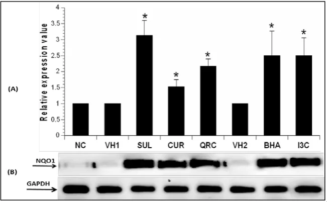
The gene expression levels of NQO1 in mice liver following administration of 50mg/kg SUL, CUR, QRC, BHA and I3C for 14 days in mice is shown in Figure 1. NQO1 gene expression in the liver was measured using quantitative real-time PCR. NQOI levels were normalized to GAPDH as housekeeping gene. Administration of 50 mg/kg SUL, CUR, QRC, BHA and I3C caused a significant increase in fold change of NQO1 expression levels (3.1-, 1.5-, 2.2-, 2.5- and 2.5-fold increase respectively) compared to controls (P<0.05).
statistically significant difference from control groups (P<0.05). (B) Agarose gel electrophoresis was also performed to determine the reliability of the melting curve analysis and to confirm the size of the PCR product.
SUL: sulforaphane, CUR: curcumin, QRC: quercetin, BHA: butylated hydroxyanisole, I3C: indole-3-carbinol, VH1: vehicle control 1, VH2: vehicle control 2, NC: normal control.
Liver NQO1 protein expression
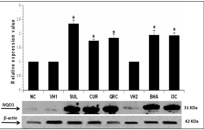
The expression levels of NQO1 protein in mice liver following administration of 50mg/kg SUL, CUR, QRC, BHA and I3C for 14 days in mice is shown in Figure 2. NQOI levels were normalized to β-actin as housekeeping protein. The band intensity obtained for NQO1 was optimized to that of β-actin and the value of fold-expression in the graph is expressed relative to controls. The results showed that SUL, CUR, QRC, BHA and I3C significantly induced NQO1 protein expression by 2.3-,
1.7-, 1.8-, 1.9- and 1.9-fold respectively, compared to controls (P< 0.05).
Figure 2. Effect of 50 mg/kg SUL, CUR, QRC, BHA and I3C on NQO1 protein expression in mice liver as assayed by Western blotting. The graph represents the average optical density of bands (mean ± SEM) from three different biological replicates of Western blot experiments, relative to controls. β-actin served as the housekeeping protein. Asterisks (*) indicate statistically significant difference from control groups (P<0.05).
SUL: sulforaphane, CUR: curcumin, QRC: quercetin, BHA: butylated hydroxyanisole, I3C: indole-3-carbinol, VH1: vehicle control 1, VH2: vehicle control 2, NC: normal control.
Discussion
In this study, we examined the effects of an equal dose (50 mg/kg) of five dietary chemicals with different chemical structures on NQO1 gene and protein expression in mice liver. Therefore, the difference in ability of several chemicals widely found in our diet to induce the expression of NQO1 could be ascertained. We discovered a significant increase in the fold change of NQO1 gene and protein expression in the dietary chemicals-treated groups, compared to control groups. We found that although all the dietary chemicals increased NQO1 expression significantly, sulforaphane induced NQO1 expression the highest.
Curcumin, a yellow pigment isolated from the rhizomes of Curcuma longa (turmeric), had received prominent attention in recent years for its varied pharmacological properties e.g. antioxidant, anti-inflammatory, antibacterial, antiviral activities, anticancer, and also as a potential treatment for Alzheimer’s disease [18-20]. It has been observed that curcumin enhances NQO1 in the livers and kidneys of mice [21,22], in murine hepatoma cells [23] and in astrocytes [24]. The α,β-unsaturated diketone moiety in the curcuminoids is a Michael reaction acceptor, which comes from the main class of phase II enzyme inducers [25]. It is highly plausible that this attribute may be responsible for the stimulation of NQO1 in the liver by curcumin.
Our study is the first in vivo study to demonstrate that quercetin (QRC) simultaneously increased the gene and protein expression of NQO1 in adult male mice liver. Our result for QRC was also in agreement with an experiment conducted to examine the impact of QRC on the expression and enzymatic activity of NQO1 in MCF-7 human breast carcinoma cells. It was discovered that when these cells were treated for 24 hours with 15 µM quercetin, the NQO1 protein levels and enzyme activity doubled, and the NQO1 mRNA expression increased by three to four fold [26]. The increase in the transcription of NQO1 as a result of the reaction to quercetin indicated that plant polyphenols could promote the transcription of phase II detoxifying systems, possibly through a mechanism that is dependent on the antioxidant response element (ARE) activation by Nrf2 [26]. It has been proposed in several studies that many phase II inducers and/or their metabolites possess electrophilic Michael reaction acceptor functionality [27,28]. Furthermore, it was discovered that the effectiveness of inducers is related to the degree of reactivity in the Michael reaction [28]. The efficacy of the inducer in the Michael reaction acceptors is significantly increased by the sole existence of orthohydroxyl substituents on the aromatic rings [29]. As many flavonoids have Michael reaction centres in their molecules, this chemical feature be might be important for their effects on phase II enzymes.
In the current study, we also examined the potential effect of indole-3-carbinol (I3C) on the induction of the NQO1 gene and protein in mice liver. As shown in Figure 1 and Figure 2, I3C was able to induce NQO1 gene and protein expression simultaneously’. Found abundantly in cruciferous vegetables, I3C has been studied as an anticancer agent for quite some time. Our data are in line with other findings. A previous in vitro study showed that I3C was able to induce detoxifying enzymes such as NQO1 in the transgenic adenocarcinoma of mouse prostate (TRAMP) cells [30]. In vivo results also showed that administration of I3C increases the expression of NQO1 in TRAMP tissues [30]. I3C had also been shown to significantly increase NQO1 expression in rat liver [31].
In our study, BHA treatment for 14 days significantly induced the gene and protein expression of NQO1 in mice livers. BHA is a synthetic phenolic antioxidant that is commonly used as a food preservative to extend the shelf life of many food products by preventing oxidative rancidity of fats. Previous studies had shown that BHA increased the NQO1 activity in the liver, kidney, lung, and the mucosa of the upper small intestine of mice, at a concentration of 7.5 g/kg for 14 days [32]. It was postulated that the metabolite of BHA, tBHQ, is able to stimulate Nrf2. Triggering of Nrf2 and the resultant up-regulation of its downstream genes in relation to tBHQ expression was assumed to be an outcome of oxidative stress (e.g. ROS formation) [33]. However, later studies concluded that Nrf2 is not activated by tBHQ itself but rather by its oxidation product, TBQ, which is the decisive inducer because of its electrophilic properties [34]. It was hypothesized that TBQ activates Nrf2 and hence up-regulates downstream protein expression in RAW264.7 cells via the covalent modification of Keap1 [35]. Quinones are able to covalently modify proteins to nucleophiles by serving as Michael acceptors [36]. Quinones could also undergo redox cycling to generate reactive oxygen species (ROS). This is clearly exemplified through BHA, whereupon it is metabolised by NADPH-dependent microsomal enzymes to produce tBHQ, which in turn goes through a redox cycle by auto-oxidation to yield TBQ, resulting in ROS production [37].
Protection from the harmful effects of carcinogenic and cytotoxic metabolites might derive from weakening of the carcinogen-activating gene signalling pathways and/or increasing the expression of the detoxification and antioxidant genes [38]. Dietary compounds containing phenol and sulphur had been found to affect the expression level of numerous detoxifying enzymes [39]. The upregulation of detoxifying enzymes, as a possible chemoprevention tactic, was first verified by applying phenolic compounds in animal samples, such as BHA and its active metabolite, tert-butylhydroquinone (tBHQ), together with 3,5-di-tertbutyl-4-hydroxytoluene (BHT), and ethoxyquin, all of which are extensively used to preserve processed foods [40, 41].
NQO1 expression is highly thought to be regulated through the activation of a cis-acting enhancer sequence known as the ARE, by the transcription factor Nrf2 [48, 49]. Nrf2 is a basic leucine zipper (bZIP) transcription factor that is well known as the master regulator of ARE-mediated gene expression. Under normal conditions, Nrf2 is present in the cytoplasm and is bound to a protein called Keap1 [50]. In the presence of oxidative stress or chemical inducers, Nrf2 is released from its binding to Keap1. Nrf2 is then able to translocate into the nucleus. Once in the nucleus, Nrf2 is able to activate and stimulate NQO1 and other ARE-dependent phase II genes expression machinery [48, 51]. It had been observed in previous studies that Nrf2-null mice showed decreased NQO1 gene expression, and that this trait was associated with increased sensitivity to benzo[a]pyrene-induced gastric neoplasia in these mice [52].
Our results showed sulforaphne induced NQO1 the highest and curcumin the lowest. It could be that sulforaphane causes the highest translocation of Nrf2 into the nucleus to activate the ARE pathway. The increase in the amount of Nrf2 translocation might be due to the ability of sulforaphane to increase the quantity of modification (e.g. chemical adduction, oxidation, nitrosylation or glutathionylation) of one or more critical cysteine residues in Keap1 that represents a likely chemico-biological trigger for the activation of Nrf2 [53]. Curcumin, quercetin, BHA and I3C could as well have affected Nrf2 translocation through the same mechanisms, but with slightly or lower potencies than sulforapane, resulting in reduced NQO1 expression. Further studies should be done to explore these mechanisms.
Apart from Keap1-dependent mechanisms, sulforaphane might also produce the highest rate of Nrf2 phosphorylation by various protein kinases (PKC, PI3K/Akt, GSK-3b, JNK), cause more interaction with other protein partners (p21, caveolin-1) and influence more epigenetic factors (micro-RNAs -144, -28 and -200a, and promoter methylation) [53], in which all these Keap1-independent mechanisms resulted in increased amounts of Nrf2 translocated into the nucleus, thus causing increased expression of downstream ARE-Nrf2 dependent genes/proteins such as NQO1. Other dietary chemicals might be less potent in inducing Nrf2 phosphorylation and other mechanisms that affect Nrf2 translocation, resulting in reduced expression of NQO1 compared to sulforaphane. These possibilities should be explored in future studies.
Conclusion
According to the findings of this study, at a dose of 50mg/kg body weight for 14 days, administration of sulforaphane has the most significant impact on the induction of the NQO1 expression in the livers of mice, followed by indole 3 carbinol, butylated hydroxyanisole, quercetin and curcumin. Also, the pattern of NQO1 gene expression correlates with the pattern of NQO1 protein expression [sulforaphane inducing liver NQO1 gene and protein expression at the highest level, followed by indole-3-carbinol and butylated hydroxyanisole (equivalent expression levels), quercetin and curcumin]. Sulforaphane can be found abundantly in cruciferous vegetables such as broccoli. The results of this study further strengthens the importance of consuming more fruits and vegetables which could potentially prove to be an affordable chemopreventive measure. The results of this study also suggests that a pharmacological dose of several well-elucidated chemicals and phytochemicals commonly found in many vegetables and fruits, especially sulforaphane, could potentially prove to be a beneficial chemopreventive tactic. Further studies should be done to conclusively support these recommendations.
Acknowledgments
This work was supported by the Ministry of Higher Education (MOHE) of Malaysia and Universiti Kebangsaan Malaysia (UKM) [grant number FRGS/1/2012/SKK03/UKM/02/2 and grant number UKM-GUP-2011-297].
References
1. Wattenberg LW (1966). Chemoprophylaxis of carcinogenesis: a review. Cancer Res 26: 1520-1526.
2. Surh YJ, Kundu JK, Na HK (2008). Nrf2 as a master redox switch in turning on the cellular signaling involved in the induction of cytoprotective genes by some chemopreventive phytochemicals. Planta Med 74: 1526-1539. 3. Siegel D, Gustafson DL, Dehn DL, Han JY, Boonchoong P, Berliner LJ et al. (2004). NAD(P)H: quinone
oxidoreductase 1: role as a superoxide scavenger. Mol Pharmacol 65: 1238-1247.
4. Dinkova-Kostova AT, Talalay P (2010). NAD (P) H: quinone acceptor oxidoreductase 1 (NQO1), a multifunctional antioxidant enzyme and exceptionally versatile cytoprotector. Arch Biochem Biophys 501: 116-23.
5. Radjendirane V, Joseph P, Lee YH, Kimura S, Klein-Szanto AJ, Gonzalez FJ et al. (1998). Disruption of the DT diaphorase (NQO1) gene in mice leads to increased menadione toxicity. J Biol Chem 273: 7382-7389.
7. Yang FY, Guan QK, Cui YH, Zhao ZQ, Rao W, Xi Z (2012). NAD(P)Hquinone oxidoreductase 1 (NQO1) genetic C609T polymorphism is associated with the risk of digestive tract cancer: a meta-analysis based on 21 case–control studies.Eur J Cancer Prev 21: 432-441.
8. Livak KJ, Schmittgen TD (2001). Analysis of relative gene expression data using real-time quantitative PCR and the 2−ΔΔCT method. Methods 25: 402-408.
9. Lowry OH, Rosebrough NJ, Farr AL, Randall RJ (1951). Protein measurement with the Folin phenol reagent. J Biol Chem 193: 265-275.
10. Talalay P (2000). Chemoprotection against cancer by induction of phase 2 enzymes. Biofactors 12: 5-11.
11. Prochaska HJ, Santamaria AB, Talalay P (1992). Rapid detection of inducers of enzymes that protect against carcinogens. Proc Natl Acad Sci USA 89: 2394-2398.
12. Zhang Y, Talalay P, Cho C-G, Posner GH (1992). A major inducer of anticarcinogenic protective enzymes from broccoli: isolation and elucidation of structure. Proc Natl Acad Sci USA 89: 2399-2403.
13. Basten GP, Bao Y, Williamson G (2002). Sulforaphane and its glutathione conjugate but not sulforaphane nitrile induce UDP-glucuronosyl transferase (UGT1A1) and glutathione transferase (GSTA1) in cultured cells. Carcinogenesis 23(8): 1399-1404.
14. Zhang Y (2000). Role of glutathione in the accumulation of anticarcinogenic isothiocyanates and their glutathione conjugates by murine hepatoma cells. Carcinogenesis 21: 1175-1182.
15. Dinkova-Kostova AT, Holtzclaw WD, Cole RN, Itoh K, Wakabayashi N, Katoh Y et al. (2002). Direct evidence that sulfhydryl groups of Keap1 are the sensors regulating induction of phase 2 enzymes that protect against carcinogens and oxidants. Proc Natl Acad Sci USA 99: 11908-11913.
16. Mcwalter GK, Higgins LG, Mclellan LI, Henderson CJ, Song L, Thornalley PJ et al. (2004). Transcription factor Nrf2 is essential for induction of NAD (P) H: quinone oxidoreductase 1, glutathione S-transferases, and glutamate cysteine ligase by broccoli seeds and isothiocyanates. J Nutr 134(12): 3499S-3506S.
17. Sharma R, Sharma A, Chaudhary P, Sahu M, Jaiswal S, Awasthi S et al. (2012). Role of 4-hydroxynonenal in chemopreventive activities of sulforaphane. Free Radic Biol Med 52: 2177-2185.
18. Aggarwal BB, Sung B (2009). Pharmacological basis for the role of curcumin in chronic diseases: an age-old spice with modern targets. Trends Pharmacol Sci 30: 85-94.
19. Goel A, Kunnumakkara AB, Aggarwal BB (2008). Curcumin as “Curecumin”: From kitchen to clinic. Biochem Pharmacol 75(4): 787-809.
20. Goel A, Aggarwal BB (2010). Curcumin, the golden spice from Indian saffron, is a chemosensitizer and radiosensitizer for tumors and chemoprotector and radioprotector for normal organs. Nutr Cancer 62: 919-930. 21. Shen G, Xu C, Hu R, Jain MR, Gopalkrishnan A, Nair S et al. (2006). Modulation of nuclear factor E2-related factor
2–mediated gene expression in mice liver and small intestine by cancer chemopreventive agent curcumin. Mol Cancer Ther 5: 39-51.
22. Iqbal M, Sharma SD, Okazaki Y, Fujisawa M, Okada S (2003). Dietary supplementation of curcumin enhances antioxidant and phase II metabolizing enzymes in ddY male mice: possible role in protection against chemical carcinogenesis and toxicity. Pharmacol Toxicol 92: 33-38.
23. Dinkova-Kostova AT, Talalay P (1999). Relation of structure of curcumin analogs to their potencies as inducers of Phase 2 detoxification enzymes. Carcinogenesis 20: 911-914.
24. Scapagnini G, Colombrita C, Amadio M, D'agata V, Arcelli E, Sapienza M et al. (2006). Curcumin activates defensive genes and protects neurons against oxidative stress. Antioxid Redox Signal 8: 395-403.
25. Jeong G-S, Oh G-S, Pae H-O, Jeong S-O, Kim Y-C, Shin M-K et al. (2006). Comparative effects of curcuminoids on endothelial heme oxygenase-1 expression: ortho-methoxy groups are essential to enhance heme oxygenase activity and protection. Exp Mol Med 38: 393-400.
26. Valerio LG Jr, Kepa JK, Pickwell GV, Quattrochi LC (2001). Induction of human NAD(P)H: quinone oxidoreductase (NQO1) gene expression by the flavonol quercetin. Toxicol lett 119: 49-57.
27. Dinkova-Kostova A (2002). Protection against cancer by plant phenylpropenoids: induction of mammalian
anticarcinogenic enzymes. Mini Rev Med Chem 2: 595-610.
28. Talalay P, De Long MJ, Prochaska HJ (1988). Identification of a common chemical signal regulating the induction of enzymes that protect against chemical carcinogenesis. Proc Natl Acad Sci USA 85: 8261-8265.
29. Dinkova-Kostova AT, Massiah MA, Bozak RE, Hicks RJ, Talalay P (2001). Potency of Michael reaction acceptors as inducers of enzymes that protect against carcinogenesis depends on their reactivity with sulfhydryl groups. Proc Natl Acad Sci USA 98: 3404-3409.
31. Krajka-Kuźniak V, Szaefer H, Bartoszek A, Baer-Dubowska W (2011). Modulation of rat hepatic and kidney phase II enzymes by cabbage juices: comparison with the effects of indole-3-carbinol and phenethyl isothiocyanate. Br J Nutr 105: 816-826.
32. Benson AM, Hunkeler MJ, Talalay P (1980). Increase of NAD(P)H: quinone reductase by dietary antioxidants: possible role in protection against carcinogenesis and toxicity. Proc Natl Acad Sci USA 77: 5216-5220.
33. Gharavi N, Haggarty S, El-Kadi S, Ayman O (2007). Chemoprotective and carcinogenic effects of tert-butylhydroquinone and its metabolites. Curr Drug Metab 8: 1-7.
34. Dinkova-Kostova AT, Wang XJ (2011). Induction of the Keap1/Nrf2/ARE pathway by oxidizable diphenols. Chem Biological Interact 192: 101-106.
35. Abiko Y, Miura T, Phuc BH, Shinkai Y, Kumagai Y (2011). Participation of covalent modification of Keap1 in the activation of Nrf2 by tert-butylbenzoquinone, an electrophilic metabolite of butylated hydroxyanisole. Toxicol Appl Pharmacol 255: 32-39.
36. Miura T, Kumagai Y (2010). Immunochemical method to detect proteins that undergo selective modification by 1,2-naphthoquinone derived from naphthalene through metabolic activation. J Toxicol Sci 35: 843-852.
37. Kahl R, Weinke S, Kappus H (1989). Production of reactive oxygen species due to metabolic activation of butylated hydroxyanisole. Toxicology 59: 179-194.
38. Cuendet M, Oteham CP, Moon RC, Pezzuto JM (2006). Quinone Reductase Induction as a Biomarker for Cancer Chemoprevention. J Nat Prod 69: 460-463.
39. Kelloff GJ, Crowell JA, Steele VE, Lubet RA, Malone WA, Boone CW et al. (2000). Progress in cancer chemoprevention: development of diet-derived chemopreventive agents. J Nutr 130: 467S-471S.
40. Wattenberg LW (1973). Inhibition of chemical carcinogen-induced pulmonary neoplasia by butylated
hydroxyanisole. J Natl Cancer Inst 50: 1541-1544.
41. Weisburger E, Evarts R, Wenk M (1977). Inhibitory effect of butylated hydroxytoluene (BHT) on intestinal carcinogenesis in rats by azoxymethane. Food Cosmet Toxicol 15:139-141.
42. Iskander K, Gaikwad A, Paquet M, Long DJ, Brayton C, Barrios R et al. (2005). Lower induction of p53 and decreased apoptosis in NQO1-null mice lead to increased sensitivity to chemical-induced skin carcinogenesis. Cancer Res 65: 2054-2058.
43. Long DJ, Waikel RL, Wang XJ, Roop DR, Jaiswal AK (2001). NAD(P)H: quinone oxidoreductase 1 deficiency and increased susceptibility to 7, 12-dimethylbenz [a]-anthracene-induced carcinogenesis in mouse skin. J Natl Cancer Inst 93: 1166-1170.
44. ] Patrick B, Gong X, Jaiswal A (2010). Disruption of NAD(P)H: quinone oxidoreductase 1 gene in mice leads to 20S proteasomal degradation of p63 resulting in thinning of epithelium and chemical-induced skin cancer. Oncogene 30: 1098-1107.
45. Begleiter A, Sivananthan K, Lefas GM, Maksymiuk AW, Bird RP (2009). Inhibition of colon carcinogenesis by post-initiation induction of NQO1 in Sprague-Dawley rats. Oncol Rep 21: 1559-1565.
46. Lu F, Zahid M, Wang C, Saeed M, Cavalieri EL, Rogan EG (2008). Resveratrol prevents estrogen-DNA adduct formation and neoplastic transformation in MCF-10F cells. Cancer Prev Res (Phila) 1(2): 135-145.
47. Montano M, Chaplin L, Deng H, Mesia-Vela S, Gaikwad N, Zahid M et al. (2006). Protective roles of quinone reductase and tamoxifen against estrogen-induced mammary tumorigenesis. Oncogene 26: 3587-3590.
48. Nioi P, Hayes JD (2004). Contribution of NAD(P)H: quinone oxidoreductase 1 to protection against carcinogenesis, and regulation of its gene by the Nrf2 basic-region leucine zipper and the arylhydrocarbon receptor basic helix-loop-helix transcription factors. Mutat Res 555: 149-171.
49. Jaiswal AK (2000). Regulation of genes encoding NAD(P)H: quinone oxidoreductases. Free Radic Biol Med 29: 254-262.
50. Itoh K, Wakabayashi N, Katoh Y, Ishii T, Igarashi K, Engel JD et al. (1999). Keap1 represses nuclear activation of antioxidant responsive elements by Nrf2 through binding to the amino-terminal Neh2 domain. Genes Dev 13: 76-86.
51. Hayes JD, McMahon M (2001). Molecular basis for the contribution of the antioxidant responsive element to cancer chemoprevention. Cancer lett 174: 103-113.
52. Ramos-Gomez M, Kwak MK, Dolan PM, Itoh K, Yamamoto M, Talalay P et al. (2001). Sensitivity to carcinogenesis is increased and chemoprotective efficacy of enzyme inducers is lost in nrf2 transcription factor-deficient mice. Proc Natl Acad Sci USA 98: 3410-3415.
53. Bryan HK, Olayanju A, Goldring CE, Park BK (2013). The Nrf2 cell defence pathway: Keap1 dependent and independent mechanisms of regulation. Biochem Pharmacol 85: 705-717.
54. Kong L, Tanito M, Huang Z, Li F, Zhou X, Zaharia A et al. (2007). Delay of photoreceptor degeneration in tubby mouse by sulforaphane. J Neurochem 101: 1041-1052.