Original Research Article
A study on clinical and epidemiological profile of patients admitted with
white patch in throat to Sir Ronald Ross Institute of Tropical and
Communicable Diseases, Hyderabad, India
Asma
1, Phani Bhushan Ivaturi
2*
INTRODUCTION
Diphtheria is a rare disease in most developed countries owing to routine children vaccination. Diphtheria is an endemic disease in India. A total of 4090 cases were reported in India in the year 2013, and about 64 deaths were reported.1
One of the most common differential diagnosis of white or grey patch in throat is diphtheria. Diphtheria is an acute infectious disease caused by the bacterium corynebacterium diphtheriae, and primarily infects the throat and upper airways. The organism produces an
exotoxin which affects myocardium and kidneys. The common clinical presentation is sore throat, low fever and swollen glands in the neck, usually giving an appearance of “bull neck”. In severe cases, the toxin attaches to myocardium causing myocarditis or peripheral neuropathy. The diphtheria toxin causes a membrane of dead tissue to build up over the throat and tonsils, which is white or grey or yellowish in color. The adherent membrane in throat makes breathing and swallowing difficult. The disease is spread through direct physical contact or from breathing in the aerosolized secretions from coughs or sneezes of infected individuals.2
ABSTRACT
Background: One of the most common differential diagnosis for a white patch in throat is diphtheria. Diphtheria is an acute infectious disease caused by toxigenic strains of corynebacterium diphtheriae. These bacilli release powerful exotoxin which is responsible for formation of white or greyish or yellowish membrane commonly over tonsils, pharynx or larynx. The objectives of the study were to determine the clinical and epidemiological factors of cases admitted with white patch in throat; to assess the microbiological confirmation rate of diphtheria among cases with white patch in throat; to identify the mortality and morbidity among study population.
Methods: A cross sectional record based retrospective study was carried out at Sir Ronald Ross Institute of Tropical & Communicable Diseases, Hyderabad. All the cases admitted in year 2016 with a white/grey/yellowish patch in throat were included in the study. The study was carried out for 2 months.
Results: A total of 230 cases were reported and 46.1% of cases were in the age group of 11-20 years. Males constituted about 48.3% and females 51.7% of study population. The most common clinical presentation was Sore throat, fever and a pseudo membrane over tonsils/pharynx. A total of 12 deaths occurred among 230 cases.
Conclusions: Deaths occurred most commonly in the younger age group and in the non-immunized. Recovery was faster and the course of illness was uncomplicated in the older age group and in completely immunized individuals.
Keywords: White patch, Diphtheria, Epidemiological factors, Exotoxin
1
Department of Community Medicine, 2Department of Otorhinolaryngology, Gandhi Medical College, Hyderabad,
Telangana, India
Received: 23 February 2018
Accepted: 21 March 2018
*Correspondence:
Dr. Phani Bhushan Ivaturi, E-mail: [email protected]
Copyright: © the author(s), publisher and licensee Medip Academy. This is an open-access article distributed under the terms of the Creative Commons Attribution Non-Commercial License, which permits unrestricted non-commercial use, distribution, and reproduction in any medium, provided the original work is properly cited.
Vaccination against diphtheria has reduced the mortality and morbidity drastically; however diphtheria is still a significant child health problem in countries with poor immunization coverage. In countries endemic for diphtheria, the disease occurs mostly as sporadic cases or in small outbreaks. Diphtheria is fatal in 5 - 10% of cases, with a higher mortality rate in young children. Treatment involves administering diphtheria antitoxin to neutralize the effects of the toxin, as well as antibiotics to kill the bacteria.2
This study was conducted to assess the clinical and epidemiological profile of clinical diphtheria such as the frequency in children and adults, seasonal variation, gender predisposition, relationship between clinical disease and immunization status, rate of complications, case fatality rate.
Objectives of study
1. To determine the clinical and epidemiological profile
of cases admitted with white patch in throat.
2. To assess the microbiological confirmation rate of diphtheria among cases with white patch in throat.
3. To identify the mortality and morbidity among study
population.
METHODS
The present study is a record based retrospective analysis of data available from January 2016 to December 2016 at Sir Ronald Ross Institute of Tropical & Communicable diseases (SRRITCD)/Govt. Fever Hospital, (tertiary care center) Hyderabad, India. Clinically suspected Diphtheria patient’s identified in Hyderabad and neighboring districts are admitted to the Fever Hospital for treatment as it is the sentinel surveillance center for treatment of all vaccine preventable diseases, including Diphtheria. Criteria for case description for probable and confirmed case was made as per World Health Organization (WHO) guidelines. All clinical cases which met the clinical criteria for diphtheria were included in the study.
Case definition as per WHO guidelines3
Clinical description
An illness characterized by laryngitis or pharyngitis or tonsillitis, and an adherent membrane of the tonsil, pharynx and/or nose.
Laboratory criteria for diagnosis
Isolation of Corynebacterium diphtheriae from a clinical specimen, or a fourfold greater rise in serum antibody (but only if both serum samples are obtained before the administration of diphtheria toxoid or antitoxin)
Case classification3
Probable: A case that meets the clinical description
Confirmed: A case that is laboratory confirmed or linked epidemiologically to a laboratory confirmed case.
The immunization status was documented as per the information given by the parents. Those who had received three primary doses at 4–6-week intervals starting at 1 month of age, followed by booster doses at 18 months and 5 years were recorded as “Immunized”. Those who had not received any dose were considered “Unimmunized”. Patients who had missed one or more of the three primary doses or booster doses were included as “Partially immunized”.
The study duration was for a period of 2 months. A pre tested and pre designed proforma was used to obtain information from medical records. The data was analyzed retrospectively with respect to demographic details, clinical features, laboratory confirmation reports, immunization status, complications and mortality using MS excel and open epi.
Treatment protocol
All patients with a clinical suspicion of diphtheria were given parenteral crystalline penicillin, L- Carnitine. Antidiphtheritic serum (ADS) will be given in a single dose as recommended depending on the site and extent of disease. Throat swab for Albert’s stain and culture will be sent in all, at the time of admission. Those patients who developed complications like airway obstruction, myocarditis were given treatment in the form of Tracheostomy and ventilator support.
RESULTS
A total of 230 cases were admitted with white patch in throat from January 2016 to December 2016 at Sir Ronald Ross Institute of Tropical and Communicable diseases (SRRITCD)/Govt. Fever Hospital, (tertiary care center) Hyderabad, India.
Table 1: Distribution of study population according to age.
Age group (in years) Number %
0-10 100 43.4
11-20 106 46.1
21-30 14 6.1
31-40 8 3.5
41-50 2 0.9
Total 230 100
around 43.4% was found in the age group of 0-10 years. Least disease burden was found in the age group of 41-50 years.
Table 2: Distribution of study population according to gender and religion.
Number %
Gender
Males 111 48.3
Females 119 51.7
Total 230 100
Religion
Hindus 101 43.9
Muslims 120 52.2
Christians 9 3.9
Total 230 100
In the present study, the disease was found to be higher among females (51.7%) as compared to males (48.3%). Highest disease burden was found among Muslims (52.2%), followed by Hindus (43.9%). Around 3.9% of cases were Christians.
Figure 1: Month wise distribution of diphtheritic cases.
In the present study, around 64.3% of cases were registered in the months of August, September and October (corresponding to rainy season). It was found that highest number of cases (23.9%) were recorded in the month of September, followed by August (22.6%) and 17.8% in October. Around 16.5% of cases were recorded in the months of November and December (corresponding to winter season). Least number of cases (0.9% each) were recorded in the months of April, May and June.
In the present study it was found that mean duration between onset of symptoms and first contact with health centre was 4-5 days.
Table 3: Distribution of study population according to clinical features at presentation.
Clinical features Number %
Fever 230 100
Throat pain/dysphagia 230 100
Membrane/white patch 230 100
Rhinorrhea 97 42.2
Bull neck 56 24.3
Stridor 10 4.3
In the present study it was found that the most common presentation in all study subjects was fever with sore throat/throat pain and white membrane in the throat. Around 42.2% of study subjects also had complaints of running nose. Bull neck was found in 24.3% of diphtheritic cases. Around 4.3% of study subjects had stridor at the time of presentation.
Figure 2: Distribution of study population according to diagnostic mode.
Figure 3: Distribution of study population according to immunization status.
In the present study using WHO case definition, clinical diagnosis of diphtheria was made in around 43.9% of study population and Laboratory confirmed diagnosis was made in 56.1% of study population. Among the laboratory confirmed cases, Albert stain was positive in
0 10 20 30 40 50 60
Number of cases
January February March April
May June July August
September October November December
43.9% 56.1%
Diagnostic mode
Clinical Cultural
3
0
.9
0
%
2
0
.4
0
%
10%
3
8
.7
0
%
Unimmunized Partially
Immunized
Completely Immunized
Unknown
Immunization status
Unimmunized Partially Immunized
16 cases constituting 6.9% of study population and culture was positive in 113 cases constituting 49.1% of total study population. Thus, the microbiological confirmation rate was 56.1% among the study population.
In this study it was found that 30.9% of study population were unimmunized and 20.4% of study population were partially immunized and 10% of study population were completely immunized. Around 38.7% of cases did not have a mention about their immunization status in their case records.
Table 4: Distribution of study population according to complications.
Complications Number %
None 190 82.6
Myocarditis 15 6.5
Respiratory stridor 16 6.9
Myocarditis and respiratory
stridor 12 5.2
Around 82.6% of study population, did not develop any complications. There were no complications among the immunized individuals. Around 6.9% of study population developed respiratory stridor and 6.5% of study population developed myocarditis. Almost 5.2% of study population developed myocarditis and respiratory stridor.
In all the 16 patients (6.9%) who developed respiratory stridor, tracheostomy was done to relieve airway obstruction. Among 15 patients who developed Myocarditis (6.5%) was treated with L carnitine and antidiphtheritic serum. The recovery rate was very less once the patient had developed complications. Among a total of 16 patients who developed complications, 12 patients succumbed to death.
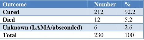
Table 5: Distribution of study population according to outcome.
Outcome Number %
Cured 212 92.2
Died 12 5.2
Unknown (LAMA/absconded) 6 2.6
Total 230 100
Table 6: Distribution of study population with complications and clinical outcome.
Complications Cured
(%)
Died
(%) Total (%)
Present 4 (25%) 12 (75) 16 (100)
Absent 208 (100) 0 (0) 208 (100)
Total 212 (94.6) 12 (5.4) 224 (100)
Using Fisher exact test of significance – p statistically highly significant <0.0001.
The cure rate in the present study was found to be 92.2% and the case fatality rate was 5.2%. Mean age of study population who had fatality was found to be 10 years. Mean duration in days between admission and discharge was found to be 7 days.
In the present study it was found that cure rate among study subjects without any complications was 100% and 25% among those who had complications like myocarditis or/and respiratory stridor. The difference was found to be statistically highly significant using Fisher exact test.
DISCUSSION
In the present study the maximum incidence of disease (46.1%) was found in the age group of 11-20 years. This findings were concurrent with a study conducted by Basavaraja et alwhere 74.1% of cases were found in >5 years of age group.4 The present study findings were different when compared to a study conducted by Singh et al,where maximum incidence (92.5%) of disease was found in 0-10 years of age group.5 In the present study only 43.4% was found in the age group of 0-10 years. The study findings also differed with a study conducted in Gujarat where maximum disease burden was found among <5 years of age.6 The shifting trend in disease occurrence can be attributed to immunization coverage.
In the present study the Mean age of study population was 12.56±6.9 years. This finding was similar to a study by Sailaja et al where median age of study subjects was 17 years.7
In the present study, the disease showed a female preponderance (51.7%) was slightly higher compared to males (48.3%). These findings were similar to a study by Maheriya et al 8 where disease burden was 63% among females and 37% among males.
The study findings in relation to religion and region distribution were similar to a study conducted by Vijay Kumar et al where Muslims (60%) and urban area residents (83%) dominated the study subjects.9
In this study maximum incidence of disease burden (64.3%) was found in rainy season (August – October) followed by winter months (November–December) – 16.5%. Similar findings were found in a study by Kumar et al.9
This findings were similar to a study conducted by Meera and Rajarao.10
The microbiological confirmation rate in present study was 56.1% and clinical diagnosis was made in 43.9% of study subjects. The present study findings were similar to a study conducted by Basavaraja et al where laboratory confirmation (Albert’s stain/ culture) was 48.3%.4
In the present study, it was found that 10% of immunized individuals developed the disease and around 30.9% of unimmunized individuals developed the disease. These findings concurred with a study by Bandichhode et al where 11.11% of immunized individuals and 58.3% of
unimmunized individuals developed the disease.11
The common complications in the present study were myocarditis and respiratory stridor. A total of 16 patients developed airway obstruction and tracheostomy was done among them to relieve the obstruction. This findings were similar to a study by Singh et al and Maheriya et al where the common complications were myocarditis and stridor due to palatal paralysis.5,8
The cure rate in the present study was found to be 92.2% and the case fatality rate was 5.2%. The case fatality rate was higher (23.67%) when compared to a present study in a study conducted by Maheriya et al and the case fatality rate was only 1.8% in a study by Kumar et al.8,9
CONCLUSION
The higher prevalence of diphtheria in age group of 11-20 years suggests the need for completing immunization with booster doses.
There was significantly higher mortality among cases who developed complications like myocarditis and respiratory stridor.
Early diagnosis and standard treatment protocol consisting of anti-diphtheritic serum (ADS), antibiotics like penicillin and L carnitine to prevent myocarditis has helped in achieving a cure rate of 92.2% in the present study.
Since, the vaccine is a toxoid and is not directed towards the organism, immunization does not prevent carrier state and hence immunization rate must be maintained at a very high level to curb outbreaks of diphtheria in community.
Limitations
As it’s a hospital based retrospective study, data related to immunization was missing in few case sheets. Subjects who left against medical advice could not be retrieved with regards to their outcomes and all the findings of this study cannot be generalized to community.
ACKNOWLEDGEMENTS
The authors are grateful to the Principal of Gandhi Medical College and Superintendent of Govt. Fever Hospital/ Sir Ronald Ross Institute of Tropical and Communicable Diseases for providing an opportunity to carry out the study. The authors are thankful to the records section in charge for her cooperation and support in carrying out this study.
Funding: No funding sources Conflict of interest: None declared
Ethical approval: The study was approved by the Institutional Ethics Committee of Gandhi Medical College
REFERENCES
1. Park K. Park’s textbook of Preventive and Social
Medicine. 24th edition. Jabalpur: Banarsi Das Bhanot; 2017: 169-171.
2. World Health Organization (WHO) - Vaccines and
diseases. Diphtheria. Available at: http://www.who. int/immunization/diseases/diphtheria/en/. Accessed on 20th February 2018.
3. World Health Organization (WHO) –recommended
surveillance standard of diphtheria: Available at: http://www.who.int/immunization/monitoring_surve illance/burden/vpd/surveillance_type/passive/diphth eria_standards/en. Accessed on 20th February 2018.
4. Basavaraja GV, Chebbi PG, Joshi S. Resurgence of
diphtheria: clinical profile and outcome - a retrospective observational study. Int J Contemp Pediatr 2016; 3:60-3.
5. Singh SN, Singh A, Chandra S. Clinical profile and
predictors of poor outcome of hospitalized diphtheria cases in children from Lucknow region of North India. Clinical Epidemiol Global Health. 2014;2(2):75-9.
6. Patel UV, Patel BH, Bhavsar BS, Dabhi HM, Doshi
SK. A retrospective study of diphtheria cases, Rajkot, Gujarat. Indian J Community Med. 2004;29(4):161-3.
7. Bitragunta S, Murhekar MV, Huntin YJ, Penumur
PP, Gupte MD. Persistence of diphtheria,
Hyderabad, India, 2003-2006. Emerg Infect Dis. 2008;14:1144–6.
8. Maheriya KM, Pathak GH, Chauhan AV, Maulik
KM, Agrawal PC. Clinical and epidemiological profile of Diphtheria in tertiary care hospital. Gujarat Med J. 2014;69(2):105-8.
9. Vijay KM, Surya Prabha ML. Epidemiological
study on admitted cases of clinical Diphtheria at Sir
Ronald Ross Institute of Tropical and
Communicable Diseases, Hyderabad, Telanagana. J Basic Clinical Res. 2015;2(2):64-67.
10. Meera M, Rajarao M. Diphtheria in Andhra
11. Bandichhode S, Jatti G, Anita M, Nandimath V. A clinical study of diphtheria cases in a pediatric population in tertiary care hospital in western Maharashtra. Indian J Child Health. 2017;3(3):251-3.