Carotid Body Tumors: Series of Case Report and
Carotid Body Tumors: Series of Case Report and
Retrospective Analysis: An Institutional Audit
Retrospective Analysis: An Institutional Audit
Rajnish Nagarkar, Shirsendu Roy, Mohammad Akheel, Vijay Palve, Prakash Pandit,
Nayana Kulkarni
Department of Oncology, Curie Manavata Cancer Centre, Nasik, Maharashtra, India
ABSTRACT
Aim: The aim was to perform a retrospective audit of rare vascular neoplasm - Carotid body tumor (CBT) in our cancer center. Materials and Methods: An institutional retrospective audit was performed in Curie Manavata Cancer Centre, Nasik, India from 30th June 2013 to 30th June 2014. Total of 5 patients were diagnosed with CBT in a period of 6 months.
Results: Out of 5 patients who were diagnosed with CBT, 2 (40%) were males and 3 (60%) were females with a ratio of 1:1.5. The types of CBT were familial in 2 patients (40%), sporadic in 2 patients (40%) and hyperplastic in 1 patient (20%). The clinical presentation of the 5 cases was, 3 patients (60%) had a painless, progressive swelling, 2 patients (40%) had pre-auricular pain, and 1 patient (20%) was identifi ed in routine follow-up. All patients underwent surgical excision of CBT with no need to pre-operative embolization. 40% of patients had cranial nerve injury and hematoma formation. 1 out of 5 patients received post-operative adjuvant radiotherapy due to large tumor size and lymphatic spread. No recurrence was found after a follow-up of 6 months. Conclusion: Surgical management of CBT remains a challenge for head and neck surgeon. Despite a reduction in stroke and mortality, there can be a signifi cant morbidity associated inadvertent adjacent cranial nerve injuries. However, our audit shows these tumors can be managed well with meticulous planning and execution of the surgery.
Key words: Carotid body tumor, paragangliomas, surgical management
INTRODUCTION
The carotid body is located at the adventia of the carotid bifurcation that controls pulmonary ventilation through afferent input by way of glossopharyngeal nerve to the medullary reticular formation. The carotid body tumor (CBT) mass usually shows a salt pepper appearance caused by vessels with single void within the tumor stroma.
CBTs also known as glomus caroticum or carotid paragangliomas belong to the family of paragangliomas and, for the most part, occur as a painless mass in the lateral aspect of the neck. CBTs are rare vascular neoplasms representing about 65% of all head and neck paragangliomas.[1] The reported incidence is
between 0.06 and 3.33 per 100,000 patients. Male and female distribution is 1:1.5 in normal altitudes and 1:8 in high altitude.[2,3] The association of CBT with
high altitude habitation and chronic hypoxemia have been documented. These tumors develop within the adventitia at the region of the carotid bifurcation. The second commonest type of cervical paraganglioma is a glomus intravaginal tumor, derived from closely adjacent paraganglionic tissue located on the vagus nerve.[3,4]
Clinically the CBT can produce a bruit of auscultation and is freely mobile in a lateral direction but fixed in cephalic-caudal direction.
Address for correspondence:
Dr. Mohammad Akheel, 4H, Block 5, VGN Laparasiene, 4th Main Road, Nolambur, Mogappair West, Chennai - 600 037, Tamil Nadu, India. Phone: +91-7058401419. E-mail: drakheelomfs@gmail.com.
Submission: 08 Dec 2014; Revision: 06 Jan 2015; Acceptance: 12 Feb 2015 Access this article online
Publisher
Website:
www.renupublishers.com
DOI:
It originates from mesodermal elements of the third arch artery and neural crest ectoderm. The carotid body is involved in chemoreceptor control of blood pressure, heart rate and respiration.
The CBT most commonly presents as a asymptomatic slow enlarging as a painless mass at anterolateral part of the neck at the level of hyoid bone with limited superior-inferior mobility near the angle of mandible during 5th-6th decade of life. The pattern
of displacement of neurovascular structure in the vicinity of the mass coupled with the clinical examination usually insufficient to confirm the diagnosis of CBT.
The carotid body is a chemoreceptor organ, reddish-brown oval structure that is located in the posteromedial aspect of the bifurcation of the carotid artery. It measures around 3-5 mm in diameter and weighs <15 mg.[5,6] The
literature states that the carotid body is located in the adventitia near the bifurcation of carotid artery. However, according to Maxwell et al.,[7] in his article he states that,
most surgeons experienced with CBT dissection maintain that it is more peripherally located, within periadventitial tissue. This distinction between the lesion and vessel wall is critical, as dissections in the deeper planes of the carotid artery are associated with higher risk for complications from vessel injury. The carotid body is an important structure in the bodies for adaptation to fluctuating concentrations of carbon dioxide, oxygen, and pH of blood. The carotid body protects the organs from hypoxic damage by releasing neurotransmitters like adrenaline, nor-adrenaline that increase the ventilatory rate when stimulated.[8,9] The structure was most likely to injure
while resecting a CBT includes the superior laryngeal nerve, vagus nerve and hypoglossal nerve.
In 1971[10] Shamblin introduce a classifi cation system of CBT based on the tumor size and involvement of carotid vessels.
Group I: Tumor is small and does not involve or encase the surrounding vessels.
Group II: Tumors are adherent or partially surround and compress the carotid vessels but are not problematic for surgical resection (may be treated with radiation) with a careful sub adventitial dissection.
Group III: Tumors have an intimate adherent relationship to the entire circumference of the carotid bifurcation, requiring partial or complete vessel resection and reconstruction.
Due to the proximity of the nerve the rate of neurological complications is higher with tumor of the higher shambling group.
There are three distinct types of CBTs; they are familial, sporadic and hyperplastic. Out of these sporadic is the commonest type of CBT with 85%, familial form with 10-15% and hyperplastic type around 5%. The hyperplastic form is very prevalent in patients with chronic hypoxia, which involves those patients who live at a high altitude.[11] This article is an institutional audit
of CBT’s treated in our tertiary care center with review of the literature.
MATERIALS AND METHODS
An institutional retrospectives audit was performed in Curie Manavata Cancer Centre, Nasik, India, which is a tertiary care cancer center from June, 30th 2013 to
June, 30th 2014. Total of 5 patients were diagnosed
with CBT in a period of 1 year.
Case 1
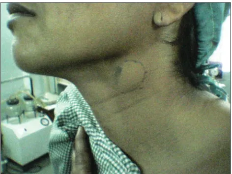
A female aged 23 years reported to our cancer center with a painless, progressive swelling on the left side of the neck for 7-8 months. Her general examination was insignificant. Local examination revealed a 3 cm × 4 cm vascular mass on the left side at carotid bifurcation region with mild pulsations [Figure 1].
Radiological examination (ultrasonography [USG] - neck) was done which revealed a 41 mm × 31 mm × 23 mm, solid, homogenous well-defined lesion at the carotid bifurcation suggestive of CBT.
Routine blood investigations were done, and the patient was taken for surgery. Transverse incision was taken on the skin crease anterior to sternocleidomastoid and sub platysmal flap raised. Sub-adventitial dissection of mass was done after careful dissecting it from the carotid bifurcation. External carotid artery was temporarily strangulated to control the bleeding [Figures 2 and 3]. Hemostasis and closure were done procedure was uneventful.
Case 2
A female aged 23 years reported to our cancer center with a painless, progressive swelling on the left side of the neck for 7-8 months. Her general examination was insignificant. Local examination revealed a 3 cm × 4 cm vascular mass on the left side at carotid bifurcation region with mild pulsations [Figure 1].
Radiological examination (USG - neck) was done which revealed a 41 mm × 31 mm × 23 mm, solid, homogenous well-defined lesion at the carotid bifurcation suggestive of CBT. Routine complete blood investigations and X-ray were done, and the patient was taken for surgery. Transverse incision was taken on the skin crease anterior to sternocleidomastoid and sub platysmal flap raised. Sub-adventitial dissection of mass was done after careful dissecting it from the carotid bifurcation. External carotid artery was temporarily strangulated to control the bleeding [Figures 2 and 3]. Hemostasis and closure were done procedure was uneventful.
RESULTS
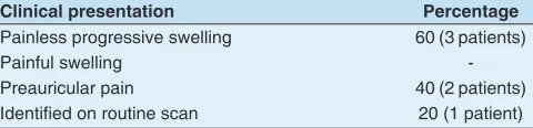
Of 5 patients who were diagnosed with CBT, 2 (40%) were males and 3 (60%) were females with a ratio of 1:1.5 [Table 1]. Table 2 reveals the types pf CBT which were familial in 2 patients (40%), sporadic in 2 patients (40%) and hyperplastic in 1 patient (20%). Table 3 reveals clinical presentation of the 5 cases.
Figure 2: Skin incision on 2nd crease on neck. Sub platysmal fl ap raised
Figure 3: Resection of the lesion. Exposure of the carotid bifurcation
Table 1: Statistics of patients
Age/sex Size
(clinical)
Primary management
Adjuvant management
23 years/Female 3 cm×4 cm Surgery Nil
30 years/Male 2 cm×3 cm Surgery Nil
28 years/Male 3 cm×2 cm Surgery Nil
40 years/Female 4 cm×5 cm Surgery Radiotherapy
36 years/Female 2 cm×2 cm Surgery Nil
Table 2: Types of CBT
Case No. Types
Case 1 Familial
Case 2 Sporadic
Case 3 Familial
Case 4 Sporadic
Case 5 Hyperplastic
CBT: Carotid body tumor
Table 3: Clinical presentation
Clinical presentation Percentage
Painless progressive swelling 60 (3 patients)
Painful swelling
-Preauricular pain 40 (2 patients)
Identifi ed on routine scan 20 (1 patient)
Table 4: Complications
Case No. Complications Recurrence
Case 1 Cranial nerve injury Nil
Case 2 Hematoma Nil
Case 3 Nil Nil
Case 4 Cranial nerve injury, hematoma Nil
3 patients (60%) had a painless, progressive swelling, 2 patients (40%) had pre-auricular pain, and 1 patient (20%) was identified in routine follow-up. All patients underwent surgical excision of CBT with no need to pre-operative embolization. Table 4 reveals complications of post-surgery. 40% of patients had cranial nerve injury and hematoma formation. 1 out of 5 patients received post-operative adjuvant radiotherapy due to large tumor size and lymphatic spread. No recurrence of carotid body tumor was observed for more than 6 months.
DISCUSSION
The anatomical description and surgical management of CBTs have existed for 100 years. The early reports described significant complications or mortalities secondary to perioperative bleeding. The earliest successful CBT surgery was performed by Scudder in 1903 in US.[12] CBT can occur in children/young adults,
but they are found more common in the middle-aged population. The average age of onset is 45 years. In most of the case, paragangliomas are inherited representing around 10-50%.[13,14] If CBT is inherited, it usually
identified in younger age group, in second to fourth decade.
CBT can occur unilateral or bilateral. About 5% CBT’s are bilateral and 95% unilateral. Usually CBT’s are benign but 5-10% of them can turn malignant.[15]
Malignant transformation is usually seen in inherited disease. In our patients, all were unilateral CBTs. Familial tumors are found to be 5.8 times more common among patients who have CBTs as compared with patients who have paragangliomas at other sites.[16] The two risk factors for the development of
CBT are chronic hypoxic stimulation and the genetic predisposition. CBT’s have a general classification of sporadic, familial and hyperplastic form. The familial type is a kind of the genetically heterogeneous entity. There are four genes encoding the subunits of succinate dehydrogenase (SDH) which is a part of Krebs cycle.[17]
Defective SDH has been postulated to cause an increase in the intracellular concentration of molecular hypoxic mediators and the vascular endothelial growth factor resulting in hyperplasia, angiogenesis, and neoplasia. Chronic hypoxic conditions in patients were residing at high altitudes or those who have respiratory problems such as pneumonia, chronic obstructive
pulmonary diseases can overburden the carotid bodies and lead to hyperplasia, hypertrophy, and neoplasia of the cells. This condition is seen in the hyperplastic type of CBTs.
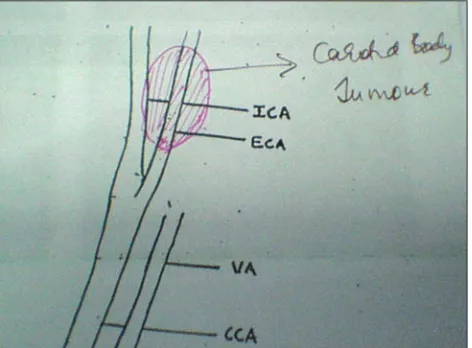
Finally, CBTs can be occasionally coupled with non-paraganglionic tumors in syndromes including MEN Type II, von Hippel–Lindau syndrome, and neurofibromatosis Type 1. CBTs clinically presents most commonly as a painless, progressive palpable mass in the anterior triangle of the neck. They are very slow-growing and can remain asymptomatic for years. The doubling time of CBTs, as estimated by Jansen et al. was 7-13 years with an average growth rate of 0.83 mm/ year.[18,19] On clinical examination, CBT is typically fixed
vertically because of its attachment to the bifurcation of the common carotid and hence it can only be moved transversely but not vertically [Figure 4]. This is called as Fontaine sign. A bruit can sometimes be felt, but however absence of a bruit does not rule out a CBT.
Approximately 10% of the cases present with transient cranial nerve palsy with paralysis of the glossopharyngeal, hypoglossal, recurrent laryngeal, or spinal accessory nerve. It can also involve sympathetic chain.[20,21] If the
tumor size is large, it can compress the carotid artery and the surrounding cranial nerves and manifest itself with pain, tongue paresis, hoarseness, winging scapula, Horner syndrome, and dysphagia. In our cases, pre-operative cranial nerve palsy was found in only one patient while post-operative in two patients.
Modern imaging and current surgical and vascular techniques have significantly improved the safety and success of this operation. Non-invasive radiological
investigative modalities utilized in the work-up of patients with suspected CBT include ultrasound, color Doppler, computerized tomography angiography and magnetic resonance angiography. Fine needle aspiration cytology (FNAC) is rarely employed because of the risk of carotid injury or hemorrhage in these highly vascular tumors, and open biopsy is clearly contraindicated due to the risk of catastrophic hemorrhage.[22] In our cases,
FNAC was not done in any patient. USG and computed tomography - neck were done in all patients as a pre-operative investigation.
Schick et al.[21] in 1980 described the use of
pre-operative embolization to decrease blood loss and subsequent transfusion rates while leading to potential reductions in tumor size by up to 25% if performed within 48 h of surgery.[23,24] If the surgery is delayed,
revascularization edema combined with a localized inflammatory response can create difficulty with the periadventitial dissection embolization is also a time consuming process associated with the inherent risks of distal migration of the embolization medium and a stroke incidence as high as 10%.[25] In our case, no
patient was subjected to embolization.
Advances in endovascular surgery suggest the possibility of carotid body resection with the feeding branches and replacement with intraluminal shunts and carotid artery reconstruction with autologous saphenous veins of an alloplastic cortex graft. However, in our cases we have dissected the tumor by sub-adventitial dissection. Post-operatively adjuvant radiotherapy is indicated where histological report demonstrates an infiltrative growth pattern.[26] In our surgical resection of CBT, one
patient received radiotherapy due to a large size and lymphatic local spread.
Embolization may facilitate surgical resection and decrease blood loss but does not reduce the rate of neurological complications.[27] Acrylic glue,
endovascular coils or ethylene vinyl alcohol (EVOH) can be utilized for pre-operative embolization. EVOH may be easier to use and may have better tumor penetration because it can be injected slowly for precise delivery into the feeding vessels 2 compared to acrylic glue, which polymerize immediately on contact with blood. Embolization can be safely performed by the treating vascular surgeon to vascularize a significant CBT with minimal risk of migration and stroke.
CONCLUSION
Surgical management of CBT remains a challenge for head and neck surgeon. Despite a reduction in stroke and mortality, there can be a significant morbidity associated inadvertent adjacent cranial nerve injuries. However, our audit shows these tumors can be managed well with meticulous planning and execution of the surgery.
REFER ENCES
1. Sajid MS, Hamilton G, Baker DM; Joint Vascular Research Group. A multicenter review of carotid body tumour management. Eur J Vasc Endovasc Surg 2007;34:127-30.
2. Georgiadis GS, Lazarides MK, Tsalkidis A, Argyropoulou P, Giatromanolaki A. Carotid body tumor in a 13-year-old child: Case report and review of the literature. J Vasc Surg 2008;47:874-880.
3. Mitchell RO, Richardson JD, Lambert GE. Characteristics, surgical management, and outcome in 17 carotid body tumors. Am Surg 1996;62:1034-7.
4. Naughton J, Morley E, Chan D, Fong Y, Bosanquet D, Lewis M. Carotid body tumours. Br J Hosp Med (Lond) 2011;72:559-64.
5. Martin H. In: Surgery of Head and Neck Tumours.New York:
Hoeber-Harper Books;1957.
6. Sevilla García MA, Llorente Pendás JL, Rodrigo Tapia JP,
García Rostán G, Suárez Fente V, Coca Pelaz A, et al. Head and
neck paragangliomas: Revision of 89 cases in 73 patients. Acta Otorrinolaringol Esp 2007;58:94-100.
7. Maxwell JG, Jones SW, Wilson E, Kotwall CA, Hall T, Hamann S,
et al. Carotid body tumor excisions: Adverse outcomes of adding carotid endarterectomy. J Am Coll Surg 2004;198:36-41.
8. Kotelis D, Rizos T, Geisbüsch P, Attigah N, Ringleb P, Hacke W, et al.
Late outcome after surgical management of carotid body tumors from a 20-year single-center experience. Langenbecks Arch Surg 2009;394:339-44.
9. Netterville JL, Reilly KM, Robertson D, Reiber ME, Armstrong WB, Childs P. Carotid body tumors: A review of 30 patients with 46 tumors. Laryngoscope 1995;105:115-26.
10. Karatas E, Sirikci A, Baglam T, Mumbuc S, Durucu C, Tutar E, et al.
Synchronous bilateral carotid body tumor and vagal paraganglioma: A case report and review of literature. Auris Nasus Larynx 2008;35:171-5.
11. Gardner P, Dalsing M, Weisberger E, Sawchuk A, Miyamoto R. Carotid body tumors, inheritance, and a high incidence of associated cervical paragangliomas. Am J Surg 1996;172:196-9.
12. Barnes L, Tse LLY, Hunt JL. Carotid body paragangliomas. In:
Pathology and Genetics of Head and Neck Tumours.Lyon:IARC;
2005. p. 364-5.
13. Baysal BE, Myers EN. Etiopathogenesis and clinical presentation of carotid body tumors. Microsc Res Tech 2002;59:256-61. 14. Isik AC, Imamoglu M, Erem C, Sari A. Paragangliomas of the head
and neck. Med Princ Pract 2007;16:209-14.
15. Ghoreishi M, Akbar-Beigi A, Tahery D, Sehhat S. Fever as the main presenting symptom of a carotid body tumor. Arch Iran Med 2008;11:214-7.
16. Lack EE. Anatomy and physiology of peripheral arterial chemoreceptors. In: Pathology of Adrenal and Extra-adrenal Paraganglia. Philadelphia: W.B Saunders; 1994. p. 1-14.
17. Jansen JC, van den Berg R, Kuiper A, van der Mey AG, Zwinderman AH, Cornelisse CJ. Estimation of growth rate
in patients with head and neck paragangliomas infl uences the
18. Day TA, Joe JK. Primary neoplasms of the neck. In: Cummings:
Otolaryngology: Head & Neck Surgery. 4th ed. St. Louis:
Elsevier-Mosby; 2005. p. 113.
19. Knight TT Jr, Gonzalez JA, Rary JM, Rush DS. Current concepts for the surgical management of carotid body tumor. Am J Surg 2006;191:104-10.
20. Arya S, Rao V, Juvekar S, Dcruz AK. Carotid body tumors: Objective criteria to predict the Shamblin group on MR imaging. AJNR Am J Neuroradiol 2008;29:1349-54.
21. Shamblin WR, ReMine WH, Sheps SG, Harrison EG Jr. Carotid body tumor (chemodectoma). Clinicopathologic analysis of ninety cases. Am J Surg 1971;122:732-9.
22. Rosa M, Sahoo S. Bilateral carotid body tumor: The role of fi
ne-needle aspiration biopsy in the preoperative diagnosis. Diagn Cytopathol 2008;36:178-80.
23. Kim H, Cho YP, Moon KM, Kwon TW. Embolic stroke after carotid artery ligation during carotid body tumor resection. Vascular 2013;21:23-6.
24. Makeieff M, Raingeard I, Alric P, Bonafe A, Guerrier B,
Marty-Ane Ch. Surgical management of carotid body tumors. Ann Surg Oncol 2008;15:2180-6.
25. Sahin MA, Jahollari A, Guler A, Doganci S, Bingol H, Karaman B,
et al. Results of combined preoperative direct percutaneous embolization and surgical excision in treatment of carotid body tumors. Vasa 2011;40:461-6.
26. Hinerman RW, Amdur RJ, Morris CG, Kirwan J, Mendenhall WM.
Defi nitive radiotherapy in the management of paragangliomas
arising in the head and neck: A 35-year experience. Head Neck 2008;30:1431-8.
27. Thakkar R, Qazi U, Kim Y, Fishman EK, Veith FJ, Malas MB. Technique and role of embolization using ethylene vinyl-alcohol copolymer before carotid body tumor resection. Clin Pract 2014;4:661.
How to cite this article: Nagarkar R, Roy S, Akheel M, Palve V, Pandit P, Kulkarni N. Carotid Body Tumors: Series of Case Report and Retrospective Analysis: An Institutional Audit. Int J Dent Med Spec 2015;2(1):24-29.