Adam Finn MA, BM, BCh, MRCP(UK)
This thesis is presented to the University of London for the degree of Doctor of Philosophy in the Faculty of Medicine
December 1993
Division of Cell & Molecular Biology Institute of Child Health
All rights reserved
INFORMATION TO ALL USERS
The quality of this reproduction is dependent upon the quality of the copy submitted.
In the unlikely event that the author did not send a complete manuscript and there are missing pages, these will be noted. Also, if material had to be removed,
a note will indicate the deletion.
uest.
ProQuest U064007
Published by ProQuest LLC(2016). Copyright of the Dissertation is held by the Author.
All rights reserved.
This work is protected against unauthorized copying under Title 17, United States Code. Microform Edition © ProQuest LLC.
ProQuest LLC
789 East Eisenhower Parkway P.O. Box 1346
bypass(CPB) to maintain systemic perfusion, a procedure which still carries significant morbidity and occasional mortality particularly in small children. CPB induces an acute inflammatory state unique in that it is iatrogenic and planned, rendering it peculiarly amenable to study. New information about leukocyte adhesion to endothelium,
associated endothelial injury, and the control of these events by inflammatory cytokines was applied to the study of these patients.
A flow cytometric assay of surface adhesion molecule expression on circulating
neutrophils was developed. The nature and timing of the inflammatory changes induced by CPB were examined in a group of paediatric patients. Consistent changes in
circulating neutrophil concentrations, Interleukin-8 (ILS) a pro-inflammatory cytokine with specific effects on neutrophil function, soluble C5b-9 a product of complement activation, and elastase a proteolytic enzyme released by neutrophils were demonstrated in plasma but changes in neutrophil adhesion molecule expression were inconsistent. Changes in the same variables in human blood in a model of CPB and the effects of soluble complement receptor 1 (sCRl), a specific, potent inhibitor of complement activation were examined. A clear pattern of changes in neutrophil adhesion molecule expression emerged in this system. Although inhibition of complement activation and ELS release was achieved, changes in neutrophil adhesion molecule expression and release of elastase were unaffected. An in vitro model of neutrophil-mediated endothelial injury, assessed immunohistochemically by staining cultured human endothelial cells, was adapted to allow flow cytometric quantitation of neutrophil adhesion and changes in expression of neutrophil adhesion molecules CDl lb/CD 18 and Leukocyte selectin. Lipopolysaccharide and the secretagogue formyl methionine leucine phenylalanine(fMLP) were used to create a spectrum of degrees of endothelial
Table of Contents
A bstract... 2
Table o f contents...4
Acknowledgements... 10
List of publications... 11
Abbreviations...12
CHAPTER 1: INTRODUCTION...14
1.1 Neutrophil-endothelial adhesion...14
1.11 The p2-integrins and the Inter Cellular Adhesion M olecules...17
1.12 The selectins...22
1.13 The role of chemoattractants...25
1.131 Interleukin 8 and the Intercrine fam ily...26
1.132 Complement activation: effects on neutrophils and endothelium... 31
1.2 Neutrophil-mediated tissue injury... 34
1.21 Neutrophil degranulation and elastase...35
1.22 The neutrophil respiratory burst...35
1.23 Endothelial injury and capillary leak... 36
1.3 Cardiopulmonary bypass... 37
1.31 The types of oxygenator used... 38
1.32 Associated clinical problems...39
1.33 Associated inflammatory changes...39
1.4 Use o f anti-adhesion molecule monoclonal antibodies in v iv o...40
1.5 Aims o f the study... 41
CHAPTER 2: MATERIALS AND METHODS... 43
2.1 Materials... 43
2.11 Reagents: Chemicals, reagents, solutions, standards and se ra... 43
2.21 Flow cytometry...45
2.22 Cytokine ELISAs - IL la , IL ip, TNFa, IL8... 49
2.23 Human umbilical cord endothelial cell culture... 50
2.24 Purification o f neutrophils from whole b lo o d ...50
2.3 Statistical methods... 51
CHAPTER 3: MEASUREMENT OF NEUTROPHH. ADHESION MOLECULE EXPRESSION IN VIVO...52
3.1 Introduction and overview...52
3.2 Indirect immunofluorescence... 54
3.21 Establishment o f saturating concentrations and incubation times o f antibodies... 56
3.22 Effects o f wash media and anti-coagulants on integrin expression... 58
3.23 Use of fMLP to modulate adhesion molecule expression...60
3.24 U se o f fixation to stabilise adhesion molecule expression prior to staining 62 3.241 Effective erythrocyte lysis and granulocyte cytometry following fixation.. 62
3.242 Effects o f paraformaldehyde fixation on P2“integrin epitope expression.... 62
3.243 Stability of CDl lb and CD18 expression with and without fixation over tim e...66
3.244 Measurement of L-selectin expression by indirect immunofluorescence ... 68
3.3 Direct immunofluorescence... 70
3.31 Establishment o f saturating concentrations o f monoclonal antibodies... 71
3.32 Use of fixation to stabilise epitope expression following staining... 71
3.321 The effect of fixation with 1%formaldehyde following staining...71
3.322 The stability of immunofluorescence with time following fixation...73
3.4 Neutrophil adhesion molecule expression in normal individuals... 73
CHAPTER 4; IN VIVO STUDIES OF PAEDIATRIC CARDIOPULMONARY
BYPASS... 79
4.1 Introduction...79
4.2 Methods...81
4.3 Controlled descriptive study of the effects of cardiopulmonary bypass on neutrophil activation pathways... 82
4.31 Methods...82
4.311 Patients and cardiopulmonary bypass techniques... 82
4.312 Sample collection... 84
4.313 Statistics...84
4.32 Results...85
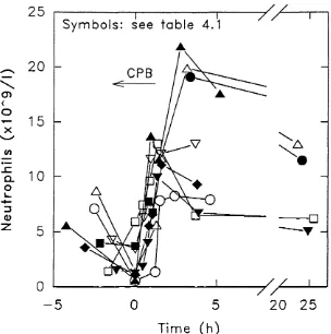
4.321 Changes in circulating neutrophil concentrations... 85
4.322 Release o f Interleukin-8...85
4.323 Absence of circulating Tumour Necrosis Factor-a and Interleukin-1...85
4.324 Release of elastase...88
4.325 Activation of complement - sC5b-9...88
4.33 Discussion... 88
4.4 Study of the relationship between chemoattractant release and neutrophil counts, degranulation and adhesion molecule expression in cardiopulmonary bypass...90
4.41 Methods... 91
4.411 Patients and cardiopulmonary bypass techniques...91
4.412 Sample collection...92
4.413 Statistics...93
4.42 Results...93
4.421 Relationship o f ILS release to neutrophil counts and elastase release...93
4.5 Study of neutrophil function during the rewarming period in
cardiopulmonary bypass...104
4.51 M etho d s...105
4.511 Patients and cardiopulmonary bypass tech n iq u es...105
4.512 Sample collection... 106
4.52 R esults...106
4.521 Emergence o f inflammatory mediators: IL8, complement & e la s ta se 106 4.522 Patterns o f change in neutrophil number and adhesion molecule expression...107
4.53 Discussion... i l l 4.6 Chapter discussion... i ll 4.7 Sum m ary...112
CHAPTER 5: EX VIVO STUDIES OF CARDIOPULMONARY BYPASS CIRCUITS... 113
5.1 Introduction... 113
5.2 M eth od s...114
5.21 Cardiopulmonary bypass circuits... 114
5.22 Samples and assays... 116
5.23 Statistics...116
5.3 R esults...117
5.31 Studies in a bubble oxygenator system and the effects o f s C R l ...117
5.311 Activation o f com plem ent...117
5.312 Changes in neutrophil c o u n ts ...117
5.313 Changes in neutrophil adhesion molecule expression...121
5.314 Release o f Interleukin-8... 128
5.322 Changes in neutrophil counts...131
5.323 Changes in neutrophil adhesion molecule expression...131
5.324 Release o f Interleukin-8...133
5.325 Release o f elastase...133
5.4 Discussion...133
5.5 Summary...138
CHAPTER 6; DEVELOPMENT OF FLOW CYTOMETRIC METHODS TO STUDY AN IN VITRO MODEL OF NEUTROPHIL-MEDIATED ENDOTHELIAL INJURY...139
6.1 Introduction...139
6.2 Methods...139
6.3 Experiments...140
6.31 Effects of neutrophil purification technique on adhesion molecule expression...140
6.32 Use of EDTA to dissociate neutrophils from endothelial cells; effects on adhesion molecule epitope expression...144
6.33 Resolution o f neutrophils and endothelial cells by flow cytometry using light scatter... 145
6.34 Development of a flow cytometric adhesion assay...151
6.35 Interpretation o f fluorescence data to quantify neutrophil adhesion molecule expression... 152
6.36 Lack of inhibition of adherence by anti-CD 1 lb and anti-L-selectin monoclonal antibodies...158
7.1 Introduction...161
7.2 Methods...162
7.21 Incubation of neutrophils with endothelial cells...162
7.22 Staining for endothelial fibronectin and heparan sulphate...164
7.23 Assessment of endothelial fibronectin and heparan sulphate morphology.. 165
7.24 Flow cytometry...165
7.25 Statistics...166
7.3 Results...166
7.31 Fibronectin and heparan sulphate morphology...166
7.32 Neutrophil adhesion...169
7.33 Neutrophil CDl lb/CD 18 expression...170
7.34 Neutrophil L-selectin expression...173
7.4 D iscu ssion...173
7.5 Summary...176
CHAPTER 8: CONCLUSIONS...177
8.1 Summary of new findings...177
8.2 Implications for future studies...179
8.3 Concluding remarks...183
APPENDIX: ASSAY METHODS DONE IN COLLABORATION... 184
A. 1 Elastase a 1-antitrypsin ELISA...184
A . 2 Complement assays...184
A.21 Terminal complement complex(sC5b-9) ELISA...184
A.22 C3a anaphylatoxin radioimmunoassay...185
Acknowledgements
I would like to thank all the members of the Division of Ceil and Molecular Biology at the Institute of Child Health and of the Cardiothoracic Unit, Great Ormond St. Hospital who have provided help and support with this work. Roland Levinsky first enabled me and encouraged me to undertake research work. Stephan Strobel provided essential supervision throughout the experimental work, analysis and writing up. Martin Elliott not only made the in vivo studies possible, but was always generous with his energy and enthusiasm. Suren Naik provided practical help in the design and execution of the early
in vivo studies. The studies with bypass circuits involved collaboration with Neil Moat, without whose help they could not have been done. He also arranged the
radioimmunoassays for C3a which were performed at the laboratories of SmithKline Beecham. The studies using the in vitro model of endothelial injury were done in collaboration with Nigel Klein, again his involvement was integral to their successful completion. He also performed the immunoassays for elastase-alantitrypsin. Cathy Rogers and Paul Morgan performed the immunoassays for sC5b-9. A number of the studies involved the collection and processing of large numbers of specimens over short periods of time, work requiring more than one pair of hands. The assistance of Naomi Rebuck was invaluable on these occasions.
I would like to thank the Child Health Research Appeal Trust, the Richard Hall Trust and the British Shoe Corporation who generously provided funds for much of this work.
I would like to thank the children and their parents who allowed me to study them and the many colleagues who also provided their blood for study.
List of publications
The following publications form part of the work presented in this thesis Copies are bound at the back of this volume.
Finn A, Naik S, Klein N, Levinsky RJ, Strobel S, Elliott M. Interleukin 8 release and neutrophil degranulation after paediatric cardiopulmonary bypass. J Thorac Cardiovasc Surg 1993;105:234-241
Finn A, Moat N, Rebuck N, Strobel S, Elliott M. Systemic inflammation during cardiopulmonary bypass: changes in neutrophil adhesive properties. Perfusion
1993;8:39-48
Finn A, Moat N, Klein N, Strobel S, Elliott M. Changes in neutrophil CDl lb/CD 18 and L-selectin expression and release of Interleukin 8 and elastase in paediatric cardiopulmonary bypass. Agents Actions 1993; 38, C44-C46.
Finn A, Rebuck N, Moat N. Neutrophil activation during cardiopulmonary bypass.(Letter) J Thorac Cardiovasc Surg 1992; 104:1746-1748
Finn A, Rebuck N, Moat N, Naik S, Klein N, Levinsky R, Strobel S, Elliott M. Cytokines and mechanisms of capillary leakage after cardiopulmonary bypass. (Letter) J Thorac Cardiovasc Surg 1993; 106: 567
Abbreviations
C..(e.g. C3a) Ca2+ CD CPB CR..(e.g.CRl) DMSO EAT ECCM EDTA E-selectin ECS FITC fMLP FN GAGs HESS Hep HS HUVEC ICAM IL LFA LED EPS E-selectin MAC Complement fragment... Calcium ion
Cluster of differentiation antigens - nomenclature system for proteins expressed on leukocyte cell surface
Cardiopulmonary bypass Complement receptor... Dimethyl sulphoxide
Elastase a 1-antitrypsin complex
Endothelial cell culture medium (see section 223) Ethylene diamine tetra acetic acid
Endothelial selectin Fetal calf serum
Fluorescein isothiocyanate
Formyl-Methionine-Eeucine-Phenylalanine Fibronectin
Glycosaminoglycans
Hanks balanced salt solution Heparin
Heparan Sulphate
Human umbilical vein endothelial cell Inter cellular adhesion molecule Interleukin
Lymphocyte function antigen Lower limit of detection
Eipopolysaccharide ( active component of endotoxin)
Mg2+ Magnesium ion
MPRM "Mono-poly resolving medium" mRNA messenger ribonucleic acid PBS Phosphate buffered saline
PE Phycoerythrin
PF Paraformaldehyde
PMN Polymorphonuclear leukocytes (neutrophils) P-selectin Platelet selectin
ROD Arginine-glycine-asparagine
sC5b-9 soluble Complement fragment C5b-9 (Terminal complement complex) sCRl recombinant soluble complement receptor 1
Chapter 1: Introduction
1.1 Neutrophil-endothelial adhesion
The earliest detailed description of the interaction of blood leukocytes with the walls of blood vessels under conditions of acute inflammation was written by Julius Cohnheim over 100 years ago (1889). Using an intra-vital microscopic preparation of the tongue of a frog, he observed "...within a multitude of capillaries....slowly and gradually the originally plasmatic zone becomes filled with innumerable colourless
corpuscles...which are driven to the periphery of the stream, and whose adhesiveness makes it difficult for them to escape from the wall once they have come into contact with it. They remain stationary at most for a time, they then advance a little, and perhaps make another short halt, and so on. Yet this does not lessen the striking contrast presented by the central column of red blood-corpuscles, flowing in an uninterrupted stream of uniform velocity But the eye of the observer hardly has time to catch all the details of the picture before it is fettered by a very unexpected occurrence. A pointed projection is seen on the external contour of the vessel wall; it pushes itself further outwards....and is transformed into a colourless rounded
hump;...this gradually withdraws itself from the vessel wall. Finally, there lies outside the vessel a ...corpuscle, of the size of a white blood cell, and having one or more nuclei Keeping pace with this exodus..of corpuscular elements there occurs an increased transudation of fluid, in consequence of which the meshes of the
mesentery...are infiltrated and swell."
This description of neutrophil margination, transendothelial migration and associated exudation of fluid remains unsurpassed.
Further advances in the understanding of the specific mechanisms underlying the
first step in the cellular immune response to inflammation and infection has been
underlined by the elucidation of the underlying defect in a group of patients with severe recurrent infections as a leukocyte adhesion deficiency (see below).
Leukocyte adhesion is now appreciated to be not only a mechanical process enabling these cells to hitch their internal cytoskeleton to the outside environment and thus move through it or stick to it, but also a process fundamental to signalling and control of other cell functions. Adhesion molecules in the cytoplasmic membrane have been shown to transmit activating signals into the cell on binding to their specific ligands. Furthermore, the converse has also been demonstrated, and signals from within the cell can modulate the adhesive function of these cell surface proteins.
Interest in this area of immunological research has been fuelled by a growing awareness that the pathophysiology of tissue injury in a wide variety of both acute and chronic inflammatory disorders is immune mediated. It has therefore been hypothesised that a fuller understanding of the processes involved may lead to new specific interventions to block or impair adhesive interactions at the crucial point and time in a disease process in order to minimise the associated injury.
N e u t r o p h i l
Circulating
1. Rolling: Selectins
2 .C h e m o ta c tlc activation
3. Transmigration:
superfamlly
E n d o t h e l i u m
Figure 1.1 Three stages in the passage of leukocytes from the circulation to the
extravascular tissues by passage through the endothelium.
which is mediated by chemotactic agents released from, or attached to the endothelial surface. This activation enables the third stage of firm or secondary adhesion and transendothelial migration to occur by promoting the function o f integrin adhesion molecules on the leukocyte surface. Important changes in the endothelial expression of ligands for the various leukocyte adhesion molecules are also critical for the evolution of these processes. The heterogeneity in timing and distribution of leukocyte traffic to the tissues are made possible by the existence of a complex variety of adhesion
molecules and mediators which bring about these processes, so that the same individual receptor may perform widely differing functions when expressed on different cell populations.
occur more slowly, often taking hours (Pober & Cotran, 1990). It is not unusual for there to be distinct adhesion molecules which bring about the same functional result (e.g. neutrophil rolling on endothelium) but which fall into these 2 different categories of response, thus inducing changes at different times and for different durations during the evolution of inflammation.
1.11 The p2~ûitegrms and the Inter Cellular Adhesion Molecules
The integrins are a family of cell surface receptors which are involved in almost every aspect of cell adhesion to other cells and to the extra-cellular matrix and associated aspects of signalling and cellular mobility (Hynes, 1987). Two areas of active research have been their role in cell migration during embryological development and leukocyte function in immunology. All members of this family are heterodimers, consisting of 2 dissimilar proteins (designated a and P chains) which are non-covalently associated and expressed on the cell surface. The a chains have approximately 1,100 amino-acid residues (130-210kDa), the P chains approximately 750 (95-130kDa) (Springer, 1990). There is considerable sequence homology between the different integrin a chains (25- 65%). Likewise, homology is observed between the different P chains (37-45%), but not between a and p. There is close similarity between integrins of different species (Pytela, 1988). Initial classifications were based on sub-families of integrins sharing the same p chain, however the picture has become increasingly complex as more proteins have been recognised and as it has become clear that a chains can associate with different p chains as well as the reverse. It is likely that integrins serve as
CD designation
Other names M.Wt Ligand(s) Expressed on:
CDlla/CD18 LFA-1, aLP2 180,95 ICAMl (=CD54), ICAM2
Lymphocytes, monocytes, granulocytes CDllb/CD18 Mac-1, Mo-1
Complement receptor 3 (CR3), aMp2
170,95 ICAMl, iC3b, fibrinogen factor X
Natural killer(NK) cells, granulocytes, monocytes,
macrophages CD11C/CD18 pl50,95.
Complement receptor 4 (CR4), aXp2
150,95 ?iC3b Granulocytes(low level) monocytes,
macrophages, some B- and T-cell tumour lines
Table 1.1 The leukocyte integrins
1986; Kishimoto et al., 1987; Fischer et al., 1988), has done much to advance understanding of the function of these proteins.
The first 2 members of this family, CDlla/CD18 and CDllb/CD18, were described initially in terms of widely differing functions before their close structural similarity became appreciated. CDlla/CD18 was shown to mediate homotypic lymphocyte adhesion (Shaw et al., 1986) through binding to a specific ligand ICAMl (for Inter Cellular Adhesion Molecule) (Rothlein et al. y 1986). CDl lb/CD 18 was first recognised
in the context of phagocytosis (Amaout et al., 1983) as a granulocyte receptor for the opsonic complement fragment iC3b (Beller et al., 1982; Wright et al., 1983) while subsequent reports have suggested that it also binds coagulation factors X and
fibrinogen (Altieri et al., 1988; Wright et al., 1988). The physiological significance of binding sites for polysaccharides such as zymosan and lipopolysaccharide on
CDl 1/CD 18 proteins, demonstrated by monoclonal antibody blocking studies (Ross et al. y 1985; Wright & Jong, 1986), remains unclear.
Neutrophil
CDTla/CD 18 C D n b /C D 1 8
M atrix
ICAM2 IC A M l
Endothelial cell
Figure 1.2 Neutrophil CDlla/CD18 adheres to ICAMl and ICAM2 on endothelial
cells. C D llb/C D lS also adheres to ICAMl and possibly also elements o f the
extracellular matrix.
expressed on the endothelial cell, but that ICAMl was only expressed significantly after stimulation of the latter with pro-inflammatory agents such as the lipopolysaccharide of bacterial endotoxin (LPS), interleukin (IL)1 and tumour necrosis factor (TNF) (Osborn,
1990; Dustin & Springer, 1988). A second ligand, structurally related to ICAMl, which bound CDlla/CD18 but which was constituitively expressed by unstimulated endothelial cells, was designated ICAM2 (Staunton et a/., 1989) (figure 1.2).
In studies which focused on the adhesion of neutrophils to endothelium, both
CDlla/CD18 and CDl lb/CD 18 were found to be involved to an approximately equal extent (Lo et ah, 1989; Tonnesen et a l , 1989). CDllb/CD18 was shown also to bind ICAMl (Smith et al, , 1989) (figure 1.2) although it may also adhere to proteins of the endothelial extracellular matrix. This is supported by the observation of binding to fibrinogen (Wright et al. , 1988) and that binding of iC3b occurs through an Arginine- Glycine-Asparagine(RGD) sequence (Wright et al. , 1987), one shared by many extracellular matrix proteins (Ruoslahti & Pierschbacher, 1986). An important difference between the nature of the surface expression of these 2 integrins by neutrophils is that, while CDU a/CD 18 expression varies little between cells or in response to activating stimuli, CDl lb/CD 18 expression increases rapidly in response to a wide variety of chemotactic stimuli including the anaphylatoxin C5a, the tri-peptide formyl-Methionine-Leucine-Phenylalanine (fMLP) and the cytokine EL8 (Berger et al. , 1984; Tonnesen et al. , 1989; Detmers et al. , 1990). This is because the specific or secondary granules of the neutrophil contain CDl lb/CD 18, and these can rapidly be mobilised to and fuse with the cytoplasmic membrane releasing the preformed protein onto the cell surface (Bainton et al. , 1987).
Altieri & Edgington, 1988). Such changes may be brought about by protein kinase C mediated phosphoryllation of the common CD 18 subunit (ChatUa et al., 1989). Thus change in expression of these adhesion molecules is probably, in itself, not the sole, or even the main controlling mechanism for integrin-mediated adhesion (Vedder & Harlan,
1988). The explanation for the mechanism for changing levels of CDl lb/CD 18 expression may lie in the crawling nature of diapedesis of the adherent neutrophil. Secondary granules may discharge their contents selectively at the leading edge of the advancing cell (exposed to the highest concentration of chemotaxin in a gradient)
(Gallin et al., 1978). CDl lb/CD 18 molecules could then roll under the advancing cell, adherent to the underlying matrix or cell surface until they reach the trailing edge where they could either be recycled into the cell or shed (Hughes et a l., 1992) (figure 1.3). The importance of CDl lb/CD 18 expression on the cell surface may therefore be, not so much in its absolute level, as in its local distribution (Anderson et al., 1986).
Advancing neutrophil
CDllb/CD18 lost or recycled a t trolling e d g e CDllb/CDlS
~ CD 1 lb/CD 18 fixed to urtderlying surface — ►
Figure 1,3 Possible role o f localised upregulation o f expression o f CDllb/CD18 at the
leading edge o f the migrating/crawling neutrophil.
With development of techniques to investigate leukocyte adherence to endothelium under conditions of flow, such as exist in vivo, it has become clear that the leukocyte integrins cannot initiate adherence at the shear stresses that normally exist in the
its granular contents (Richter et al., 1990) and to produce reactive oxygen intermediates ( the "respiratory burst") (Shappell et al., 1990). These 2 potent anti-microbial
mechanisms may also induce host tissue injury (see section 1.2 below) if they occur inappropriately or as an unavoidable side-effect of effective combat of infection (figure
1.4).
ADHEREMT NEUTROPHIL
pj Integrin respiratory
I burst degranulaflon CD lib/CD 18 ICAMl oxygen
intermediates e.g. elastase
INJURY
Figure 1.4 Adherence via CDllb/CD18 appears to have important neutrophil priming
effects fo r the release o f both reactive oxygen intermediates and degranulation.
1.12 T he selectins
Recently the 3 members of a further family of adhesion molecules, the selectins, have been shown to be vital in the initial stages of leukocyte adhesion. Their nomenclature has recently been simplified (see table 1.2). These 3 molecules were all characterised at around the same time (Tedder et al., 1989; Bevilacqua et al., 1989; Johnston et al. , 1989) and were originally named after the distinct character of their protein structure (Springer, 1990): LEC-CAMs (CAM - cell adhesion molecule)
• L - Lectin - N-terminal 117-120 amino acid sugar-binding domain
• B - Epidermal growth factor-like 34-40 amino acid domain
Name
Other names M.Wt. Expressed on Ligand
L-selectin LEC-CAM, LAMl, LEG AMI, human equivalent of mouse Mel 14 homing receptor
74** 90-100*
Leukocytes **lymphocytes *neutrophils
?Fucosylated CHO moiety
E-selectin ELAM-1 115 Endothelial cells sLex ?? on
L-selectin
P-selectin PADGEM,
GMP-140
140 Platelets (a granules). Endothelial cells (Weibel-Palade bodies)
sLex
Table 1,2 The selectins
P-selectin was first described as a protein rapidly mobilised to the surface on
stimulation from the a-granules of platelets (Stenberg et al., 1985). It is also stored in the Weibel-Palade bodies which are the secretory apparatus of endothelial cells (Hattori
et a l , 1989a). Stimulation of endothelial cells with agonists such as histamine (Hattori
et al., 1989a) and the terminal complement complex (Hattori et al., 19896) rapidly induces a transient expression of surface P-selectin (lasting 5-10 min) and P-selectin- dependant neutrophil adhesion (Geng et al., 1990). P-selectin can mediate neutrophil rolling under conditions of flow and provide the conditions for chemoattractant-induced upregulation of integrin adhesive function (Lawrence & Springer, 1991).
E-selectin, like ICAMl, is expressed on endothelial cells in vitro following stimulation with IL l, TNF and endotoxin (Bevilacqua et al., 1987). Expression peaks after around 4 hours and subsequently declines towards baseline levels (Bevilacqua et al., 1989) unlike ICAMl which continues to rise. It has been shown to mediate a pathway of neutrophil adhesion to endothelial cells in vitro distinct from ICAMl (Luscinskas et al.,
1989; Dobrina et a l., 1989).
Sialylated derivatives of the Lewis x oligosaccharide (sLex) have been shown to be ligands on neutrophils for both E-selectin (Walz et al., 1990; Phillips et al., 1990; Goelz et al., 1990 and others) and P-selectin (Policy et al., 1991). Recently it has been suggested that E-selectin and L-selectin are involved in the same adhesive interaction - perhaps adhering one to the other (Kishimoto et al., 1991). However sLex is widely distributed over the neutrophil cytoplasmic membrane, only a small proportion being associated with L-selectin. Recent evidence suggesting that neutrophil (and not lymphocyte) L-selectin may present sLex to both E- and P-selectins, being
preferentially localised to microvillous processes on the cell surface (Picker et al.,
The chemotactic stimuli which induce rapid increase in expression of CD 1 lb/CD 18 (see above) also induce equally rapid loss of L-selectin expression from neutrophils
(Kishimoto et al., 1989). This process appears to be an enzyme-mediated cleavage of the extracellular part of the protein but its control is poorly understood (Spertini et al. , 1991). It is possible that these reciprocal changes in adhesion molecule expression may reflect a transition from the rolling to the fully adherent state of the neutrophil.
However, it is not clear why L-selectin loss might be necessary to achieve this. It is also possible that loss of L-selectin from the surface of circulating neutrophils could prevent them from marginating and thus becoming adherent (Hasslen et al., 1991).
1.13 The role of chemoattractants
A large and chemically disparate group of substances have potent effects on neutrophil adhesive function and motility and have been alluded to above as chemoattractants. These include lipids such as leukotriene B4 and platelet activating factor, formyl- peptides such as fMLP which are thought to mimic bacterial products, and proteins such as the complement fragment C5a and some members of a large and recently
described cytokine family called the intercrines or chemokines which include interleukin 8(IL8). The ability to migrate up soluble gradients of these substances has been
observed for some time. More recent observations of their modulating affects on adhesive function, and in particular their effects on integrins, suggests that their physiological effects may become manifest at the second activation phase between selectin-induced rolling and integrin-induced firm adherence and transendothelial migration. Thus soluble chemotactic, or fixed haptotactic (Colditz & Movat, 1984) (figure 1.5) gradients of chemoattractants may co-ordinate the function of the adhesion molecules discussed above and thus regulate the passage of leukocytes out of the vascular compartment.
the later IL l- and TNF- induced phase of adherence which involves endothelial ICAM l
and E-selectin. The relative contribution of products of complement activation, and in
particular C5a, to these 2 phases is less clear. In this section, the cytokine IL8 and
complement activation products, the chemoattractants examined in the studies presented
in this thesis, are reviewed further.
Neutrophfl
Concentration of LB
Figure 1.5 Neutrophil migration, using integrin adhesion molecules, may be co
ordinated by fixed (i.e. surface bound or "haptotactic") gradients o f chemotactic agents
such as IL8. They may be bound to proteoglycans or specific proteins.
1.131 Interleukin 8 and the Intercrine family
The recently named intercrine (or chemokine) family of cytokines (Oppenheim et al. ,
1991) are a group of basic, heparin-binding polypeptides with 20-50% amino-acid
sequence homology and molecular weights in the range 8-lOkd. Their genes share many
common characteristics including rapidly inducible mRNA expression. The family can
be divided into a and |3 subfamilies based on the chromosomal location of their genes
and the sequence pattern of the 4 disulphide bridge-forming cysteine residues common
to all members (see table 1.3). In the a subfamily the first 2 of these bridge-forming
cysteines are separated by one aminoacid whereas in the P subfamily they are adjacent;
they are therefore also designated "C-X-C" and "C-C" respectively.
The first intercrines to be identified and characterised were platelet factor 4 (PF4)
(Walz et al., 1977; Deuel et al., 1977) and (3-thromboglobulin (pTG) (Begg et al. ,
1978) which are released from the «-granules of stimulated platelets. IL8 was first
& 6), a neutrophil chemoattractant in psoriatic scales (Schroder & Christophers, 1986; Schroder et al., 1987), and a factor activating neutrophils to release enzymes (Walz et al., 1987). It was subsequently cloned (Matsushima et al., 1988; Lindley et al., 1988). The name IL8 has now superseded the various function-defined names that have been used for this cytokine.
Intercrine a subfamily Intercrine p subfamily
Chromosome 4(ql2-21) 17(q 11-32)
Cysteines 1 & 2 configuration
C-X-C (where X=aminoacid) C-C
Members include:
IL8, Platelet factor 4,
PBP/pThromboglobulin/CTAP/NAP2 yIP-10, GRO/melanoma growth stimulating activity, ENA-78
JE/MCP-1, M IP -la, M IP-ip, RANTES, 1-309 HC14
Table 1,3 The human intercrine (or chemokine) family o f cytokines
IL8 is formed as a 99 aminoacid precursor intra-cellularly, while N-terminal truncated active forms of 79, 77, 72 and 69 residues have been isolated (Yoshimura et al., 1989; Schroder et al., 1990; Gimbrone et al., 1989) in supernatants from activated
monocytes. The 77-residue form is readily cleaved to the 72-residue form by thrombin (Hebert et al., 1990). The protein has a globular three-dimensional structure and forms dimers at high concentrations (Clore et al., 1990).
The protein is not glycosylated and is a stable molecule which is resistant to proteolytic digestion and withstands extremes of pH and temperature.
Among cytokines only TNF and ILl have been shown to cause ILS production by inducing ILS mRNA expression (Matsushima et al., 19SS; Thornton et al., 1990; Larsen et a l., 19S96; Sica et al., 1990). This increase in transcription is rapid, being seen within 1 hour of stimulation and reaching a peak at around 3 hours before falling. Bacterial endotoxin also stimulates release of ILS (Schroder & Christophers, 19S9; Yoshimura et al., 19S76) as do a number of mitogens and other potent activators of cellular functions.
ELS can be produced by a wide variety of nucleated cell types. Initial reports were of release by monocytes (Yoshimura et al., 19S76), but it is now clear that T lymphocytes (Gregory et a l , 19SS), alveolar macrophages (Strieter et al., 1990), fibroblasts (Mielke
et al., 1990), kératinocytes (Larsen et al., 19S9Z?), chondrocytes (Van Damme et al.,
1990), hépatocytes (Thomton et al., 1990), endothelial cells (Schroder &
Christophers, 19S9) and even neutrophils themselves (Bazzoni et al., 1991) can do the same.
ILS interacts with neutrophils by binding to specific receptors which have been
characterised and are distinct from receptors for other cytokines and chemotactic agents such as IL l, TNF, fMLP, C5a and platelet activating factor (Grob et al., 1990;
Samanta et al., 19S9). ILS appears rapidly to down regulate expression of its receptors on the surface of neutrophils, a process which appears to be accompanied by
receptor-associated pertussis toxin-sensitive guanosine triphosphate binding protein (Thelen et a l , 1988).
A large number of in vitro effects of IL8 have been reported, sometimes leading to apparently contradictory interpretations of its function. To the neutrophil-specific chemotactic activity (Yoshimura et a l , 19876) and induction of enzyme release (Walz
et a l , 1987) that originally defined the cytokine, has been added evidence of
stimulation of the neutrophil respiratory burst by naturally occurring IL8 (Thelen et a l ,
1988) although this has not been demonstrated with the recombinant protein. IL8 also promotes neutrophil-mediated inhibition of growth of the yeast Candida albicans (Djeu
et a l , 1990). These activation effects and reports of increased neutrophil adherence to endothelial monolayers (Carveth et a l , 1989) appeared to conflict with others
describing an inhibitory effect on adhesion to endothelium (Gimbrone et a l , 1989) and suggesting an anti-inflammatory role. The explanation may lie in the reciprocal effects that IL8 has, in common with other chemoattractants, on neutrophil adhesion molecule expression and function (increasing GDI 1/CD 18 and reducing L-selectin) (Detmers et a l , 1990; Kishimoto et a l , 1989). In vitro systems or conditions favouring the different modes of adherence would thus lead to conflicting results.
A central role for IL8 in the regulation of neutrophil transendothelial migration has recently been demonstrated (Huber et a l, 1991). IL8 has also been shown bound to endothelium in the microvasculature in immunohistochemical studies (Rot, 1992). These findings have lead to the proposition that IL8 may bind to specific sites on
thence across the endothelial monolayer by controlling changes in expression and function of neutrophil L-selectin and CD 1 lb/CD 18.
Most reports focus on the potent effects of ILS on neutrophils; however it also has effects on other cell types including basophils, eosinophils and some mononuclear leukocytes (Leonard et al. , 1990a; Dahinden et al. , 1989; Larsen et al. , 1989a).
Injection of IL8 sub-cutaneously into animals induces a rapid and specific infiltration by neutrophils without the rapid characteristic weal and flare reaction seen with other chemoattractants such as C5a. A characteristic plasma leak also develops which is neutrophil dependant (Rampart et al. , 1989; Colditz et al. , 1990) and which is greatly enhanced by concomitant injection of a vasodilating agent (Colditz, 1990). Intravenous injection of IL8 in animals induces a neutrophilia and may also lead to neutrophil
sequestration in the lungs with associated pulmonary injury (Rot, 1991). Two reports of experimental sepsis in primates have demonstrated rises in plasma IL8 preceded by rises in TNF and ILl and associated in one study with a rise in plasma neutrophil elastase (van Zee et al. , 1991; Redl et al. , 1991).
1.132 Complement activation: effects on neutrophils and endothelium
In contrast to the cytokines discussed above, the elucidation of the complement cascade dates back almost 100 years to the work of Buchner, Bordet and Ehrlich (Bouldan, 1910). Activation of this complex set of at least 25 serum proteins is now known to have anaphylactic and chemotactic effects, to enhance elimination of foreign particles and the final product can cause cell activation or lysis (Morgan, 1990). In many cases these effects are brought about by the interaction of complement fragments with specific surface receptors on cells.
Complement activation is usually subdivided for descriptive purposes into the classical and alternative activation pathways, each of which culminates in the cleavage of C5 and leads to the subsequent membrane attack pathway. The former two pathways are
summarised in simplified form in table 1.4 which demonstrates that both result in release of C3a and C5a.
Classical Alternative
1. Contact of C lq with IgG/IgM bound to antigen
Spontaneously generated C3b contacts "activating surface"
2. Activation involving C1,C4 & C2 leads to:
Activation involving Factor D, C3 & Factor B leads to:
3. Formation of membrane bound C3/C5 convertases: C4bC2a, C3bBb respectively
4. Convertase cleaves C3 forming C3a & C3b; some C3b becomes membrane bound 5. C5 binds to membrane-bound C3b
6. Convertase cleaves C5 forming C5a & C5b; C5b binds C6 to initiate membrane attack pathway
C3a and C5a are 77 & 74 aminoacid peptides cleaved enzymatically from the N- terminal ends of the a-chains of their parent molecules during complement activation. Known as anaphylatoxins for their ability to stimulate release of vasoactive amines from mast cells and basophils, these peptides also have chemotactic activity for neutrophils in vitro. However, C3-derived peptides probably have no significant in vivo
chemoattractant activity, while rapid cleavage of the C-terminal Arginine residue from both C3a and C5a by carboxypeptidase N probably means that C5a-desArg is the most important complement derived chemoattractant in vivo. C5a also causes rapid changes in neutrophil adhesion molecule expression (see sections 1 . 1 1 and 1 . 1 2 above) and can promote the respiratory burst and degranulation (see below) (Sacks et al. , 1978).
Specific receptors for C5a, which are expressed by myeloid cells including neutrophils, have been well characterised (Gerard & Gerard, 1991). Not only do they mediate these cells' responses to C5a and C5a desArg, but also its rapid removal from the circulation by binding, internalising and degrading the peptide. For this reason, plasma levels of C5a may be poor indicators of recent complement activation.
Complement receptor 1 (CRl) is a receptor for C3b and C4b which is expressed on the surface of erythrocytes, neutrophils and other cells. Thus it binds the C3/C5
convertases of the classical and alternative pathways (table 1.4) causing their
inactivation and protecting these cells from the damaging effects of local complement activation (Morgan, 1990). Administration of a recombinant form of human CRl, which lacks the trans-membrane and cytoplasmic domains of the protein, has been shown to reduce infarction size in a rat model of myocardial ischaemia (Weisman et al. , 1990) and to reduce neutrophil-mediated pulmonary vascular injury in models of immune complex-induced alveolitis and cutaneous thermal injury (Mulligan et al. ,
A down regulating mechanism affecting membrane-bound C3b, an essential component in both activation pathways, is the action of Factor I which cleaves a small fragment C3f and renders the molecule inactive with respect to C5 binding. The residual
fragment iC3b is the ligand first associated with CDllb/CD18 (see section 1.11) and is thought thereby to be important in the processes of opsonization and phagocytosis by neutrophils (Rosen & Law, 1989).
C6bC6C7--- ►
sC6M(rCO
C5M„(MAC)
Rcsma membrane
Figure 1,6 The events o f the terminal pathway o f complement activation occur partly
bound to the cell plasma membrane and partly in the fluid phase. Thus both soluble and
membrane-bound forms o f C5b-9 are formed, the former a useful plasma marker o f
activation.
In contrast to the serial enzymatic cleavage steps which characterise the activation pathways, the membrane attack pathway is a sequential assembly of C5b, C6, C7, C8 and C9 into a complex protein which can insert into and interfere with the structure and function of the membrane of the target cell. This complex probably consists of single molecules of the first 4 components (C5 to C8) and 1-12 molecules of C9 and is commonly referred to as the membrane attack complex (MAC).
S.C5b-7 complex subsequently binds C8 and C9 to form an inactive fluid phase complex (S)3C5bC6C7C8(C9)2or3- This complex is usually designated sC5b-9 and referred to as the terminal complement complex. Since, unlike the anaphylatoxins, this complex is neither rapidly degraded nor bound to specific cell surface receptors, it remains in the plasma as a useful marker of recent complement activation. For this reason, assays for this complex have been used extensively in the studies described in this thesis.
Membrane bound C5b-7 binds C8 and (C9)„ to form MAC which inserted into the cell membrane of leukocytes, including neutrophils, may activate, lyse or cause
sequestration of these cells in the lung (Salama et al., 1988). MAC also induces upregulation of P-selectin expression by endothelial cells (Hattori et al. , 1989Z?).
1.2 Neutrophil-mediated tissue iiyury
There is much evidence to suggest that neutrophils may inflict injury on host tissues as well as killing invading micro-organisms and that this may constitute a significant part of the pathophysiology of many inflammatory disorders. This has been particularly suggested in the context of pulmonary injury (Donnelly & Haslett, 1992) although other organ systems have also been implicated. Infusion of plasma containing activated
1.21 Neutrophil degranulation and elastase
The neutrophil contains a large array of inflammatory mediators within cytoplasmic granules. These are released in a process referred to as degranulation by fusion of the granules either with the cytoplasmic membrane or with the membrane of a phagosome in which the cell has engulfed a foreign cell or particle. The granules can be divided morphologically into 2 main groups: azurophil (primary) which are larger, denser and peroxidase-positive (approx. a third) and specific (secondary) which are peroxidase- negative (approx. two thirds). The contents of specific granules include receptors such as CDllb/CD18 and the fMLP receptor. The azurophil granules are particularly rich in cationic proteins and proteolytic enzymes including elastase (Spitznagel et al. , 1974).
Neutrophil elastase is a highly potent serine protease released upon degranulation or disintegration of the cells. Endothelial functional integrity could be damaged by adherent neutrophils' release of this enzyme as it can degrade collagen, elastin,
proteoglycan and fibronectin (Egbring et al. , 1986) which form much of the endothelial extracellular matrix and basement membrane. Elastase has also been implicated in an in vitro model of neutrophil mediated endothelial injury (Smedly et al., 1986). Free circulating elastase is rapidly bound to the protease inhibitor a 1-antitrypsin, and elevated levels of this complex, a marker of neutrophil degranulation, have been detected in patients with acute inflammatory states such as sepsis (Speer et al. , 1986) and the haemolytic uraemic syndrome (Fitzpatrick et al. , 1992) and following CPB (Hind et al. , 1988; Wachtfogel et al. , 1987; Faymonville et al. , 1991).
1.22 The neutrophil respiratory burst
progressive severe bacterial and fungal infections. CD 1 lb/CD 18 has recently been shown to regulate generation of reactive oxygen products by neutrophils (Shappell et al., 1990) (figure 1.4). Anti-CD 18 antibodies can inhibit neutrophil adherence to and hydrogen peroxide-mediated injury of isolated cardiac myocytes (Entman et a l., 1990) and accumulation of neutrophils in the heart in a model of ischaemia and reperfusion (Dreyer et a l., 1991). Thus the mechanisms of adhesion described above, which are the main focus of this thesis, may influence oxidative mechanisms of neutrophil-inflicted host tissue injury.
1.23 Endothelial injury and capillary leak
The endothelial cell monolayer which lines the vascular compartment controls the passage of plasma and its components and blood cells into the extravascular spaces as well as providing a non-thrombogenic surface to circulating blood. The increased permeability seen in acute inflammation may occur as a result of change in shape or injury to the cells themselves, alterations in the matrix which surrounds them or in the junctions between them. Cytokines can directly alter endothelial cell properties and thus reversibly increase permeability (Brett et al. , 1989) perhaps by modifications to
endothelial cytoskeletal integrity (Shasby et al., 1982) which may also alter passage of leukocytes (Paterson et al., 1989). Neutrophils can also increase endothelial
permeability (Shasby et al., 1983) and previous in vitro studies have demonstrated gross endothelial cell injury (Smedly et al., 1986; Varani et al., 1988) which might be
expected to cause a less reversible vascular leak. Although the precise pathophysiology of this injury remains unclear, both the release of proteases (Smedly et al., 1986;
Harlan et al., 1981) and of reactive oxygen intermediates (Sacks et al., 1978; Shasby et a l, 1983) appear to be involved, perhaps acting synergistically (Varani et al., 1989).
The normal endothelial luminal surface carries a negative charge which electrostatically repels other negatively-charged molecules such as albumin, a mechanism which
glycosaminoglycans, in particular heparan sulphate, which can be visualised as a matrix on the endothelial surface (Klein et a l, 1993) (figure 7.1). Exposure to activated neutrophils can also disrupt this matrix (Klein et a/., 1992). Fibronectin is another important component of the endothelial extracellular matrix which may play a role in leukocyte adhesion. Disruption of the fibronectin matrix may contribute to endothelial dysfunction (Forsyth & Levinsky, 1990). Observation of changes in these components of the endothelial matrix could provide a more sensitive index of neutrophil-mediated endothelial injury. The adhesion events discussed above precede such changes and may play a role in the control of neutrophil-mediated endothelial injury (figure 1.4).
1.3 Cardiopulmonary bypass
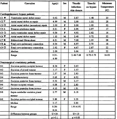
Cardiopulmonary bypass(CPB) has now been in routine use for 20 years in many centres (Utley, 1990). The technique involves the diversion of the venous blood returning to the heart through a pump and oxygenator and returning it to the aorta so that the job of both heart and lungs is done mechanically (figure 1.7). Circulation through the chambers of the heart is stopped by diverting the blood from the vena cavae. The coronary circulation is stopped by clamping the ascending aorta above the coronary ostia and proximal to the tube returning blood from the oxygenator; thus surgery to either or both of these areas can be performed. The only residual circulation to the lungs is via the systemic bronchial collaterals (which may be considerable in patients with antecedent congenital reduction of pulmonary blood flow) and any such blood returning to the left atrium is removed by suction into the bypass circuit. In most centres, (including the centre where these studies were performed), mechanical
ventilation of the lungs is discontinued during bypass, so that once the chest is opened the lungs collapse, further impeding any pulmonary blood flow. The heart is usually cooled by perfusion of the coronary circulation with an ice-cold solution and
The temperature to which the patient is cooled is broadly a function of the expected
length of the operation and range in the studies in this thesis from 32“C for atrial septal
defect repairs to 18°C for more complex procedures.
Pump
V9HOUSUne
Pump
Venous
Une
SVC:Superior Vena Cava
IVC:lnferior Vena Cava
CS: Coronary Sinus
RA: Right Alrlum
RV: Right Ventricle
PA: Pulmonary Artery
PViPuimonary Vein
LA: Left Atrium
IV: Left Ventricle
AA: Aortic Arch
Normal blood flow
Blood flow on
bypass
Figure 1.7 Schematic diagram o f the heart and great vessels indicating the pattern of
normal blood flow and the points where this flow is interrupted to achieve
cardiopulmonary bypass during cardiac surgery.
1.31 The types of oxygenator used
Early oxygenators worked on the principle of bubbling air/oxygen mixtures through the
blood. Such devices are still widely used in adult coronary artery surgery but carry the
risk o f macro-air embolism, and the certainty of micro-air embolism (Blauth et al. ,
1988). They have been replaced in paediatric cardiac surgery in the UK with hollow-
fibre or "membrane" oxygenators which remove the direct air-blood interface by
passing the gas, as the name suggests, through a network of hollow fibres made of
that such membrane systems induce less activation of acute inflammatory pathways (van Oeveren et a l, 1985; Cavarocchi et a l, 1986; Videm et al., 1989; Nilsson et al. ,
19906; Stahl et al., 1991) and may result in less associated organ dysfunction (Nilsson
e t a l , 1990c).
1.32 Associated clinical problems
These and many other refinements of CPB technique have greatly improved the clinical outlook for patients undergoing cardiac surgery. Nevertheless, there is still a significant incidence of "post pump" complications most commonly manifest in the brain (Shaw et a l., 1987; Smith, 1988), the lungs (Emhardt et al., 1991) and the performance of the heart itself (Nilsson et al., 1990c). These tend to be more common in infants than older children (Kirklin et al., 1983; Tanaka et al., 1986) and are thus being seen more
frequently with the trend towards definitive corrective operations in the neonatal period (Sethia, 1992). As well as an incidence of neurological handicap (Shaw et al., 1987), these problems produce a short term requirement for increased intensive support
(Kanter et al., 1986) which itself carries with it a risk of complications and a substantial cost. Leakage of fluid from capillaries into the extra-vascular space has been observed after CPB (Breckenridge et al., 1970; Maehara et al., 1991; Brans et al., 1981; Smith
et a l., 1987). In practice this is hard to quantify in vivo but it appears to occur to some extent universally and may be related to the pathophysiology of the various clinical complications of CPB. However, the relative importance of the contact of blood with artificial surfaces, haemodilution, cooling and rewarming, administration of drugs and ischaemia and reperfusion of the cardiac and pulmonary vascular beds in causing these problems remains unclear.
1.33 Associated inflammatory changes
metabolism (arachidonic acid pathways) (Greeley et al., 1988). The clinical importance of complement activation has been emphasised in some studies (Moore et al., 1988; Kirklin et al., 1983; Miyamoto et al., 1989) and questioned in others (Tennenberg et al., 1990; Nilsson et al., 1990c). The closely related effects on circulating blood cells have also been investigated (reviewed in Royston, 1990) and include injury to and changes in the deformability of red and white cells, consumption and activation of platelets and activation of neutrophils. Bacterial endotoxin(LPS) can be detected in the blood of patients immediately following CPB (Kharazmi et al., 1989; Andersen & Baek, 1992; Nilsson et al., 1990a) and could also be triggering elements of the acute inflammatory response.
During CPB, neutrophils have both an increased propensity to release oxidative products (van Oeveren et al., 1985) and to degranulate, releasing proteolytic enzymes (Hind et ah, 1988; Colman, 1990). Reperfusion with neutrophil-free blood was shown to protect the ischaemic heart and lungs against injury in dogs (Engler et al., 1986a; Engler et a l., 1986&) and humans (Breda et al., 1989; Bando et al., 1990) and removal of leukocytes and platelets by plasmapheresis prior to CPB has been shown to
ameliorate blood loss and pulmonary function (Davies et al., 1992) . Neutrophils sequester in the lungs of patients following CPB (Howard et al., 1988) and there is evidence that they play a role in post-operative lung injury (Ratliff et al., 1973) perhaps by release of reactive oxygen intermediates (Braude et al., 1986) and degranulation. Although there is a growing body of evidence for a role for neutrophils in acute lung injury (Donnelly & Haslett, 1992), the exact pathophysiology of damage to this and other organs post-CPB remains poorly understood.
1.4 Use of anti-adhesion molecule monoclonal antibodies in vivo
The critical role of CD11/CD18 adhesion molecules in acute inflammatory tissue
several animal models of acute inflammation including intestinal ischaemia-reperfusion in the cat (Hernandez et al. , 1987), myocardial ischaemia-reperfusion in the dog using an anti-CD 11b antibody (Simpson et a l , 1988) and ischaemia-reperfusion in the rabbit ear (Vedder et al. , 1990) and lung (Morgan et al. , 1990) and in haemorrhagic shock and resuscitation in the rabbit, using anti-CD 18 antibodies (Vedder et al., 1988). Anti- CD lla/C D 18 antibodies, but not antibodies to CD 11b or L-selectin, have been shown to prevent the development of cerebral malaria in Plasmodium-infected mice (Falanga & Butcher, 1991; Grau et a l, 1991).
In humans, anti-CD lla/C D 18 monoclonal antibodies have been used in an attempt to improve engraftment of transplanted bone marrow in conjunction with
immunosuppressive chemotherapy (Fischer et al., 1988). However the use of monoclonal antibodies to inhibit neutrophil interactions has not yet been reported.
1.5 Aims of the study
Since cardiopulmonary bypass is a planned procedure, the inflammation it causes can be studied from before its onset. Patients are invariably intensively supported and
monitored throughout the procedure and for at least 24 hours thereafter enabling studies to be performed without extra intervention or inconvenience to the patient. Furthermore the performance of intra-thoracic surgery provides unique access, during this period, to the major vessels carrying blood to and from the pulmonary, cardiac and systemic vasculatures. Since differences in the composition of blood entering and leaving these organs can be studied, more can be inferred about the changes occurring within them than sampling of peripheral blood would allow.
Studies of isolated oxygenator circuits allow a far greater standardization of variables than can be achieved in vivo. This allows accurate observation of changes in systems, such as the regulation of neutrophil adhesion molecule expression, that are volatile and easily disrupted. The approach also allows experimentation with specific interventions which could not be used directly in the patient, in a whole blood model which contains many of the complexities and regulatory pathways of the intact vascular system.
The endothelium is an inaccessible tissue. Although there has recently been a return to Cohnheim's methods of intra-vital microscopy to perform studies of leukocyte adhesion this approach does not lend itself to detailed study of endothelial injury and cannot be used in humans. The used of tissue culture to propagate endothelial cells is widely used and offers the best current approach to modelling damage to this tissue.
Chapter 2: Materials and methods
2.1 Materials
2.11 Reagents: Chemicals, reagents, solutions, standards and sera
Bovine serum albumin Sigma
Citric acid BDH
Coliagenase type II Sigma
DMEM (Dulbecco’s Modification of Eagle's Medium) Gibco Ethylene diamine tetra acetic acid (EDTA) Sigma
FACS lysing solution Becton Dickinson
Fetal calf serum (PCS) Gibco
Formaldehyde BDH
Formyl-Methionine-Leucine-Phenylalanine (fMLP) Sigma
Gelatin (porcine) Sigma
Glutamine BDH
Gold conjugated poly-l-lysine (Cationic gold) Biocell Hanks Balanced Salt Solution + /- calcium, magnesium Gibco
Hartmann's solution Gibco
Hepes Gibco
Hydrogen peroxide Sigma
Limulus amoebocyte lysate assay Kabi-Vitrium
Lipopolysaccharide (E.coli 0128:B12) Sigma
Mono-poly resolving medium Flow
o-phenylenediamine dihydrochloride Sigma
Paraformaldehyde BDH
Penicillin Glaxo
Phosphate buffered saline tablets Sigma
Preservative-free heparin, l(XX)u/ml Sigma
Recombinant soluble CRI RPMI medium
Silver enhancer Sodium azide
Sodium hydrogen phosphate Streptomycin Sulphuric Acid SmithKline Beecham Gibco Biocell Sigma BDH Evans Medical BDH
2.12 Monoclonal (m) antibodies and antisera
Foetal calf serum Gibco
Goat (Fab)2 anti-mouse IgG conjugated to FITC: [4350,TAGO] TCS Mouse m anti-CD 11a (MHM24): [M782]
Mouse m anti-CD 11a conjugated to FITC (25-3-1): [lOTl6] Mouse m anti-CD llb(44): [CBL145]
Mouse m anti-CD 11b conjugated to FITC(44): [CBL145] Mouse m anti-CD 18 (MHM23): [M783]
Mouse m anti-fibronectin (cell attachment domain): Mouse m anti-human negative control: IgGl [X927] Mouse m anti-keratin: [M615], (used as negative control) Mouse m anti-L-selectin (Leu8): [BD7440]
Mouse m anti-L-selectin (TQl) conjugated to phycoerythrin: Rabbit anti-mouse IgG conjugated to FITC:(TAGO)
Sheep anti-human elastase
Peroxidase-conjugated sheep anti-human a 1 antitrypsin
Dako
The Binding Site Cymbus Cymbus Dako Boehringer Dako Dako Becton Dickinson Coulter TCS
The Binding Site The Binding Site
2.13 Suppliers
BDH, Poole, UK
Becton Dickinson, Oxford, UK
Binding Site, Birmingham, UK Boehringer, Mannheim, Germany City University, London, UK Coulter Electronics, Luton, UK Cymbus, Southampton, UK Dako, High Wycombe, UK Evans Medical, West Sussex, UK
Flow Cytometry Standards Corp., NC, USA Flow Laboratories, Herts, UK
Gibco, Paisley, UK Glaxo, Middlesex, UK Greiner, Germany
Kabi-Vitrium, Uxbridge, UK Sigma, Poole, UK
SmithKline Beecham, Herts., UK TCS, Botolph Claydon, UK
2.2 Methods
2.21 Flow cytometry
logarithmic mode. Both linear and logarithmic scales are divided electronically, for acquisition purposes, into 1024 equally sized channels.
Forward and orthogonal light scatter and green and red fluorescence data from each experimental sample were then acquired. In most studies, this information was obtained for 2000 events (leukocytes) from each sample. In some later experiments (see chapters 6 & 7), events were acquired over a fixed 10 second period in order to obtain additional information about leukocyte concentrations in the samples under analysis. Data were stored as files initially on the hard disk of the dedicated computer attached to the flow cytometer and then on 600Ü Hewlett Packard tape cartridges(HP88140LC). In the initial studies, analysis was performed using the FACScan Research software program on the flow cytometer computer. In later studies, data were transferred on floppy diskettes and read and analysed on an IBM compatible desktop computer using the Datamate flow cytometry software (Applied Cytometry Systems, Sheffield, UK).
For each experiment, an analysis gate was created to encompass the granulocyte cluster and exclude other cell populations (e.g. lymphocytes and monocytes (figure 2. la)). A new gate was created for each experiment to compensate for variation in the light scattering properties of granulocytes from different individuals and for day to day variations and adjustments to the cytometer light scatter detectors. The gate was then verified visually to include the entire granulocyte cluster for all the samples in the experiment. Blood counts were routinely done and the differential count showed that granulocytes were >90% neutrophils in all (and >95% in most) of the patients and volunteers studied. The gated population was then analysed on a fluorescence histogram of the appropriate wavelength to the fiuorochrome on the antibodies used for staining (green - fluorescein, red - phycoerythrin). In most experiments this histogram
(a)
i i f Mi Bw« cta2azo*f^ocn(b)
(c)
(K-xmi f«c ~
il Wl OWqaON.042
UlLiL
0<->102UU ->
BZ.
Figure 2,1 (a) Forward/orthogonal light scatter dot plot o f 2000 events from a
suspension o f leukocytes prepared from venous blood, showing gating o f the granulocyte
cluster. Fluorescence histograms o f gated granulocyte populations from whole blood
(a)
N R 1 0 J A 2 n 2 8 / N R 1 0 J A 2 NeutrophilsT O T A L = 2 0 0 0 G A T E D = 5 8 1
A F 1 0 J A 2 . 0 1 4 / A F 1 0 J A 2
(b)
Neutrophils
Endothelial cells
I_Hü t T O T A L = 3 3 0 0 G A T E D = 3 3 GO
Figure 2.2 Fluorescence histograms of granulocytes stained fo r L-selectin showing (a) 2
distinct populations o f neutrophils with separate analysis windows and (b) granulocytes
population was demonstrated; this occurred in 2 ways. Either there were 2 populations of neutrophils, identical in their light scattering properties but heterogeneous in their expression of the epitope under study (figure 2.2a) or a different population of cells partially overlapped with the neutrophil forward/orthogonal scatter cluster (figures 2.2b). In the former case subsidiary analysis windows were created for each distinct population and the median channel number used whereas in the latter case, in which there was often some overlap of the fluorescence histograms of the populations and in which the granulocytes were invariably more numerous, the mode channel number was used instead.
The average fluorescence channel number for each sample was then converted mathematically into a relative fluorescence value as seen on the logarithmic fluorescence scale using the formula:
relative fluorescence = 1 0"(channel number/number channels per decade) These values were then used to represent the average level of expression of the epitopes under study on the neutrophils' surface.
2.22 Cytokine ELIS As - I L la , IL lp , TN Fa, EL8
Enzyme linked immunoassays (ELISAs) for IL la , IL ip, TN Fa and IL8 were
performed using kits (Quantikine, R & D Systems, Minneapolis, USA) according to the manufacturer's instructions. Samples were diluted lin2 in appropriate diluent, and incubated for 2 hours at room temperature in wells coated with monoclonal anti cytokine antibody. Wells were washed, and then incubated similarly with polyclonal anti-cytokine antibody conjugated to horseradish peroxidase. After further washes, a substrate solution containing tetramethylbenzidine and H2O2 was added; the reaction was stopped after 20 minutes with 2M H2SO4 and plates were read using a