R E S E A R C H A R T I C L E
Open Access
A hereditary spastic paraplegia mutation in
kinesin-1A/KIF5A disrupts neurofilament transport
Lina Wang, Anthony Brown
*Abstract
Background:Hereditary spastic paraplegias are a group of neurological disorders characterized by progressive distal degeneration of the longest ascending and descending axons in the spinal cord, leading to lower limb spasticity and weakness. One of the dominantly inherited forms of this disease (spastic gait type 10, or SPG10) is caused by point mutations in kinesin-1A (also known as KIF5A), which is thought to be an anterograde motor for neurofilaments.
Results:We investigated the effect of an SPG10 mutation in kinesin-1A (N256S-kinesin-1A) on neurofilament transport in cultured mouse cortical neurons using live-cell fluorescent imaging. N256S-kinesin-1A decreased both anterograde and retrograde neurofilament transport flux by decreasing the frequency of anterograde and retrograde movements. Anterograde velocity was not affected, whereas retrograde velocity actually increased. Conclusions:These data reveal subtle complexities to the functional interdependence of the anterograde and retrograde neurofilament motors and they also raise the possibility that anterograde and retrograde neurofilament transport may be disrupted in patients with SPG10.
Background
Hereditary spastic paraplegias are a group of neurologi-cal disorders characterized by progressively increasing lower-extremity weakness and spasticity [1]. The pri-mary cause appears to be distal degeneration of the longest ascending and descending axons in the spinal cord, though the explanation for this selective vulner-ability is not known. To date, at least 41 spastic paraple-gia gene loci have been mapped (termed SPG1 through SPG 41) and 17 genes have been identified [2]. The inheritance can be autosomal dominant, autosomal recessive, or X-linked. One of the autosomal dominant forms, SPG10, is caused by mutations in kinesin-1A, also known as KIF5A, which is a member of the kine-sin-1 family of motor proteins.
Kinesin-1 motor proteins are heterotetramers com-posed of two heavy chains and two light chains [3]. There are three kinesin-1 heavy chain genes in mam-mals: kinesin-1A, B and C (also known as KIF5A, B and C) [4]. Kinesin-1A and kinesin-1C are neuron specific, whereas kinesin-1B ("conventional kinesin”) is expressed
ubiquitously [5-7]. Little is known about the cargoes of kinesin-1A, though potential cargoes and interactors include HAP-1 (huntingtin associated protein-1) [8], DISC-1 (disrupted in schizophrenia protein-1) and the
NUDEL/LIS1/14-3-3εcomplex [9], Grb2 (growth factor
receptor bound protein-2) [10], and b-dystrobrevin
[11,12].
Neurofilaments are the intermediate filaments of neu-rons. They are heteropolymers of variable composition and subunit stoichiometry, typically composed of the low, medium and high molecular weight neurofilament triplet proteins (NFL, M and H), as well as peripherin and/or alpha-internexin [13]. Live-cell imaging of neuro-filament transport in cultured neurons and
computa-tional modeling studies of neurofilament transportin
vivo have demonstrated that neurofilaments move along
axons in a rapid, intermittent and bidirectional manner [14-17].
Studies on kinesin-1A knockout mice have suggested that kinesin-1A may be an anterograde motor for neu-rofilaments in axons [18]. In support of this proposal, we observed a 75% reduction in the frequency of neuro-filament movement in cultured neurons from kinesin-1A knockout mice [19]. Interestingly, both anterograde * Correspondence: brown.2302@osu.edu
Center for Molecular Neurobiology and Department of Neuroscience, The Ohio State University, Columbus, OH 43210, USA
and retrograde movement was affected and movement in both directions could be rescued by kinesin-1A. Par-tial rescue was also observed with kinesin-1B and kine-sin-1C, though with successively decreasing efficacy. In addition, headless kinesin-1A and kinesin-1C each inhibited both anterograde and retrograde neurofilament transport in a dominant-negative manner in wild type neurons. Because dynein is thought to be the retrograde motor for axonal neurofilaments, we investigated the effect of dynein inhibition on neurofilament transport. Disruption of dynein function by using RNA interfer-ence, dominant- negative approaches, or a function-blocking antibody also inhibited both anterograde and retrograde neurofilament movement. These data suggest that kinesin-1A is the principal but not exclusive antero-grade motor for neurofilaments, that there may be some functional redundancy among the kinesin-1 isoforms with respect to neurofilament transport, and that the activities of the anterograde and retrograde neurofila-ment motors are tightly coupled.
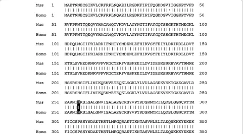
Since kinesin-1A appears to be a motor for neurofila-ments, we have investigated the effect of an SPG10 mutation in this motor on neurofilament transport in cultured neurons. Of the 16 different SPG10 mutations that have been identified to date, 15 reside in the kine-sin motor domain and 14 of these are missense
mutations [20-26]. A particular hot spot for these muta-tions is in the vicinity of the microtubule and nucleotide binding sites. In the present study, we have focused on the N256S mutation, which results in the substitution of a highly conserved asparagine residue in the switch II loop/helix motif of the microtubule binding site [20] (Figure 1). We show that expression of N256S-kinesin-1A disrupts both anterograde and retrograde neurofila-ment neurofilaneurofila-ment transport in cultured mouse cortical neurons, raising the possibility that neurofilament trans-port may also be disrupted in patients with SPG10.
Results
To study the effect of N256S-kinesin-1A on neurofila-ment transport, we transfected cultured mouse cortical neurons with GFP-tagged neurofilament protein M (GFP-NFM) with or without mutant or wild type mouse kinesin-1A. The purpose of the wild type kinesin-1A was to control for possible effects due to overexpression of the motor. Mouse cortical neurons exhibit gaps in the axonal neurofilament array similar to those that we have observed in neurons from superior cervical ganglia [14-19], but the gaps in the cortical neurons are longer and more numerous, making these cells particularly sui-table for studies of neurofilament movement (Figure 2). The GFP-NFM fusion protein incorporates throughout
all the neurofilaments in these cells, permitting all neu-rofilaments to be detected. Additional files 1 & 2 in the Supplementary Data are examples of movies showing the abundant neurofilament movement that can be observed in these neurons.
To track the movement of the GFP-tagged neurofila-ments, we observed gaps by epifluorescence microscopy and acquired time-lapse movies using one second expo-sures at four second time intervals. Each movie was exactly 15 minutes in length. Ninety six percent of the moving structures were filamentous in shape, ranging from 1.3μm to 44.5μm in length and diffraction limited in width. The average length was 8.6μm, which is com-parable to what we observed in previous studies on neu-rons from mouse superior cervical ganglia [19-27]. We defined transport frequency as the number of filaments
that moved at least 50 pixels (6.55μm) per 15-minute
movie. N256S-kinesin-1A reduced the average neurofila-ment transport frequency significantly, from 4.5 to 1.4 filaments/hour in the anterograde direction (p < 0.001) and from 3.2 to 2.0 filaments/hour in the retrograde direction (p = 0.047; Figure 3). Expression of wild type kinesin-1A reduced the average neurofilament transport frequency from 4.5 to 3.7 filaments/hour anterogradely and from 3.2 to 3.1 filaments/hour retrogradely, but these effects were not statistically significant (p = 0.26 and p = 0.92, respectively). Thus N256S-kinesin-1A impaired neurofilament transport in both anterograde and retrograde directions in these axons. Additional files 3, 4 & 5 in the Supplementary Data are examples of the movies obtained in these experiments.
To analyze the motility defect in more detail, we tracked the movement of each filament through succes-sive frames of the time-lapse movies. We defined ante-rograde and retante-rograde transport flux as the total distance moved in the corresponding direction by all the filaments in each 15-minute movie. Since each movie contained a single axon, we expressed the fluxes in units
of μm/axon/hour. N256S-kinesin-1A reduced the
aver-age neurofilament transport flux from 149 to 46 μm/
axon/hour in the anterograde direction (p < 0.001) and
from 116 to 90μm/axon/hour in the retrograde
direc-tion (p = 0.007; Figure 3). Expression of wild type
kine-sin-1A reduced the transport flux from 149 to 109μm/
axon/hour anterogradely and from 116 to 105μm/axon/
hour retrogradely, but these reductions were not statisti-cally significant (p = 0.13 and p = 0.69, respectively). Thus, in addition to decreasing the frequency of neuro-filament movement, N256S-kinesin-1A also decreased the total extent of neurofilament movement.
To determine whether N256S-kinesin-1A also affected the velocity or persistence of neurofilament movement,
we measured the velocity, distance and duration of each bout of movement for each moving filament. We defined a bout as a period of uninterrupted movement between two pauses or between a pause and a reversal. N256S-kinesin-1A decreased the average bout velocity from 0.27μm/s to 0.24 μm/s in the anterograde direc-tion, but this was not statistically significant (p = 0.053; Figure 4). In contrast, N256S-kinesin-1A increased the average retrograde bout velocity from 0.32 to 0.40μm/s, and this was statistically significant (p < 0.001). N256S-kinesin-1A also increased the average retrograde bout
distance from 3.7 μm to 5.7 μm (p < 0.001) and the
average retrograde bout duration from 11 seconds to 15 seconds (p < 0.001), but without any significant effect
on average anterograde bout distance (3.2μm, p = 0.36) or average anterograde bout duration (19 seconds, p = 0.98). Expression of wild type kinesin-1A had no signifi-cant effect on average retrograde bout velocity (0.30 μm/s, p = 0.76), average retrograde bout distance (3.4 μm, p = 0.57), or average retrograde bout duration (14 seconds, p = 0.32). Expression of wild type kinesin-1A did decrease the average anterograde bout velocity from 0.27μm/s to 0.23μm/s (p = 0.006) and average antero-grade bout distance from 3.4 μm to 3.0μm (p = 0.043), but without any significant effect on average anterograde bout duration (15 seconds, p = 0.31). Thus, N256S-kine-sin-1A reduced the anterograde flux by decreasing ante-rograde frequency without affecting anteante-rograde
velocity. N256S-kinesin-1A also reduced the retrograde flux in spite of an increase in retrograde velocity because the increase in retrograde velocity was not suffi-cient to compensate for the decrease in retrograde fre-quency (summarized in Figure 5).
We have shown previously that neurofilaments are delivered to distal axons by anterograde movement and retrieved by retrograde movement [27]. Thus it is possi-ble that the decrease in retrograde flux described above
could be explained by a depletion of neurofilaments from distal axons as a secondary consequence of the disruption of anterograde movement. To test this hypothesis, we transfected cultured cortical neurons with GFP-NFM, either with or without N256S-kinesin-1A, and then fixed and processed the cells for immuno-fluorescence microscopy after 10-11 days in culture using antibodies specific for GFP and neurofilament protein M (NFM). Since axonal neurofilament distribu-tion is discontinuous in these neurons, we also stained for tubulin and actin, which are present along the entire length of each axon. Cells expressing N256S-kinesin-1A were identified by their GFP expression. To quantify neurofilament content, we measured the fluorescence
intensity of NFM in distal axons, extending 100 μm
proximally from the base of the growth cone. There was no statistically significant difference in the neurofilament content of axons expressing N256S-kinesin-1A com-pared to axons expressing no exogenous kinesin-1A (Figure 6). Thus, disruption of neurofilament transport by N256S-kinesin-1A does not appear to deplete distal axons of neurofilaments, and therefore the reduction in retrograde neurofilament flux cannot be a secondary consequence of the disruption of anterograde neurofila-ment moveneurofila-ment. The absence of a reduction in neurofi-lament content in distal axons expressing N256S-kinesin-1A is probably due to the impairment of both anterograde and retrograde neurofilament movement, which would be expected to impair both delivery and departure of neurofilaments from these axonal regions.
Microtubules in axons are widely accepted to be orientated exclusively with their plus-ends distal, and kinesin-1 motors are known to move cargoes exclusively toward the plus-ends of these polymers. Thus the dis-ruption of retrograde neurofilament movement by N256S-kinesin-1A suggests that this mutant also dis-rupts minus-end directed neurofilament movement, which is thought to be mediated by dynein [28-31]. However, another possibility, albeit unlikely, is that microtubule polarity in cultured mouse cortical axons is not entirely or predominantly plus-end distal. To test this hypothesis, we transfected cultured cortical neurons with the microtubule plus-end tracking protein EB1 tagged with yellow fluorescent protein (YFP-EB1) and imaged the movement of YFP-EB1 comets. Additional file 6 in the Supplementary Data is an example of a movie showing YFP-EB1 comet movement in these cells. Using kymograph analysis, we tracked 192 comets in 23 axons. 190 comets moved anterogradely and 2 comets moved retrogradely (Figure 7). The average comet velocity was 0.13 μm/s, which is consistent with published estimates of the rate of microtubule growth in cells [32]. These data confirm that microtubules in these axons are indeed almost exclusively plus-end distal
(especially considering that the plus-end proximal microtubules could represent microtubules that looped back on themselves). Thus the disruption of retrograde neurofilament transport by N256S-kinesin-1A appears to be due to disruption of minus-end directed move-ment by this mutant plus-end directed motor.
Discussion
The SPG10 form of hereditary spastic paraplegia is an autosomal dominant disease caused by mutations in the kinesin-1A motor protein. Since there is evidence that kinesin-1A is a motor for neurofilaments, we investi-gated the effect of an SPG10 mutant, N256S-kinesin-1A, on neurofilament transport in cultured neurons. Expres-sion of N256S-kinesin-1A in cultured mouse cortical neurons impaired neurofilament transport in both ante-rograde and retante-rograde directions, but unexpectedly this was due primarily to a decrease in the frequency, not the velocity of movement. A limitation of our experi-mental approach is that transient transfection of the mutant motor does not permit quantification or regula-tion of the expression level relative to the endogenous wild type motor. Therefore, to control for possible effects of over-expression, we also characterized neurofi-lament movement in neurons transfected with wild type
kinesin-1A motor. Simply over-expressing wild type kinesin-1A had a small effect on anterograde bout velo-city and anterograde bout distance, but no effect on average frequency or flux in either the anterograde or retrograde direction. Thus the effects of the N256S-kinesin-1A on neurofilament transport were due to the N256S mutation and were not an artifact of over-expression.
The impairment of both anterograde and retrograde neurofilament movement in these experiments is nota-ble because kinesin-1A is an anterograde motor in axons, but this result is consistent with recent evidence from our laboratory showing that the anterograde and retrograde neurofilament motors are interdependent [19]. In that study, we found that both anterograde and retrograde neurofilament movement were impaired in neurons from kinesin-1A knockout mice, and that expression of wild type kinesin-1A rescued the move-ment in both directions. In addition, expression of a headless dominant negative kinesin-1A construct in wild type neurons impaired both anterograde and retrograde neurofilament movement, and disruption of dynein function by using RNA interference, dominant negative approaches, or a function-blocking antibody also inhib-ited both anterograde and retrograde neurofilament
Figure 6N256S-kinesin-1A does not deplete neurofilaments from distal axons. Comparison of the neurofilament content of distal axons from cortical neurons expressing GFP-NFM alone (control) or GFP-NFM plus N256S-kinesin-1A. (A) Immunofluorescence microscopy for
movement. Thus there is functional coupling between kinesin-1A and dynein motors in the bidirectional trans-port of neurofilaments along microtubules in axons.
The mechanism by which microtubule motors of opposing directionality interact to regulate bidirectional cargo transport is not yet understood. Two favored models are the tug-of-war model and the coordination model [33]. In the tug-of-war model, the direction of movement is the result of a dynamic competition between opposing motors that are bound and active at the same time. In the coordination model, the opposing motors interact so that only motors of one directionality are bound or active at one time. In their simplest form, these two models have quite different predictions: in the
tug-of-war model, impairment of motors of one direc-tionality should increase the velocity and frequency of movement in the opposite direction, whereas in the coordination model it should not. In fact, according to the coordination model, manipulations or mutations that disrupt the coordination could also cause the motors to interfere with each other, leading to impair-ment of moveimpair-ment in both directions [34]. Many labs have reported reciprocal inhibition of both directions of movement after disruption of motors of one directional-ity, which is consistent with the coordination model (see [19] for citations to these studies).
In the present study, we found that expression of N256S-kinesin-1A reduced the frequency and flux of
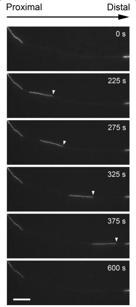
Figure 7Microtubules in cortical neuron axons are plus-end distal. Cortical neurons were transfected with YFP-EB1, which is a protein that binds to the growing plus ends of microtubules. Axons were imaged for 2 minutes at 2 second intervals in order to determine the direction of microtubule growth. (A) A typical kymograph showing a number of YFP-EB1 comets moving anterogradely. The horizontal dimension represents distance and the vertical dimension represents time. This kymograph was generated from the movie named Additional file 6 in the
neurofilament movement in both anterograde and retro-grade directions and increased bout velocity, distance and duration in the retrograde direction. It is unclear how to interpret these data mechanistically. The decrease in both anterograde and retrograde frequency and flux suggests a coordination mechanism, but the increase in retrograde bout velocity, distance and dura-tion suggests a tug-of-war. Thus it is likely that the mechanism is more complex, perhaps combining fea-tures of both models. For example, Lipowsky and collea-gues have shown that tug-of-war models that account for the load-dependence of the interaction between motors and their tracks can generate bouts of persistent anterograde and retrograde movement, depending on the relative numbers of bound motors [35,36]. To test such hypotheses in the case of neurofilaments it will be necessary to record neurofilament movement with much higher spatial and temporal resolution than we have done in the present study, and also to measure the forces acting on the moving filaments, which is cur-rently not possible because neurofilaments are too small to be optically trapped.
Studies in vitro suggest that the N256S-kinesin-1A
mutant is a defective motor and that it may act as a dominant-negative disruptor of kinesin-1A transport. Mutation of the homologous amino acid residue to a lysine in the motor domain of the yeast kinesin-14 motor kar3 (N650K-kar3) or in the motor domain of the fungal kinesin-14 motor ncd (N600K-ncd), prevents microtubule-stimulated activation of the motor ATPase by uncoupling nucleotide and microtubule binding [37]. Both N650K-kar3 and N600K-ncd are capable of bind-ing and hydrolyzbind-ing ATP, but in contrast to wild type kinesin, they bind tightly to microtubules in both the ATP-bound and ADP-bound states. In microtubule
glid-ing assaysin vitro, N600K-ncd motors bind
microtu-bules but do not translocate them. In yeast cells, N650K-kar3 exhibits a dominant negative effect over wild-type kar3 [38].In vitro, N256S-kinesin-1A is unable to generate single-motor processive motion in microtu-bule gliding and bead motility assays, resulting in decreased gliding and transport velocities [39]. However, in contrast to the N to K mutations in N650K-kar3 and N600K-ncd, the N256S mutation in kinesin-1A did not bind microtubules in rigor. When N256S-kinesin-1A was mixed with wild type kinesin-1A in microtubule gliding and particle motility assaysin vitro, the mutant kinesin appeared to exert a dominant inhibitory effect on the transport velocity. Given these observations, however, it is surprising that the disruption of neurofila-ment transport by N256S-kinesin-1A in our study was due primarily to a decrease in the frequency. There was a slight decrease in average velocity (from 0.27 to 0.24
μm/s), but this was not statistically significant.
Moreover, this effect was probably an artifact of over-expression, since we observed a similar decrease in cells expressing wild type kinesin-1A. Thus the behavior of the motor in motility assays in vitrocannot necessarily predict its effect on cargo transportin vivo.
Neurofilaments accumulate abnormally and exces-sively in many neurodegenerative diseases, including amyotrophic lateral sclerosis, giant axonal neuropathy, and Charcot Marie Tooth disease [40,41]. Several stu-dies have suggested that these accumulations may arise due to perturbations in axonal transport [42-44]. In sup-port of this idea, slowing of axonal transsup-port has been reported in mouse models of SOD-mediated amyo-trophic lateral sclerosis [45] and is an early event in the progression of this disease [46]. Disruption of axonal transport by over-expression of dynamitin in neurons also results in accumulations of neurofilaments [47], and mutations in dynein subunits are one cause of motor neuron disease in humans [48,49]. Defects in axonal transport have also been reported in mouse models of spastin-mediated hereditary spastic paraplegia (SPG4) [50,51], and swellings containing vesicular and cytoske-letal proteins, including neurofilament proteins, have been reported in humans with this disease [50,51].
While it is clear that the axonal transport of neurofila-ments is impaired in many neurodegenerative diseases, the role of neurofilament accumulations in the disease progression has been controversial. Over-expression of the human high molecular weight neurofilament protein (NFH) in mice causes a slowing of neurofilament trans-port, accumulations of axonal neurofilaments, and motor neuron degeneration [42-52]. However, the sig-nificance of these studies is unclear because over-expression of mouse NFH has no pathological effects [53]. One approach to test the role of neurofilaments in mouse models of neurodegenerative diseases has been to cross the mice with neurofilament L knockout mice, which lack neurofilament polymers. Using this approach, it has been shown that the absence of neurofilaments in neurons slows the progression of disease dramatically in mouse models of superoxide dismutase (SOD)-mediated ALS [54,55]. On the other hand, similar experiments using a transgenic mouse expressing an NFH b -galacto-sidase fusion protein, which aggregates and sequesters neurofilaments in neuronal cell bodies, showed no sig-nificant alteration of disease pathology in mouse models of dystonia musculorum and SOD-mediated ALS, though there was some prolongation of neuronal survi-val and some delay of axon loss [56].
transport of neurofilaments and other cargoes and lead to focal accumulations and depletions of axonal neurofi-laments. Studies with transgenic mice expressing NFH
b-galactosidase fusion protein suggest that neurofila-ments are not essential for the toxicity associated with the administration of these substances [57], whereas stu-dies on the Quiverer (Quv) quail, which lack NFL, sug-gest that they are [58]. Perhaps the conflicting nature of these reports may be due to differences between the ani-mal models used. For example, the NFHb-galactosidase transgenic mice have perikaryal neurofilament accumu-lations whereas the NFL knockout mice and the Qui-verer quail do not. Whatever the explanation, however, it does seem clear that the accumulation of neurofila-ments can be an exacerbating factor in at least some circumstances.
Neurofilaments are unlikely to be the sole cargo for kinesin-1A in neurons so it is possible that deficiencies in the movement of other cargoes may contribute to the disease progression in SPG10. Moreover, while neurofi-lament accumulations have been described in patients with SPG4 (see above), there have been no ultrastruc-tural studies on nerves of patients with SPG10. Thus it is presently unclear whether neurofilament accumula-tions are a feature of this disease. In the present study we did not observe local neurofilament accumulations in axons of neurons expressing N256S-kinesin-1A. How-ever, it is unclear to what extent such observations in short term cultures can predict the long-term effects of SPG10 mutaions on neurofilament organization in vivo. For example, it is quite possible that subtle changes in neurofilament organization or distribution in short-term cultures of neonatal cultured neurons might be magni-fied over longer time scales in mature neurons in vivo. Either way, the fact that kinesin-1A appears to be a neu-rofilament motor and that N256S-kinesin-1A disrupts the bidirectional transport of neurofilaments in cultured neurons suggest that patients with the SPG10 form of hereditary spastic paraplegia may well have neurofila-ment transport abnormalities which may contribute to the disease progression, and this warrants further investigation.
Conclusions
Mutations in kinesin-1A, which is a putative antero-grade motor for axonal neurofilaments, cause the SPG10 form of hereditary spastic paraplegia. We investigated the effect of an SPG10 point mutation in kinesin-1A on neurofilament transport in cultured mouse cortical neu-rons. We showed that this mutant disrupts both antero-grade and retroantero-grade neurofilament transport, raising the possibility that neurofilament transport may also be disrupted in patients with SPG10.
Materials and methods Molecular Cloning
Mouse kinesin-1A cDNA (Genbank accession No. BC058396, I.M.A.G.E. clone 6824963) was obtained from American Type Culture Collection (Manassas, VA) and then subcloned into pEGFP-C1 (Clontech, Moun-tain View, CA) lacking the EGFP sequence, as previously described [19]. The N256S-kinesin-1A plasmid construct was generated using a QuikChange Site-Directed Muta-genesis Kit (Stratagene, La Jolla, CA) with forward pri-mer 5’-GGC AAA GAA TAT CAG CAA GTC GCT
GTC GGC CC and reverse primer 5’-GGG CCG ACA
GCG ACT TGC TGA TAT TCT TTG CC. The wild type and mutant kinesin-1A constructs were tagged with cMyc at their N-terminus with forward primer 5’ -CTA GCT CCG GAA TGG AGC AGA AGC TGA TCA GCG AGG AGG ACC TGG AG and reverse
pri-mer 5’-TCG ACT CCA GGT CCT CCT CGC TGA
TCA GCT TCT GCT CCA TTC CGG AG. The result-ing cMyc-N256S-kinesin-1A and cMyc-kinesin-1A con-structs were confirmed by DNA sequencing of their open reading frames. The EGFP-mNFM plasmid con-struct, which encodes the codon-optimized F64L/S65T variant of green fluorescent protein attached to the amino terminus of mouse neurofilament protein M, was described by Yan et al. [59]. The YFP-EB1 plasmid con-struct was provided by Chen Gu [60].
Cell culture
Cortical neurons were cultured using the glial sandwich technique of Banker [61]. To prepare glial cultures, the cerebral cortices of 3 to 5 P0 mice were dissociated in phosphate buffered saline (PBS; Invitrogen, Carlsbad, CA) containing 0.25% [w/v] trypsin (Worthington Bio-chemical Corp., Lakewood, NJ), 1% [w/v] DNase-I (Sigma, St. Louis, MO) and 0.54mM EDTA (Sigma) and the cells were cultured in plastic dishes at 37°C/5% CO2 in glial medium, which consisted of Minimum Essential Medium (Invitrogen) supplemented with 10% [v/v] horse serum (Invitrogen), 0.7% [w/v] glucose (Sigma)
and 16 μg/ml gentamicin (Invitrogen). The cells were
(Invitrogen) supplemented with 2% [v/v] B-27 Supple-ment Mixture (Invitrogen), 0.27% [w/v] glucose, 2 mM glutamine (Invitrogen), 37.5 mM NaCl (Sigma), 5% [v/v] fetal bovine serum (FBS; Thermo Scientific, Waltham,
MA), 16μg/ml gentamicin, and 2.5 μM cytosine
arabi-noside (AraC; Sigma). After two days, the plating med-ium was replaced with culturing medmed-ium, which was identical to the plating medium except that it lacked serum. Every four days, half the medium was removed and replaced with fresh medium.
Transfection
The dissociated cortical neurons were transfected by electroporation prior to plating using an Amaxa Nucleo-fector™(Lonza Inc., Walkersville, MD) with the mouse neuron nucleofection kit (VPG-1001) and program
O-05. The volume of the cell suspension was 100 μl and
the cell density ranged from 4 × 10⁶to 6 × 10⁶ cells/ml. For the experiments on neurofilament movement and
distribution, we used 2 μg EGFP-mNFM construct
either alone or in addition to 2μg N256S-kinesin-1A or 2 μg wild type kinesin-1A construct. For the
microtu-bule polarity experiments, we used 2 μg YFP-EB1
construct.
Live-cell imaging
To image neurofilament movement, cortical neurons were observed after 8 to 12 days in culture by epifluor-escence microscopy on a Nikon TE300 inverted micro-scope (Nikon, Garden City, NY) using a 100x Plan Apo VC 1.4NA oil immersion objective. The observation medium consisted of Hibernate-E (BrainBits, Springfield, IL) supplemented with 2% [v/v] B27 Supplement Mix-ture, 0.3% [w/v] glucose, 1 mM L-glutamine, 37.5 mM
NaCl, and 10 μg/ml gentamicin. The temperature on
the microscope stage was maintained using an Air Stream incubator (Nevtek, Williamsville, VA). A layer of dimethylpolysiloxane fluid (Sigma, 5 centistokes) was floated over the observation medium to prevent eva-poration. For time-lapse imaging, the exciting light from the mercury arc lamp was attenuated 12-fold using neu-tral density filters, and images were acquired with one second exposures at four second intervals using a Micromax 512BFT cooled CCD camera (Roper
Scienti-fic, Trenton, NJ) and MetaMorph™software (Molecular
Devices, Sunnyvale, CA). All movies were 15 minutes in length. It was necessary to adjust the focus occasionally during movie acquisition to correct for focus drift. To image YFP-EB1 comets, cortical neurons were observed after 8-10 days in culture by epifluorescence microscopy using a Nikon TE2000 microscope. For these experi-ments, the exciting light from the mercury arc lamp was attenuated 4-fold using neutral density filters, and images were acquired with one second exposures at two
second intervals for two minutes using a CoolSNAP HQ cooled CCD camera and 2 × 2 pixel binning (Roper Scientific). For publication, the movies were saved in QuickTime format using the H.264 video codec.
Motion analysis
Neurofilament movement was analyzed by tracking the position of the leading or trailing ends of the filaments in successive frames of the time-lapse image movies using MetaMorph™software. All objects greater than or equal to 10 pixels (1.31μm) in length were analyzed if they moved a total distance of at least 50 pixels (6.55 μm) and could be tracked through at least three succes-sive frames of the movie. To calculate the frequency of movement, we classified each neurofilament as antero-grade or retroantero-grade based on its preferred direction of motion and then counted the number of anterograde and retrograde moving filaments per movie. Thus for each movie we obtained two frequency measurements, one anterograde and one retrograde. Ninety seven per-cent of the filaments exhibited a preferred direction of movement, defined as moving at least 70% of their time in the same direction. The remaining 3% of the fila-ments, which spent more than 30% of the time moving in the opposite direction, were each considered to repre-sent separate anterograde and retrograde moving events. To calculate the flux, we grouped all the filaments in each 15-minute movie together and measured the total anterograde and retrograde distance moved. Thus for each movie we obtained two flux measurements, one anterograde and one retrograde. For the analysis of bout velocity, bout duration and bout distance, we defined a bout of movement to be a phase of uninterrupted move-ment between two pauses or between a pause and a reversal. Thus each bout velocity represents the bout distance divided by the bout duration. Bouts in which the filament was moving at the start or end of the movie were ignored because their true duration could not be assessed. Statistical comparisons were performed using the Mann-Whitney test.
Immunofluorescence microscopy
Actin was visualized by including Alexa-568 phalloidin (Invitrogen, 1:20) in the secondary antibody mixture. Coverslips were mounted using ProLong Gold Antifade reagent (Invitrogen). Images were acquired on a Nikon TE2000 microscope with a 40x Plan Apo 1.0 NA oil immersion objective and a CoolSNAP HQ cooled CCD camera. The epifluorescent illumination was attenuated 4-fold using neutral density filters, and images were acquired with 100 millisecond exposures. To quantify the fluorescence in the distal axon, we measured the fluorescence intensity in the most distal 100μm of each axon, extending proximally from the base of the growth cone.
Additional material
Additional file 1: Movie showing abundant neurofilament movement in a neuron expressing GFP-NFM. An example of the abundant movement that can be observed in cultured cortical neurons expressing GFP-NFM. Proximal is left and distal is right. Width of field of view: 67μm. Time compression: 40:1
Additional file 2: Movie showing abundant neurofilament movement in a neuron expressing GFP-NFM. An example of the abundant movement that can be observed in cultured cortical neurons expressing GFP-NFM. This axon is branched, and filaments can be observed to move from the parent axon into the daughter axons and vice versa. Proximal is left and distal is right. Width of field of view: 67
μm. Time compression: 40:1
Additional file 3: Movie showing neurofilament movement in a neuron expressing GFP-NFM. A gap in the axonal neurofilament array of a cortical neuron expressing GFP-NFM. A single neurofilament moves through the gap. Proximal is left and distal is right. Width of field of view: 67μm. Time compression: 40:1
Additional file 4: Movie showing neurofilament movement in a neuron co-expressing GFP-NFM and N256S-kinesin-1A. A gap in the axonal neurofilament array of a cortical neuron co-expressing GFP-NFM and N256S-kinesin-1A. Some jiggling movement is apparent at the edges of the gap, but no filaments move into the gap. Proximal is left and distal is right. Width of field of view: 67μm. Time compression: 40:1
Additional file 5: Movie showing neurofilament movement in a neuron co-expressing GFP-NFM and wild type kinesin-1A. A gap in the axonal neurofilament array of a cortical neuron co-expressing GFP-NFM and wild type kinesin-1A. A neurofilament moves through the gap and then a shorter filament enters that gap. Proximal is left and distal is right. Width of field of view: 67μm. Time compression: 40:1
Additional file 6: Movie showing microtubule plus-end“comets”in a neuron expressing YFP-EB1. Microtuble plus-end“comets”in the axon of a cortical neuron expressing YFP-EB1. Proximal is left and distal is right. Note that all the comets move anterogradely, confirming the plus-end distal orientation of the microtubules in these axons. A kymograph generated from this movie is shown in Figure 7A. Width of field of view: 42μm. Time compression: 20:1
Acknowledgements
We thank Chen Gu (Ohio State University) for the YFP-EB1 construct and Atsuko Uchida in the Brown lab for the wild type KIF5A cDNA construct. This project was funded by NIH grant RO1-NS38526 to A.B., with additional support provided by NIH grant P30-NS0457580.
Authors’contributions
LW performed the experiments, analyzed the data, and prepared the figures. AB conceived of the study and worked closely with LW to design the
experiments and to analyze and interpret the data. Both authors participated in the writing, and both authors read and approved the final manuscript.
Competing interests
The authors declare that they have no competing interests.
Received: 8 September 2010 Accepted: 18 November 2010 Published: 18 November 2010
References
1. Fink JK:Hereditary spastic paraplegia.Curr Neurol Neurosci Rep2006,
6(1):65-76.
2. Salinas S, Proukakis C, Crosby A, Warner TT:Hereditary spastic paraplegia: clinical features and pathogenetic mechanisms.Lancet Neurol2008,
7(12):1127-1138.
3. Hirokawa N, Noda Y, Tanaka Y, Niwa S:Kinesin superfamily motor proteins and intracellular transport.Nat Rev Mol Cell Biol2009,10(10):682-696. 4. DeBoer SR, You Y, Szodorai A, Kaminska A, Pigino G, Nwabuisi E, Wang B,
Estrada-Hernandez T, Kins S, Brady ST,et al:Conventional kinesin holoenzymes are composed of heavy and light chain homodimers. Biochemistry2008,47(15):4535-4543.
5. Niclas J, Navone F, Hom-Booher N, Vale RD:Cloning and localization of a conventional kinesin motor expressed exclusively in neurons.Neuron
1994,12(5):1059-1072.
6. Navone F, Niclas J, Hom-Booher N, Sparks L, Bernstein HD, McCaffrey G, Vale RD:Cloning and expression of a human kinesin heavy chain gene: interaction of the COOH-terminal domain with cytoplasmic microtubules in transfected CV-1 cells.J Cell Biol1992,117(6):1263-1275.
7. Xia C, Rahman A, Yang Z, Goldstein LS:Chromosomal localization reveals three kinesin heavy chain genes in mouse.Genomics1998,52(2):209-213. 8. Twelvetrees AE, Yuen EY, Arancibia-Carcamo IL, MacAskill AF, Rostaing P,
Lumb MJ, Humbert S, Triller A, Saudou F, Yan Z,et al:Delivery of GABAARs to synapses is mediated by HAP1-KIF5 and disrupted by mutant huntingtin.Neuron2010,65(1):53-65.
9. Taya S, Shinoda T, Tsuboi D, Asaki J, Nagai K, Hikita T, Kuroda S, Kuroda K, Shimizu M, Hirotsune S,et al:DISC1 regulates the transport of the NUDEL/LIS1/14-3-3epsilon complex through kinesin-1.J Neurosci2007,
27(1):15-26.
10. Shinoda T, Taya S, Tsuboi D, Hikita T, Matsuzawa R, Kuroda S, Iwamatsu A, Kaibuchi K:DISC1 regulates neurotrophin-induced axon elongation via interaction with Grb2.J Neurosci2007,27(1):4-14.
11. Macioce P, Gambara G, Bernassola M, Gaddini L, Torreri P, Macchia G, Ramoni C, Ceccarini M, Petrucci TC:Beta-dystrobrevin interacts directly with kinesin heavy chain in brain.J Cell Sci2003,116(Pt 23):4847-4856. 12. Ceccarini M, Torreri P, Lombardi DG, Macchia G, Macioce P, Petrucci TC:
Molecular basis of dystrobrevin interaction with kinesin heavy chain: structural determinants of their binding.J Mol Biol2005,354(4):872-882. 13. Perrot R, Berges R, Bocquet A, Eyer J:Review of the multiple aspects of
neurofilament functions, and their possible contribution to neurodegeneration.Mol Neurobiol2008,38(1):27-65.
14. Wang L, Ho C-L, Sun D, Liem RKH, Brown A:Rapid movement of axonal neurofilaments interrupted by prolonged pauses.Nat Cell Biol2000,
2(3):137-141.
15. Brown A, Wang L, Jung P:Stochastic simulation of neurofilament transport in axons: the“stop-and-go”hypothesis.Mol Biol Cell2005,
16(9):4243-4255.
16. Trivedi N, Jung P, Brown A:Neurofilaments switch between distinct mobile and stationary states during their transport along axons.J Neurosci2007,27:507-516.
17. Jung P, Brown A:Modeling the slowing of neurofilament transport along the mouse sciatic nerve.Phys Biol2009,6(4):046002.
18. Xia CH, Roberts EA, Her LS, Liu X, Williams DS, Cleveland DW, Goldstein LS:
Abnormal neurofilament transport caused by targeted disruption of neuronal kinesin heavy chain KIF5A.J Cell Biol2003,161(1):55-66. 19. Uchida A, Alami NH, Brown A:Tight functional coupling of kinesin-1A
and dynein motors in the bidirectional transport of neurofilaments.Mol Biol Cell2009,20(23):4997-5006.
21. Fichera M, Lo Giudice M, Falco M, Sturnio M, Amata S, Calabrese O, Bigoni S, Calzolari E, Neri M:Evidence of kinesin heavy chain (KIF5A) involvement in pure hereditary spastic paraplegia.Neurology2004,
63(6):1108-1110.
22. Blair MA, Ma S, Hedera P:Mutation in KIF5A can also cause adult-onset hereditary spastic paraplegia.Neurogenetics2006,7(1):47-50.
23. Lo Giudice M, Neri M, Falco M, Sturnio M, Calzolari E, Di Benedetto D, Fichera M:A missense mutation in the coiled-coil domain of the KIF5A gene and late-onset hereditary spastic paraplegia.Arch Neurol2006,
63(2):284-287.
24. Schule R, Kremer BP, Kassubek J, Auer-Grumbach M, Kostic VS, Klopstock T, Klimpe S, Otto S, Bosch S, van de Warrenburg BP,et al:SPG10 is a rare cause of spastic paraplegia in European families.J Neurol Neurosurg Psychiatry2008,79(5):584-587.
25. Tessa A, Silvestri G, de Leva MF, Modoni A, Denora PS, Masciullo M, Dotti MT, Casali C, Melone MA, Federico A,et al:A novel KIF5A/SPG10 mutation in spastic paraplegia associated with axonal neuropathy.J Neurol2008,255(7):1090-1092.
26. Goizet C, Boukhris A, Mundwiller E, Tallaksen C, Forlani S, Toutain A, Carriere N, Paquis V, Depienne C, Durr A,et al:Complicated forms of autosomal dominant hereditary spastic paraplegia are frequent in SPG10.Hum Mutat2009,30(2):E376-385.
27. Uchida A, Brown A:Arrival, reversal and departure of neurofilaments at the tips of growing axons.Mol Biol Cell2004,15:4215-4225.
28. Shah JV, Flanagan LA, Janmey PA, Leterrier J-F:Bidirectional translocation of neurofilaments along microtubules mediated in part by dynein/ dynactin.Mol Biol Cell2000,11:3495-3508.
29. Helfand BT, Loomis P, Yoon M, Goldman RD:Rapid transport of neural intermediate filament protein.J Cell Sci2003,116(Pt 11):2345-2359. 30. Wagner OI, Ascano J, Tokito M, Leterrier JF, Janmey PA, Holzbaur EL:The
interaction of neurofilaments with the microtubule motor cytoplasmic dynein.Mol Biol Cell2004,15(11):5092-5100.
31. He Y, Francis F, Myers KA, Yu W, Black MM, Baas PW:Role of cytoplasmic dynein in the axonal transport of microtubules and neurofilaments.J Cell Biol2005,168(5):697-703.
32. Gildersleeve RF, Cross AR, Cullen KE, Fagen AP, Williams RC Jr:Microtubules grow and shorten at intrinsically variable rates.J Biol Chem1992,
267(12):7995-8006.
33. Welte MA:Bidirectional transport along microtubules.Curr Biol2004,
14(13):R525-537.
34. Gross SP:Hither and yon: a review of bi-directional microtubule-based transport.Phys Biol2004,1(1-2):R1-11.
35. Muller MJ, Klumpp S, Lipowsky R:Bidirectional transport by molecular motors: enhanced processivity and response to external forces.Biophys J
2010,98(11):2610-2618.
36. Muller MJ, Klumpp S, Lipowsky R:Tug-of-war as a cooperative mechanism for bidirectional cargo transport by molecular motors.Proc Natl Acad Sci USA2008,105(12):4609-4614.
37. Song HB, Endow SA:Decoupling of nucleotide- and microtubule-binding sites in a kinesin mutant.Nature1998,396(6711):587-590.
38. Hoyt MA, He L, Totis L, Saunders WS:Loss of function of Saccharomyces cerevisiae kinesin-related CIN8 and KIP1 is suppressed by KAR3 motor domain mutations.Genetics1993,135(1):35-44.
39. Ebbing B, Mann K, Starosta A, Jaud J, Schols L, Schule R, Woehlke G:Effect of spastic paraplegia mutations in KIF5A kinesin on transport activity. Hum Mol Genet2008,17(9):1245-1252.
40. Al-Chalabi A, Miller CC:Neurofilaments and neurological disease.Bioessays
2003,25(4):346-355.
41. Perrot R, Eyer J:Neuronal intermediate filaments and neurodegenerative disorders.Brain Res Bull2009,80(4-5):282-295.
42. Collard J-F, Côté F, Julien J-P:Defective axonal transport in a transgenic mouse model of amyotrophic lateral sclerosis.Nature1995,375:61-64. 43. De Vos KJ, Grierson AJ, Ackerley S, Miller CC:Role of axonal transport in
neurodegenerative diseases.Annu Rev Neurosci2008,31:151-173. 44. Chevalier-Larsen E, Holzbaur EL:Axonal transport and neurodegenerative
disease.Biochim Biophys Acta2006,1762(11-12):1094-1108.
45. Zhang B, Tu P, Abtahian F, Trojanowski JQ, Lee VM:Neurofilaments and orthograde transport are reduced in ventral root axons of transgenic mice that express human SOD1 with a G93A mutation.J Cell Biol1997,
139(5):1307-1315.
46. Williamson TL, Cleveland DW:Slowing of axonal transport is a very early event in the toxicity of ALS-linked SOD1 mutants to motor neurons.Nat Neurosci1999,2(1):50-56.
47. LaMonte BH, Wallace KE, Holloway BA, Shelly SS, Ascano J, Tokito M, Van Winkle T, Howland DS, Holzbaur EL:Disruption of dynein/dynactin inhibits axonal transport in motor neurons causing late-onset progressive degeneration.Neuron2002,34(5):715-727.
48. Puls I, Jonnakuty C, LaMonte BH, Holzbaur EL, Tokito M, Mann E, Floeter MK, Bidus K, Drayna D, Oh SJ,et al:Mutant dynactin in motor neuron disease.Nat Genet2003,33(4):455-456.
49. Hafezparast M, Klocke R, Ruhrberg C, Marquardt A, Ahmad-Annuar A, Bowen S, Lalli G, Witherden AS, Hummerich H, Nicholson S,et al:Mutations in dynein link motor neuron degeneration to defects in retrograde transport.Science2003,300(5620):808-812.
50. Tarrade A, Fassier C, Courageot S, Charvin D, Vitte J, Peris L, Thorel A, Mouisel E, Fonknechten N, Roblot N,et al:A mutation of spastin is responsible for swellings and impairment of transport in a region of axon characterized by changes in microtubule composition.Hum Mol Genet2006,15(24):3544-3558.
51. Kasher PR, De Vos KJ, Wharton SB, Manser C, Bennett EJ, Bingley M, Wood JD, Milner R, McDermott CJ, Miller CC,et al:Direct evidence for axonal transport defects in a novel mouse model of mutant spastin-induced hereditary spastic paraplegia (HSP) and human HSP patients.J Neurochem2009,110(1):34-44.
52. Cote F, Collard JF, Julien JP:Progressive neuronopathy in transgenic mice expressing the human neurofilament heavy gene: a mouse model of amyotrophic lateral sclerosis.Cell1993,73(1):35-46.
53. Marszalek JR, Williamson TL, Lee MK, Xu ZS, Hoffman PN, Becher MW, Crawford TO, Cleveland DW:Neurofilament subunit NF-H modulates axonal diameter by selectively slowing neurofilament transport.J Cell Biol1996,135(3):711-724.
54. Williamson TL, Bruijn LI, Zhu Q, Anderson KL, Anderson SD, Julien JP, Cleveland DW:Absence of neurofilaments reduces the selective vulnerability of motor neurons and slows disease caused by a familial amyotrophic lateral sclerosis-linked superoxide dismutase 1 mutant.Proc Natl Acad Sci USA1998,95(16):9631-9636.
55. Lobsiger CS, Garcia ML, Ward CM, Cleveland DW:Altered axonal architecture by removal of the heavily phosphorylated neurofilament tail domains strongly slows superoxide dismutase 1 mutant-mediated ALS.Proc Natl Acad Sci USA2005,102(29):10351-10356.
56. Eyer J, Cleveland DW, Wong PC, Peterson AC:Pathogenesis of two axonopathies does not require axonal neurofilaments.Nature1998,
391(6667):584-587.
57. Stone JD, Peterson AP, Eyer J, Oblak TG, Sickles DW:Neurofilaments are nonessential to the pathogenesis of toxicant-induced axonal degeneration.J Neurosci2001,21(7):2278-2287.
58. Hirai T, Mizutani M, Ochiai K, Umemura T, Itakura C:Distal axonopathy does not occur without neurofilament accumulation in gamma-diketone neuropathy: comparative studies of normal and neurofilament-deficient quail.Acta Neuropathol (Berl)1999,97(6):552-556.
59. Yan Y, Jensen K, Brown A:The polypeptide composition of moving and stationary neurofilaments in cultured sympathetic neurons.Cell Motil Cytoskel2007,64:299-309.
60. Gu C, Zhou W, Puthenveedu MA, Xu M, Jan YN, Jan LY:The microtubule plus-end tracking protein EB1 is required for Kv1 voltage-gated K+ channel axonal targeting.Neuron2006,52(5):803-816.
61. Kaech S, Banker G:Culturing hippocampal neurons.Nat Protoc2006,
1(5):2406-2415.
doi:10.1186/1750-1326-5-52
Cite this article as:Wang and Brown:A hereditary spastic paraplegia
mutation in kinesin-1A/KIF5A disrupts neurofilament transport.Molecular