R E S E A R C H
Open Access
Surface area-dependence of gas-particle
interactions influences pulmonary and
neuroinflammatory outcomes
Christina R. Tyler
1, Katherine E. Zychowski
1, Bethany N. Sanchez
1, Valeria Rivero
1, Selita Lucas
1, Guy Herbert
1,
June Liu
2, Hammad Irshad
2, Jacob D. McDonald
2, Barry E. Bleske
3and Matthew J. Campen
1*Abstract
Background:Deleterious consequences of exposure to traffic emissions may derive from interactions between carbonaceous particulate matter (PM) and gaseous components in a manner that is dependent on the surface area or complexity of the particles. To determine the validity of this hypothesis, we examined pulmonary and neurological inflammatory outcomes in C57BL/6 and apolipoprotein E knockout (ApoE−/−) male mice after acute and chronic exposure to vehicle engine-derived particulate matter, generated as ultrafine (UFP) and fine (FP) sizes, with additional exposures using UFP or FP combined with gaseous copollutants derived from fresh gasoline and diesel emissions, labeled as UFP + G and FP + G.
Results:The UFP and UFP + G exposure groups resulted in the most profound pulmonary and neuroinflammatory effects. Phagocytosis of UFP + G particles via resident alveolar macrophages was substantial in both mouse strains, particularly after chronic exposure, with concurrent increased proinflammatory cytokine expression of CXCL1 and TNFα in the bronchial lavage fluid. In the acute exposure paradigm, only UFP and UFP + G induced significant changes in pulmonary inflammation and only in the ApoE−/−animals. Similarly, acute exposure to UFP and UFP + G increased the expression of several cytokines in the hippocampus of ApoE−/−mice includingIl-1β,IL-6,Tgf-βandTnf-αand in the hippocampus of C57BL/6 mice includingCcl5,Cxcl1,Il-1β, andTnf-α. Interestingly,Il-6andTgf-βexpression were decreased in the C57BL/6 hippocampus after acute exposure. Chronic exposure to UFP + G increased expression of
Ccl5, Cxcl1, Il-6, andTgf-βin the ApoE−/−hippocampus, but this effect was minimal in the C57BL/6 mice, suggesting compensatory mechanisms to manage neuroinflammation in this strain.
Conclusions:Inflammatory responses the lung and brain were most substantial in ApoE−/−animals exposed to UFP + G, suggesting that the surface area-dependent interaction of gases and particles is an important determinant of toxic responses. As such, freshly generated UFP, in the presence of combustion-derived gas phase pollutants, may be a greater health hazard than would be predicted from PM concentration, alone, lending support for epidemiological findings of adverse neurological outcomes associated with roadway proximity.
Background
Epidemiological studies have identified that traffic emis-sions, or near-roadway exposures, are often associated with greater risk for cardiopulmonary and neurological morbidity than are other metrics of air pollution [1, 2]. While concentrations of exhaust components are highest
at the source and become dilute with transport away from the roadway, there is also the likelihood that freshly generated particulate matter (PM) may have a greater toxicity than aged PM. Findings from recent toxicological studies suggest that an interaction may occur between particulate and gaseous components of vehicle exhausts that potentiates cardiopulmonary tox-icity, although it was unclear if this effect was due to the additive toxicity of the two components or if PM toxicity was modified by the presence of adsorbed species [3]. Re-cent innovations in diesel exhaust reduction technology * Correspondence:[email protected]
1Department of Pharmaceutical Sciences, The University of New Mexico College of Pharmacy, Albuquerque, NM, USA
Full list of author information is available at the end of the article
effectively lower PM emissions from vehicles by filtration, but gaseous components may still be as high or higher. These components can then interact with background PM or resuspended road dust during operation. Thus, proved understanding of gas-particle interactions is im-portant for effective protection of human health.
The cardiovascular effects of vehicle engine-derived pollutants have been established in the literature, espe-cially as they relate to diesel emissions [4–8] and, to a lesser extent, gasoline engine emissions [9]. More re-cently, however, compelling studies have noted a
correl-ation between PM exposure and the onset of
neurodegenerative disorders, such as Alzheimer’s disease (AD) [10]. Recent in vivo and in vitro studies have re-ported exposure to particulate matter induces adverse neurological outcomes, including neuroinflammation assessed by oxidative stress and cytokine production, as-sociated with impaired cognitive function and neuro-pathology reminiscent of neurodegenerative disorders including AD and Parkinson’s disease [11–15]. The mechanism by which this toxicity in the brain occurs is still unknown; however, neuroinflammation, particularly priming of the brain’s resident immune cells, microglia, resulting in both detrimental and protective functions under pathological conditions, as measured by cytokine production, may underlie several cognitive and neurode-generative disorders [16]. Additionally, chronic systemic inflammation resulting from cardiovascular disease (CVD) may be associated with an increased risk for de-veloping neuroinflammation potentially leading to neu-rodegenerative disorders [17].
Inhaled particulate matter induces cardiovascular and respiratory inflammation [18–22]. Interactions between combustion-source particulate matter and associated gaseous components may potentiate toxicity, resulting in greater systemic inflammation and, potentially, neuroin-flammation, thus increasing the risk for development of neurodegenerative disorders, like AD. We have previ-ously demonstrated interactions between gaseous and particulate components in driving cardiovascular effects [3, 23]; however, it was unclear whether such interac-tions were influenced more by particulate morphology and surface area or chemical composition. In the present study, we hypothesize that smaller PM, with a higher surface area per mass, will have a greater interaction with gaseous co-pollutants and lead to exacerbated pul-monary and systemic toxicity. To assess this, we devel-oped a complex exposure paradigm that allows for the vapor phase of mixed engine emissions to be combined with carbonaceous PM distinctly in ultrafine particle (UFP) or fine particle (FP) modes, which were then used for exposures in a sensitive model of vascular disease, the apolipoprotein E-deficient mouse (ApoE−/−). To our knowledge, this is the first report to provide toxicological
evidence demonstrating pulmonary and neuroinflamma-tory outcomes that were determined by PM surface area and presumably physicochemical interactions with ad-sorptive gaseous species.
Methods
Development of atmosphere combinations
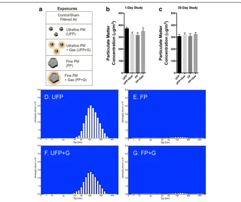
The exposure atmospheres are described in Fig. 1a, which included UFP, UFP with gaseous copollutants (UFP + G), FP, and FP with gaseous copollutants (FP + G). These core atmospheres were derived from mixed vehicle exhaust (MVE), which consisted of a combin-ation of both diesel engine exhausts (DEE) and gasoline engine exhausts (GEE), as previously described [3, 24]. Target exposure mass concentration was 300 μg PM/m3 for all exposure groups. A description of the aerosol generation technique for each of the individual atmos-pheric components or mixtures is described below. Ex-hausts from a gasoline engine (GEE) and a diesel engine (DEE) were generated and characterized as previously reported. The exhausts were extracted via eduction pumps into a 2 m3 mixing chamber where they were combined and subsequently routed to one of several separate inhalation chambers.
Generation of the base MVE atmosphere
DTC-1, Dyne Systems Co., LLC, Germantown, WI) that was controlled by a custom software program (Cell As-sistant, Dyne Systems Co.). The engine was fueled with gasoline obtained from a local station in Albuquerque, NM. Crankcase oil (10 W-30, Pennzoil Products Com-pany, Houston, TX) and oil filter (Duraguard PF52, AC Delco, Detroit, MI) were changed every 122 h (equiva-lent to 3000 miles) of engine operation.
The MVE was then created by combining a dilution of GEE (diluted approximately 10:1 with filtered air) with a similar dilution of DEE to achieve a target of 300 μg PM/m3. Relative PM contributions to the MVE atmosphere reflected a ratio of GEE to DEE of
approximately 1:5; however, GEE accounted for the ma-jority of carbon monoxide and non-methane volatile or-ganic compounds [3, 24]
Ultrafine particulate with gases (UFP + G) and ultrafine particulate without gases (UFP)
UFP + G atmosphere was generated using the MVE dir-ectly from a 1-m3mixing chamber containing DEE and GEE. UFP atmosphere was generated by passing the
MVE from 1-m3 mixing chamber through a Harvard
denuder [23]. This denuder is a parallel plate opposite flow diffusion denuder with gaseous removal efficiency of 80–90%.
Fig. 1Atmospheric characterization. (a) Visual representation of each exposure group demonstrating the relevant size and complexity of ultrafine particulate matter (UFP), ultrafine particulate matter combined with vehicular gases (UFP + G), fine particulate matter (FP), and fine particulate matter combined with gases (FP + G). The particulate matter target concentration (300μg/m3) was reached for each exposure group in both the
Fine Particulate with gases (FP + G) and fine particulate without gases (FP)
FP + G atmosphere was generated using the deposits from the exhaust line of the diesel engine. The deposits were packed in the delivery cup of a Wright Dust Feeder (WDF; CH Technologies, USA) and delivered to the in-halation exposure chamber using HEPA-filtered com-pressed air. The output from the WDF was passed through a cyclone with a cut-point of 2.5μm (URG, Inc., Chapel Hill, NC) to remove large particulates from the aerosol. HEPA filtered gases from 1 m3mixing chambers containing MVE were pumped into the exposure cham-ber. FP without gases atmosphere was generated using a similar technique as described above for FP + G; no HEPA filtered MVE gases were provided in the exposure chamber.
Atmosphere monitoring and characterization
Test atmosphere characterization was conducted as previ-ously described [25–27]. In brief, sample collection strat-egies were developed to capture and measure gas, semivolatile, and particle phases for a broad spectrum of chemical classes. Gases were analyzed by chemilumines-cence (NOx) and infrared spectroscopy (CO). Particulate mass concentration was measured via gravimetrical analysis on 47-mm glass fiber filters (GE Whatman, Pittsburgh, PA). Particle size distribution was measured with a Fast Mobility Particle Sizer (FMPS, TSI, St. Paul, MN) for the ~10–500 nm size range and an Aerodynamic Particle Sizer (TSI, St Paul, MN) to measure the 0.5–20μm size range.
Whole-body inhalation exposures to atmospheres
C57BL/6 and ApoE−/− male mice obtained from the Jackson Laboratory aged 6–8 weeks were pair-housed and maintained in controlled environment (30–60% rela-tive humidity, 20–24 °C) on a 12-h light:dark cycle with ad libitum access to water and standard chow (2016C Harlan Global Certified Rodent Chow for C57BL/6 mice). ApoE−/− mice were fed high-fat chow (TD.88137 Harlan, 21% fat, 0.2% cholesterol) for the entire period of study to model previously demonstrated vulnerability to vascular inflammatory conditions [5, 28]. Exposure chambers were also monitored for temperature and hu-midity to ensure adherence to AAALAC standards. Ani-mals were randomized into the following groups: Filtered air (FA, control), UFP, UFP + G, FP, FP + G, using 8 mice per strain per group; in one group (ApoE
−/−
mice in the FP + G group), four mice had to be ex-cluded from post-exposure biological analyses for pre-existing health reasons. Animals were placed in 2-m3 whole-body rodent inhalation chambers (Lab Products, Inc., Maywood, NJ) and chow was removed during the daily exposures. Animals received either one 6-h whole body inhalation exposure (acute exposure) or 30 days of
6-h whole body inhalation exposure (chronic exposure) to 300 μg/m3of the atmospheres (described above). All procedures were approved by the Lovelace Respiratory Research Institute’s Animal Care and Use Committee and conform to the Guide for the Care and Use of La-boratory Animals published by the US National Insti-tutes of Health (NIH Publication No. 85–23, revised 1996). Animal body weights were monitored weekly throughout the study. All mice were fasted overnight prior to necropsy. Animals were euthanized via intraper-itoneal injection of barbiturate-based sedative (Eutha-sol™). The following tissues were rapidly dissected, placed in liquid nitrogen, and stored at−80 °C until use: cerebrum (meninges, brain stem, cerebellum, OB re-moved), lung lobes, whole aorta (collected from ascend-ing aorta before abdominal bifurcation), liver, heart (upper), heart (lower), and abdominal adipose tissue.
Bronchoalveolar lavage (BALF) collection
BALF was collected via whole lung lavage with 800 μl Dulbecco’s PBS. After centrifugation, BALF supernatant was snap frozen in liquid nitrogen and stored at−80 °C until use. An aliquot of BALF supernatant was assayed for lactate dehydrogenase (LDH) and total protein as previously described [29].
BALF cells were used to prepare Cytospin slides for cell differential counting and histological scoring of par-ticulate uptake. After fixation, the slides were stained with a modified Wright-Giemsa method, and subjected to qualitative scoring by a blind observer using the fol-lowing scoring system under light microscope: 0, no par-ticles; 1, scattered cells with intra-cellular parpar-ticles; 3, slightly increased numbers of cells with particles, may not be easily detected on casual examination; 5, number of cells with particles is substantially greater than normal and can be easily noted upon examination; 7, the abun-dance of cells with particles is marked, and the intra-cellular particles are prominent or have severe cytosolic characters. The representative cell images were taken
with an Olympus CCD camera through an oil
immersion objective (×100).
Cytokine analysis in BALF
well) was added and allowed to react for 2 h at RT. Plates were washed 3× with buffer containing 1X PBS and 0.05% Tween 20. Detection antibody was added to each well and allowed to react for 1 h at RT. Plates were washed as previously noted and Read Buffer was added to each well. Plates were analyzed on MSD QuickPlex SQ 120 instrument (MSD, AI0AA-0); Discovery Work-bench (v. 4.0) software calculated cytokine concentra-tions using a linear regression analysis of the standard curve.
Transcription profiling in brain
Tissue preparation
The hippocampus and rest of cortex were dissected from the stored brains, placed in RNAlater (LifeTechnologies, AM7020), and immediately homogenized for 3 consecutive rounds using MagNA Lyzer beads (Roche, 03358941001) and homogenizer set at 6000 rpm for 60 s increments.
RNA isolation and qPCR
Total RNA was isolated from homogenized hippocampal tissue using the RNeasy Mini Kit (Qiagen, 74104). One microgram of high quality (260/280 = ~2.0 and 260/230 = ~1.0) RNA was converted cDNA using Applied Biosys-tem’s High Capacity cDNA Reverse Transcription Kit (LifeTechnologies, 4368814). Primer validation was per-formed for all TaqMan primers (LifeTechnologies) used in this study; genes of interest were matched with house-keeping genes of the same efficiency. Quantitative PCR was performed with 25 ng cDNA from hippocampal tis-sue; qPCR conditions were as follows: 2 min @ 50 °C, 10 min @ 95 °C, and 45 cycles of 15 s @ 95 °C and 60 s @ 60 °C. No template controls and RT- controls for each gene and each sample were used for each qPCR plate. Genes of interest (GOI) include: Ccl2 (C-C motif
che-mokine ligand 2; Mm00441242_m1), Ccl5 (C-C motif
chemokine ligand 5; Mm01302427_m1), Cxcl1
(chemo-kine C-X-C motif ligand 1; Mm04207460_m1), Et-1
(endothelin 1; Mm00438656_m1), Il-6 (interleukin 6;
Mm00446190_m1), Il-1β (interleukin 1 beta;
Mm00434228_m1), Tgf-β (transforming growth factor beta 1; Mm01178820_m1),Tnf-α(tumor necrosis factor alpha; Mm00443258_m1), Vcam-1 (vascular cell adhe-sion molecule 1; Mm01320970_m1). Housekeeping
genes (HKG) included B2m (beta-2-microglobulin;
Mm00437762_m1), Hprt(hypoxanthine
phosphoribosyl-transferase Mm03024075_m1), and Tbp (TATA-box
binding protein; Mm01277042_m1).
qPCR analysis
All data was run in duplicate; only samples with thresh-old cycle values (CT) under 35 were used for analysis. Average GOI CT values were normalized to average HKG CT values for associated HKGs. Subsequent ΔCT
values were assessed using the comparative CT method (ΔΔCT), and results are expressed as fold change.
Statistical analysis
One-way ANOVA (SPSS, v.19) with post-hoc analysis was performed as needed and corrected for multiple comparisons with Bonferroni, n= 6–8 per group for all analysis, except where indicated in the results section. Statistical significance was set at 95% confidence. Quan-tification of accumulation of particulate matter within resident alveolar macrophages was analyzed using the Kruskal-Wallis test for nonparametrics for both expos-ure paradigms in both strains of animals as each distri-bution failed the normality assumption of the one-way ANOVA.
Results
Characterization of inhalation exposure atmospheres
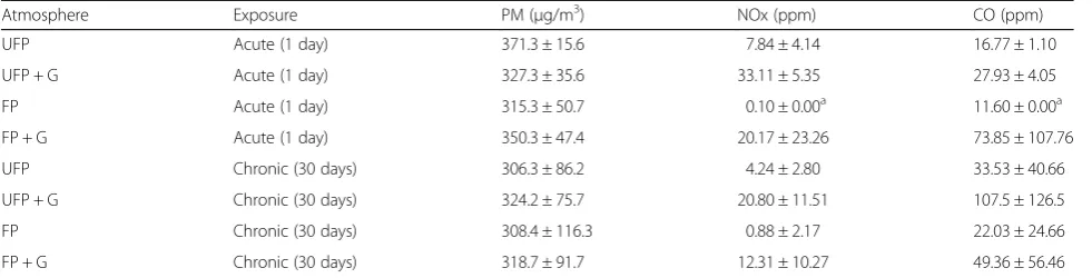
Concentrations of particulate matter, carbon monoxide, and nitrogen oxide gases for each of the test atmospheres during the acute and chronic exposures are provided in Table 1 and Fig. 1b, c. Extensive characterization of gases and particulate matter for this exposure paradigm is pro-vided in previous reports [3, 23]. PM distribution in the ul-trafine range is shown in Fig. 1d–g, with UFP atmospheres exhibiting comparable distributions (UFP mmad = 147.1 ± 1.3 nm; UFP + G mmad = 142.1 ± 1.3 nm), while FP atmo-spheres were largely devoid of detectable PM in the ultra-fine range, despite close matching of the overall mass concentrations. Fine PM was approximately 10 times larger, within the low micron range; typical number median aero-dynamic diameter (NMAD) and mass median aeroaero-dynamic diameter (MMAD) for FP and FP + G atmospheres, as measured by an Aerodynamic Particle Sizer, ranged from 1.5 to 3.0μm ± 1.3–1.6μm (geometric standard deviation).
Body weight changes, pulmonary inflammation and macrophage uptake of PM
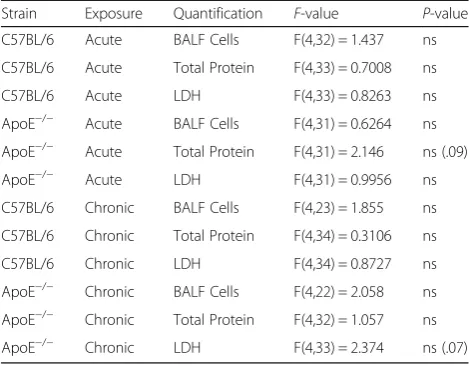
Mice in all groups exhibited significant increases in body weight during the course of the chronic 30-day exposure (Fig. 2a, b); however, when normalized to starting body weights, the UFP + G exposed C57BL/6 and ApoE−/− mice exhibited a clear reduction in this overall growth compared to control (Fig. 2c, d). Assessment of BALF for markers of inflammation, including total cell counts, LDH activity, and total protein, indicated no significant response to either acute (Fig. 3a–f ) or chronic (Fig. 4a– f ) exposure to PM or PM + G in either mouse strain. Additionally, we did not see significant influx of neutro-phils, lymphocytes or eosinophils in any group. All one-way ANOVA analyses are provided in Table 2 for bronchial lavage fluid assessments.
pattern of PM uptake. Visualization of resident lung macrophages after chronic exposure indicated significant phagocytosis of PM in both strains of mice for all atmo-spheres, which was qualitatively scored by a blinded op-erator (Fig. 5a–c). Scoring of PM uptake after acute exposures indicated moderate, but significant accumula-tion of phagocytized PM in the UFP + G and FP + G groups in C57BL/6 animals (Fig. 5d, p< .0001) and sig-nificant accumulation of the UFP and UFP + G particles in ApoE−/−animals (Fig. 5e,p< .0001). Chronic exposure resulted in more dense accumulation of particulate mat-ter and an apparent potentiation of uptake in both strains of mice exposed to UFP + G. Significant accumu-lation of UFP (p< .0001), UFP + G (p< .0001), and FP (p
< .001) particles occurred in the C57BL/6 animals (Fig. 5f ), while ApoE−/−animals had significant accumu-lation of particles in all chronic exposure groups (Fig. 5g). Table 3 provides all one-way ANOVA values and post-hoc analyses for the quantification of accumulation of particulate matter. Thus, chronic co-exposure of gases and UFP led to clearly potentiated phagocytosis of PM in comparison to other exposures, despite identical mass concentrations of PM and no measurable increase in lavagable cells in any group.
Cytokine induction in lung lavage
A single exposure to the MVE-derived atmospheres in C57BL/6 mice caused no obvious induction of cytokines
Table 1Test atmosphere concentrations of particulate matter (PM), CO, and NOx
Atmosphere Exposure PM (μg/m3) NOx (ppm) CO (ppm)
UFP Acute (1 day) 371.3 ± 15.6 7.84 ± 4.14 16.77 ± 1.10
UFP + G Acute (1 day) 327.3 ± 35.6 33.11 ± 5.35 27.93 ± 4.05
FP Acute (1 day) 315.3 ± 50.7 0.10 ± 0.00a 11.60 ± 0.00a
FP + G Acute (1 day) 350.3 ± 47.4 20.17 ± 23.26 73.85 ± 107.76
UFP Chronic (30 days) 306.3 ± 86.2 4.24 ± 2.80 33.53 ± 40.66
UFP + G Chronic (30 days) 324.2 ± 75.7 20.80 ± 11.51 107.5 ± 126.5
FP Chronic (30 days) 308.4 ± 116.3 0.88 ± 2.17 22.03 ± 24.66
FP + G Chronic (30 days) 318.7 ± 91.7 12.31 ± 10.27 49.36 ± 56.46
a
Only one measurement was captured for gas concentrations in the FP chamber per day
Fig. 2Assessment of body weights in C57BL/6 and ApoE−/−mice. Body weight was determined weekly throughout the chronic exposure
(30-day) paradigm. Body weight increased throughout the chronic exposure for (a) C57BL/6 male mice and (b) ApoE−/−male mice, aged 6–8 weeks.
in the lung (Fig. 6a, b). Interestingly, interferon-γ
(IFN-γ) was significantly reduced in the lavage of UFP + G-ex-posed C57BL/6 mice (p< .05). Conversely, ApoE−/−mice had increased expression of TNFα in the lung lavage after exposure to either UFP (p< .01) or UFP + G (p < .001) and no change in IFN-γ expression (Fig. 6c, d). Numerous other assayed cytokines were unaltered by acute exposure in either strain (data not shown). After
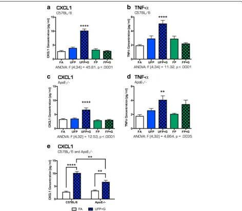
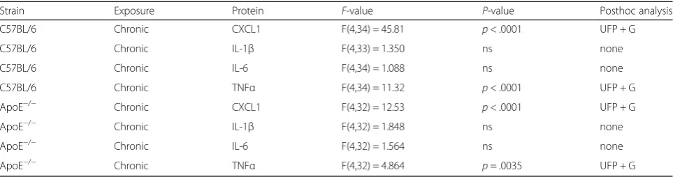
30 days of exposure, a clear picture emerged with both strains exhibiting a robust and selective increase in CXCL1 and TNFαprotein expression only in the UFP + G group, (Fig. 7a–c, p< .0001, d, p< .001). CXCL1 in-creased expression was significantly greater in C57BL/6 mice than in ApoE−/−mice (Fig. 7e,p< .001), with a sig-nificant main effect of exposure (F(1,26) = 84.11, p < .0001), main effect of animal strain (F(1,26) = 6.534,p Fig. 3Assessment of bronchial lavage fluid for cellular infiltration after acute (1-day) exposure to atmospheres. Acute exposure to UFP, UFP + G, FP, and FP + G does not induce cellular infiltration into the pulmonary system in the C57BL/6 mice as indicated by no change in bronchial lavage fluid (BALF) assessment of (a) total cell counts, (b) lactate dehydrogenase (LDH) activity, and (c) total protein (albumin). A similar lack of effect was observed in ApoE−/−mice for (d) total cell counts, (e) lactate dehydrogenase (LDH) activity, and (g) total protein (albumin).n= 7–8 per group, *p < .05; **p< .01; ***p< .001
Fig. 4Assessment of bronchial lavage fluid for cellular infiltration after chronic (30-day) exposure to atmospheres. Chronic exposure to UFP, UFP + G, FP, and FP + G does not induce cellular infiltration into the pulmonary system in the C57BL/6 mice as indicated by no changes in bronchial lavage fluid (BALF) assessment of (a) total cell counts, (b) lactate dehydrogenase (LDH) activity, or (c) total protein (albumin). A similar lack of effect was observed in ApoE−/−mice for (d) total cell counts, (e) lactate dehydrogenase (LDH) activity, and (g) total protein (albumin).n= 7–8 per
< .0168), and a significant interaction between exposure and strain, (F(1, 26) = 11.41, p< .0023). Table 4 provides all one-way ANOVA values and posthoc analyses for quantification of cytokine protein expression in the lung lavage after acute and chronic exposures in both strains of mice. Table 5 provides 2-way ANOVA values and posthoc analyses for CXCL1 expression in the lung lavage after chronic exposure to UFP+G in both strains of mice.
Acute exposure and neuroinflammatory effects in the hippocampus
To determine the effect of particulate matter size and com-position on the potential for neuroinflammation, quantifica-tion of mRNA expression of several cytokines in the hippocampus in response to exposures was performed. Despite minimal impacts on pulmonary outcomes, acute exposure induced significant changes in the expression of several cytokines in the hippocampus of both C57BL/6 and ApoE−/−animals suggesting that particulate matter induces neuroinflammation even after one day of exposure (Fig. 8a–f). In C57BL/6 mice, acute exposure to UFP alone resulted in increased cytokine expression ofCcl5(Fig. 8a,p < .05), Cxcl1 (Fig. 8b, p< .01),Tgf-β(Fig. 8e, p< .05), and
Table 2One-way ANOVA statistical analysis of BALF cells, total protein, and LDH
Strain Exposure Quantification F-value P-value
C57BL/6 Acute BALF Cells F(4,32) = 1.437 ns
C57BL/6 Acute Total Protein F(4,33) = 0.7008 ns
C57BL/6 Acute LDH F(4,33) = 0.8263 ns
ApoE−/− Acute BALF Cells F(4,31) = 0.6264 ns
ApoE−/− Acute Total Protein F(4,31) = 2.146 ns (.09)
ApoE−/− Acute LDH F(4,31) = 0.9956 ns
C57BL/6 Chronic BALF Cells F(4,23) = 1.855 ns
C57BL/6 Chronic Total Protein F(4,34) = 0.3106 ns
C57BL/6 Chronic LDH F(4,34) = 0.8727 ns
ApoE−/− Chronic BALF Cells F(4,22) = 2.058 ns
ApoE−/− Chronic Total Protein F(4,32) = 1.057 ns
ApoE−/− Chronic LDH F(4,33) = 2.374 ns (.07)
Fig. 5Particulate matter uptake in bronchial macrophages after acute and chronic exposure to atmospheres. Representative images of macrophages in the bronchial lavage fluid for (a) animals exposed to filtered air, (b) animals exposed to UFP, and (c) animals exposed to UFP + G. Images demonstrate the rubric for particulate matter uptake scoring on a scale of 0–5. Acute (1-day) exposure resulted in significant uptake of particulate matter in the (d) UFP + G and FP + G exposure groups in C57BL/6 animals and in the (e) UFP and UFP + G exposure groups in in ApoE−/−animals.
Tnf-α (Fig. 8f, p< .05) in the hippocampus. UFP + G and FP + G exposure, however, only increased hippocampal mRNA expression ofIl-1β(Fig. 8c,p< .05), while reducing expression of Il-6 (Fig. 8d, p< .0001). Interestingly, acute exposure to FP and FP + G also resulted in decreased expression ofIl-6 (Fig. 8d,p< .0001) andTgf-β(Fig. 8e,p < .05 andp< .0001, respectively) in the C57BL/6 mice.
Different results were found for acute exposures in ApoE−/−mice, which appeared to be more prone to broad inflammatory responses to most exposures. Ccl5 and Cxcl1 expression did not appear to be increased by any atmosphere after a single exposure (Fig. 9a, b). Acute posure to UFP alone resulted in significantly increased ex-pression of Il-1β (Fig. 9c, p< .05), Il-6 (Fig. 9d, p< .05), Tgf-β(Fig. 9e,p< .05) andTnf-α (Fig. 9e, p< .05). While
Il-6 and Tgf-β expression were decreased in the
hippocampus of C57BL/6 mice for most atmospheres, ApoE−/−mice exhibited opposite trends withIl-6elevated in UFP (p< .05), UFP + G (p< .05) and FP (p< .001) groups (with no difference in FP + G, Fig. 9d) andTgf-β el-evated in UFP (p< .05) and FP (p< .05) exposures and un-changed by addition of gases (Fig. 9e). HippocampalTnf-α was uniformly elevated by all single exposures in ApoE−/− mice, approximately 2-fold (Fig. 9f,p< .05).
Chronic exposure and neuroinflammatory effects in the hippocampus
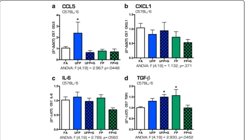
After 30 days of exposure to the four atmospheres, few inflammatory effects were noted in C57BL/6 mice. Chronic exposure to UFP resulted in significantly increased expres-sion of Ccl5 (Fig. 10a, p< .05), with no changes in Cxcl1 norIL-6cytokine expression (Fig. 10b–c). UFP + G and FP
Table 3One-way ANOVA statistical analysis of particulate matter uptake in lung
Strain Exposure F-value P-value Posthoc analysis
C57BL/6 Acute Kruskal-Wallis, 36.00 p< .0001 UFP + G, FP + G (Dunn’s)
ApoE−/− Acute Kruskal-Wallis, 25.24 p< .0001 UFP, UFP + G (Dunn’s)
C57BL/6 Chronic F(4,33) = 32.84 p< .0001 UFP, UFP + G, FP
ApoE−/− Chronic F(4,31) = 34.87 p< .0001 UFP, UFP + G, FP, FP + G
Fig. 6Cytokine protein expression profile in bronchial lavage fluid after acute (1-day) exposure to atmospheres. After 1-day of exposure to the atmospheres, bronchial lavage fluid (BALF) was obtained from animals and assessed for cytokine protein expression for (a) TNF-αin C57BL/76 BALF, (b) IFN-γin C57BL/6 BALF, (c) TNF-αin ApoE−/−BALF, and (d) IFN-γin ApoE−/−BALF. No change in (a) TNF-αexpression was observed in
C57BL/6 animals, while (b) IFN-γexpression was significantly reduced in the UFP + G exposure group in the same animals. In ApoE−/−animals, (c)
exposures led to significant increased expression of hippo-campal Tgf-β (Fig. 10d, p< .05). No other inflammatory gene changes, including Ccl1 and Et1 (data not shown), were observed in hippocampal tissues from the WT mice.
In contrast, the 30-day chronic exposure paradigm in-duced a greater and more consistent pattern of inflam-matory responses in the hippocampus of ApoE−/− animals. Assessment of the same cytokine profiles re-vealed significantly increased expression ofCcl5(Fig. 11a, p< .05),Cxcl1(Fig. 11b,p< .05,p< .01),Il-6(Fig. 11c,p < .05, p< .001) and Tgf-β (Fig. 11d, p< .01, p< .0001), with the greatest changes in cytokine expression result-ing from the UFP + G exposure atmospheres, and a
smaller but notable increase from exposure to UFP alone. Similar to C57BL/6 animals, no change in mRNA expression of Ccl2 was determined (data not shown). Table 6 includes all one-way ANOVA data values for the quantification of mRNA expression for all cytokines measured in the hippocampus of C57BL/6 and ApoE−/− mice after acute and chronic exposures. These results suggest that C57BL/6 mice exhibit healthy adaptive responses to minimize neuroinflammatory effects of vehicle-derived pollutant exposure, while the hypercho-lesterolemic mice are impaired in such homeostatic compensatory mechanisms, leading to measureable neuroinflammation.
Fig. 7Cytokine protein expression profile in bronchial lavage fluid after chronic (30-day) exposure to atmospheres. After 30-days of exposure to the atmospheres, bronchial lavage fluid (BALF) was obtained from animals and assessed for cytokine protein expression for the (a) CXCL1 in C57BL/6 BALF, (b) TNF-αin C57BL/6 BALF, (c) CXCL1 in ApoE−/−BALF and (d) TNF-αin ApoE−/−BALF. The most significant changes were induced by exposure to UFP
+ G: (a) CXCL1 expression in C57BL/6 animals, (b) TNF-αexpression in C57BL/6 animals, (c) CXCL1 expression in ApoE−/− animals, and (d) TNF-α
expression in ApoE−/−were all significantly increased in after chronic exposure to UFP + G. (e) CXCL1 expression was significantly increased in C57BL/6
Chronic exposure to MVE and MVE + gases alters expression of genes involved in cerebral vasculature
To determine if the size and composition of particulate matter impacted the expression of vascular inflamma-tory response genes in the brain, mRNA expression of Vcam1 and Et1 was assessed. Acute exposures to vehicle-derived pollutants did not alter the expression of Vcam1 in the hippocampus of C57BL/6 mice (Fig. 12a). In ApoE−/− mice, acute exposure to FP only caused a 50% increase in Vcam1 (Fig. 12b, p < .0001). Chronic exposure to UFP + G and FP in-creased Vcam1 expression in C57BL/6 mice (Fig. 12c, p< .05) but had no effect on Vcam1 expression in the hippocampus of ApoE−/− mice (Fig. 12d). No effect of chronic exposure was determined on the mRNA ex-pression of Et1 in either strain of animal (Additional file 1: Figure S1). See Table 6 for all ANOVA and F values for Vcam1 and Et1 expression after acute and chronic exposure in both animal strains.
Discussion
Recent epidemiological studies have linked traffic-related air pollution to adverse neurological outcomes [2, 30] with several toxicological reports demonstrating that PM exposure has the capacity to alter neural development [31, 32], induce neuroinflammation [33], impair cogni-tion [34], and potentially induce neurodegeneracogni-tion [35]. However, there is still much debate regarding such find-ings owing to inconsistent results and a lack of under-standing as to how inhalation exposures lead to
neurological deficits. The present study adds substan-tially to the biological plausibility of this phenomenon by providing evidence that neuroinflammatory outcomes of exposure, as assessed via cytokine production, are most pronounced with smaller PM, particularly in the presence of vehicle engine co-pollutants, a likely conse-quence of a high PM surface area for semivolatile com-pound adsorption. Furthermore, ApoE−/−mice exhibited clear vulnerability to long-term exposure to ultrafine PM recombined with gases (UFP + G exposure group), suggesting impairment of homeostatic compensatory mechanisms fully intact in C57BL/6 mice with potential neuroprotective function. Which specific aspects of the ApoE−/−mouse model on a high fat diet that contributed to this neuroinflammatory vulnerability remain uncertain. Lastly, data presented here provide an intriguing link to pulmonary effects, with clearly enhanced PM phagocytosis by alveolar macrophages and induction of proinflamma-tory cytokines in the airways without any indication of neutrophil influx or lung injury. Thus, this study suggests a potential crosstalk between pulmonary and neurological systems in response to inhaled toxicants with clear dem-onstration of exacerbated toxicological effects when PM is combined with gaseous co-pollutants.
Ample evidence supports a unique and enhanced toxicity from UFP compared to larger PM [36]. Vehicle-derived UFP are generated in the presence of gaseous co-pollutants, including carbon monoxide, oxides of nitrogen, and semivolatile and volatile hydrocarbons [25, 26, 37]. Nu-merous epidemiological findings highlight a role for road-way proximity or vehicle pollution-specific components as potent drivers of health effects [38, 39], strongly suggesting that freshly-generated PM may have enhanced toxicity. We recently found that, indeed, combined gas and PM mix-tures drove more profound vascular toxicity in ApoE−/− mice [23]. As the role of PM surface interactions with gas-eous species remained uncertain, we developed the present exposure paradigm to compare different sizes of PM of the same chemical composition in a head-to-head manner. While our findings suggest that certain biological outcomes can be induced by all respirable PM, the most consistent
Table 4One-way ANOVA statistical analysis of protein expression in BALF
Strain Exposure Protein F-value P-value Posthoc analysis
C57BL/6 Chronic CXCL1 F(4,34) = 45.81 p< .0001 UFP + G
C57BL/6 Chronic IL-1β F(4,33) = 1.350 ns none
C57BL/6 Chronic IL-6 F(4,34) = 1.088 ns none
C57BL/6 Chronic TNFα F(4,34) = 11.32 p< .0001 UFP + G
ApoE−/− Chronic CXCL1 F(4,32) = 12.53 p< .0001 UFP + G
ApoE−/− Chronic IL-1β F(4,32) = 1.848 ns none
ApoE−/− Chronic IL-6 F(4,32) = 1.564 ns none
ApoE−/− Chronic TNFα F(4,32) = 4.864 p= .0035 UFP + G
Table 5Two-way ANOVA statistical analysis of CXCL1 protein expression in BALF
Strain Exposure Protein F-value P-value Posthoc analysis
C57BL/6 Chronic CXCL1 Interaction: F(1,26) = 11.41
p= .0023 UFP + G
Strain: F(1, 26) = 6.534
p= 0.0168
Exposure: F(1,26) = 84.11
and severe pulmonary effects and neuroinflammatory consequences were clearly manifested in the UFP + G group, consistent with the hypothesis that co-pollutant gases adsorb onto the surface of PM, and greater PM surface area permits enhanced adsorption, leading to
more deleterious outcomes. Indeed, the size of UFPs results in deeper deposition in the lung after inhal-ation and subsequent locinhal-ation within and beyond the lung epithelial barrier with potential uptake into cells resulting in greater potential toxicity [40].
Fig. 8Cytokine expression profile in the C57BL/6 hippocampus after acute (1-day) exposure to atmospheres. After 1-day of exposure to atmospheres, cytokine mRNA expression was assessed in the hippocampus of the C57BL/6 mouse brain for the following: (a) Ccl5, (b) Cxcl1, (c) IL-1β, (d) IL-6, (e) Tgf-β, and (f) Tnf-α. Exposure to UFP significantly altered mRNA expression of (a) Ccl5, (b) Cxcl1, (c) IL-1β, (e) Tgf-β, and (f) Tnf-α. Exposure to UFP+G or FP+G significantly increased mRNA expression of (c) IL-1β. Exposure to UFP + G, FP and FP + G significantly decreased mRNA expression of (d) IL-6 and (e) Tgf-β.n= 7–8 per group, *p< .05; **p< .01; ***p< .001; ****p< .0001
Fig. 9Cytokine expression profile in the ApoE−/−hippocampus after acute (1-day) exposure to atmospheres. After 1-day of exposure to
atmospheres, cytokine mRNA expression was assessed in the hippocampus of the ApoE−/−mouse brain for the following: (a) Ccl5, (b) Cxcl1, (c)
Among the more interesting outcomes from this novel study design, we observed dramatic differences in macro-phage phagocytosis of PM in the UFP + G chronic (30 day) exposure group, concurrent with increased TNFα and CXCL1 protein expression in the lavage but not increased cellularity or total protein. Severe accumulation of PM did not occur after a single day of exposure; however, UFP + G exposure led to as much PM uptake as any of the other exposures. Macrophage activation resulting from diesel PM uptake has been shown to cause release of highly toxic fac-tors that activate endothelial cells more so than PM expos-ure can induce directly, though these factors have yet to be characterized [41]. Mutlu and colleagues demonstrated a role for macrophage-derived cytokines, specifically IL-6, in driving systemic platelet activation [42]. Thus, such phago-cytosis and macrophage activation appears mechanistically linked to systemic outcomes. Why the combination of UFP and co-pollutant gases leads to greater phagocytosis that the same amount of denuded UFP or same mass concen-tration of FP with gases is unclear. However, freshly gener-ated elemental carbon-based PM has been shown to have greater potential to induced oxidative stress in macro-phages in vitro[43]; this study further indicated that total surface area of different PM samples (all largely comprised of elemental carbon) was a key driver of all oxidative
outcomes. A major challenge for understanding the mech-anistic basis of the enhanced phagocytosis relates to the need for freshly-generated UFP from the MVE system. Collection of PM from fresh exhaust for later exposure leads to the loss of volatile components that may well be the causative factors. Further study of this phenomenon will, however, provide important information regarding the potency of near-roadway pollution and the pathogen-esis of PM-induced systemic disease.
ofIl-6andTgf-βafter acute exposures in the hippocampus of C57BL/6 mice was observed. As the balance between pro- and anti-inflammatory processes in the brain is highly dynamic and temporally regulated, especially with IL-6 [48], compensatory mechanisms were potentially already underway 24 h post-acute exposure. TGF-β has been shown to be neuroprotective and reduce brain injury in response to ischemia [49]; thus, lack of TGF-βsignaling could indicate that the exposure groups were altering the balance between the pro- and anti-inflammatory cascades in the C57BL/6 hippocampus. After chronic exposure, in-creased Tgf-β expression in the C57BL/6 animals in re-sponse to UFP + G may indicate anti-inflammatory protective response, perhaps derived from microglia, to control the pro-inflammatory cascade that occurs earlier in the exposure paradigm. Thus, C57BL/6 mice exhibit healthy adaptive responses to minimize neuroinflamma-tory effects of vehicle-derived pollutant exposure. In sharp contrast to the C57BL/6 animals, the hypercholesterol-emic ApoE−/−mice displayed measurable neuroinflamma-tion via increased cytokine expression, suggesting impaired homeostatic compensatory mechanisms after 30 days of exposure. Anti-inflammatory responses includ-ing increasedCxcl1andTgf-βexpression, potentially from
microglia, in the hippocampus may have been combating increased proinflammatory cytokine expression ofIl-6and Ccl5, though IL-6 for its part has been implicated in both pro and anti-inflammatory responses in the brain and may be paramount for healthy aging of the brain [50]. It should be noted that“activation” of microglia, while not directly measured in this study, results in the production of a number of pro- and anti-inflammatory cytokines depend-ing on the environmental niche [51]. Microglia can have divergent responses depending on their localization, hence increased pro-inflammatory and anti-inflammatory cyto-kines, like IL-6 and TGF-β, in the same temporal range, as in the hippocampus of ApoE−/−mice after chronic expos-ure. While we cannot definitively state that this cytokine profile is directly from microglia, it is likely that neuroin-flammation is occurring after chronic exposure in the ApoE−/− mice, and this could underlie altered microglia activity eventually culminating in neuronal death and neu-rodegeneration. These effects will be determined in future studies. For now, we can conclude that as observed after acute exposure in the C57BL/6 animals, in ApoE−/− ani-mals, the greatest response in the cytokine profile was elic-ited by the UFP and UFP + G groups, even after chronic exposure, suggesting that particulate matter size and the Fig. 11Cytokine expression profile in the ApoE−/−hippocampus after chronic (30-day) exposure to atmospheres. After 30 days of exposure to
atmospheres, cytokine mRNA expression was assessed in the hippocampus of the ApoE−/−mouse brain for the following: (a) Ccl5, (b) Cxcl1, (c)
recombination of smaller particulate matter with gases in-duces a greater inflammatory response than larger particles (even when larger particles are combined with gases).
The small, highly complex exposure groups generated for this study appropriately model the type of near-roadway air pollution to which many people are exposed. While several studies have assessed the neuroinflamma-tory potential of exposure to PM, these studies have fo-cused on larger sized particles, PM2.5 and PM10, without assessment of particulate matter recombined with gases for modeling typical exposure paradigms in humans. For example, long-term, chronic exposure to PM2.5 increases proinflammatory cytokine expression of TNF-αand IL-1β in the rat hippocampus that is concurrent with decreased dendritic arborization, deficits in learning and memory,
and depressive-like behaviors [34]. Studies assessing UFP exposure have shown various outcomes including oxida-tive stress, IL-1αand TNF-αexpression in the brain [52], altered MAPK signaling, and elevated GFAP indicative of astrocyte activation in ApoE−/− mice [53]. Early postnatal exposure to UFP results in differential microglial re-sponses dependent on sex and region of the brain, with some areas, such as the hippocampus, responding to UFP via increased microglial activation [31, 54], suggesting en-hanced vulnerability to UFP exposure during
neurodeve-lopment. UFP exposure exacerbates natural aging
mechanisms in the brain via translocation to brain regions resulting in chronic neuroinflammation concurrent with neurodegenerative outcomes [55–57]. While further characterization of the specific microglial response, the
Table 6One-way ANOVA statistical analysis of qPCR gene expression in hippocampus
Strain Exposure Gene F-value P-value Posthoc analysis
relative to control
C57BL/6 Acute Ccl5 F(4,32) = 5.808 p< .0012 UFP
C57BL/6 Acute Cxcl1 F(4,27) = 8.175 p< .0001 UFP
C57BL/6 Acute IL-1β F(4,32) = 3.404 p= 0.0199 UFP + G, FP + G
C57BL/6 Acute IL-6 F(4,33) = 52.50 p< .0001 UFP + G, FP, FP + G
(decreased)
C57BL/6 Acute Tgf-β F(4,33) = 27.02 p < .0001 UFP (increased), UFP
+ G, FP, FP + G (decreased)
C57BL/6 Acute Tnf-α F(4,33) = 3.909 p= .0105 UFP
C57BL/6 Acute Vcam-1 F(4,32) = 1.205 ns none
ApoE−/− Acute Ccl5 F(4,30) = 0.2138 ns none
ApoE−/− Acute Cxcl1 F(4,30) = 4.377 p= 0.0066 none
ApoE−/− Acute IL-1β F(4,29) = 2.766 p= 0.05 UFP, UFP + G
ApoE−/− Acute IL-6 F(4,31) = 6.408 p< 0.001 UFP, UFP + G, FP
ApoE−/− Acute Tgf-β F(4,31) = 3.978 p= .0102 UFP, FP
ApoE−/− Acute Tnf-α F(4,30) = 4.556 p= .0054 UFP, UFP + G, FP, FP + G
ApoE−/− Acute Vcam-1 F(4,30) = 8.388 p< .0001 FP
C57BL/6 Chronic Ccl5 F(4,19) = 2.967 p= .0448 UFP
C57BL/6 Chronic Ccl2 F(4,18) = 1.398 ns none
C57BL/6 Chronic Cxcl1 F(4,19) = 1.132 ns none
C57BL/6 Chronic Et1 F(4,19) = 1.866 ns none
C57BL/6 Chronic IL-6 F(4,19) = 2.789 ns (p= .0560) none
C57BL/6 Chronic Tgf-β F(4,19) = 2.930 p= .0452 UFP + G, FP
C57BL/6 Chronic Vcam-1 F(4,19) = 2.962 p= .0465 UFP + G, FP
ApoE−/− Chronic Ccl5 F(4,32) = 3.248 p= .0241 UFP + G
ApoE−/− Chronic Ccl2 F(4,29) = 1.422 ns none
ApoE−/− Chronic Cxcl1 F(4,27) = 12.02 p< .0001 UFP, UFP + G
ApoE−/− Chronic Et1 F(4,32) = 1.78 ns none
ApoE−/− Chronic IL-6 F(4,32) = 16.45 p< .0001 UFP, UFP + G
ApoE−/− Chronic Tgf-β F(4,32) = 27.87 p< .0001 UFP, UFP + G
potential for neurodegeneration, and the mechanism by which the exposures induce neuroinflammation (poten-tially via translocation of the particles) is required, this study is the first to demonstrate neuroinflammatory out-comes using exposure models that include recombination with gases to most accurately model traffic-related air pol-lution. Furthermore, ApoE−/−mice fed high fat chow were used to model individuals with increased cardiovascular disease (CVD) risk; our results suggest that common traf-fic emission exposures, whether acute or chronic, could result in more profound neuroinflammation in at-risk populations, such as those with CVD, who may lack the appropriate compensatory mechanisms to combat pro-inflammatory cascades in the brain. Based on these data, we can speculate that long-term exposure to common traffic emissions (UFP + G) would likely result in even greater exacerbation of neuroinflammation, potentially resulting in behavioral manifestations and neurodegenera-tion, possibly modeling neurological deficits observed in humans.
Conclusion
We developed an exposure model to determine the con-tribution of particle size and particle complexity to
pulmonary and potential neuroinflammatory outcomes after acute and chronic exposures in C57BL/6 and ApoE
−/−
animals. Our results showed that chronic exposure to ultrafine carbonaceous particles mixed with vehicular emissions exerted greater effects than UFP alone, or fine particles mixed with the gas phase mixture, suggesting that the surface area of particulates and the interaction of surface-adhering gaseous components enhances pul-monary and systemic toxicity. This outcome implicates freshly generated UFP as inherently more toxic than PM that has aged and lost surface-adhered volatile gases, and further suggests that near-roadway PM may have greater health effects than would be predicted from con-centration alone. Future analysis into the potential pathogenesis of neuroinflammatory outcomes by inhaled pollutants and the role of cardiovascular disease as a contributing factor will help resolve important mechan-istic questions not addressed by the present study.
Additional file
Additional file 1: Figure S1. Et-1 mRNA expression in the hippocampus of C57BL/6 and ApoE−/−mice after acute and chronic exposure to atmospheres. Expression of Et-1 mRNA was determined after
chronic exposures in the (a) hippocampus of C57BL/6 mice and (b) hippocampus of ApoE−/−mice, with no significant effects induced by any atmospheres. (TIFF 754 kb)
Acknowledgements
Not applicable.
Funding
The work was supported by the National Institutes of Health (NIH ES014639 to MJC) and by the Environmental Protection Agency, Assistance Agreement RD-83479601-0 (Clean Air Research Centers). The views expressed in this document are those of the authors. Mention of trade names or commercial products does not constitute endorsement or recommendation for use by the United States EPA.
Availability of data and materials
The datasets supporting the conclusions of this article are included within the article and its supplementary files.
Authors’contributions
CRT, KEZ, BEB, and MJC designed experiments and interpreted data. CRT and MJC analyzed data and wrote the manuscript. CRT and GH conducted experiments. BNS and VR aided in preparation of samples. JL, HI, and JDM designed and performed the exposures and the analysis of particulate matter size and distribution. All authors approved the final manuscript.
Competing interests
The authors declare that they have no competing interests.
Consent for publication
Not applicable.
Ethics approval and consent to participate
Not applicable.
Author details
1Department of Pharmaceutical Sciences, The University of New Mexico College of Pharmacy, Albuquerque, NM, USA.2Lovelace Respiratory Research Institute, Albuquerque, NM, USA.3Department of Pharmacy Practice & Administrative Sciences, The University of New Mexico, Albuquerque, NM, USA.
Received: 2 October 2016 Accepted: 25 November 2016
References
1. Hoffmann B, Moebus S, Möhlenkamp S, Stang A, Lehmann N, Dragano N, et al. Residential exposure to traffic is associated with coronary atherosclerosis. Circulation. 2007;116(5):489–96.
2. Volk HE, Hertz-Picciotto I, Delwiche L, Lurmann F, McConnell R. Residential proximity to freeways and autism in the CHARGE study. Environ Health Perspect. 2011;119(6):873–7. doi:10.1289/ehp.1002835.
3. Vedal S, Campen MJ, McDonald JD, Kaufman JD, Larson TV, Sampson PD, et al. National particle component toxicity (NPACT) initiative report on cardiovascular effects. Res Rep. 2013;178:238.
4. Bai N, Kido T, Suzuki H, Yang G, Kavanagh TJ, Kaufman JD, et al. Changes in atherosclerotic plaques induced by inhalation of diesel exhaust.
Atherosclerosis. 2011;216(2):299–306. doi:10.1016/j.atherosclerosis.2011.02. 019.
5. Campen MJ, Lund AK, Knuckles TL, Conklin DJ, Bishop B, Young D, et al. Inhaled diesel emissions alter atherosclerotic plaque composition in ApoE(−/ −) mice. Toxicol Appl Pharmacol. 2010;242(3):310–7. doi:10.1016/j.taap.2009.
10.021.
6. Hansen CS, Sheykhzade M, Moller P, Folkmann JK, Amtorp O, Jonassen T, et al. Diesel exhaust particles induce endothelial dysfunction in apoE−/−mice.
Toxicol Appl Pharmacol. 2007;219(1):24–32. doi:10.1016/j.taap.2006.10.032. 7. Hazari MS, Haykal-Coates N, Winsett DW, Krantz QT, King C, Costa DL, et al.
TRPA1 and sympathetic activation contribute to increased risk of triggered cardiac arrhythmias in hypertensive rats exposed to diesel exhaust. Environ Health Perspect. 2011;119(7):951–7. doi:10.1289/ehp.1003200.
8. Mills NL, Tornqvist H, Robinson SD, Gonzalez M, Darnley K, MacNee W, et al. Diesel exhaust inhalation causes vascular dysfunction and impaired endogenous fibrinolysis. Circulation. 2005;112(25):3930–6. doi:10.1161/ CIRCULATIONAHA.105.588962.
9. Lund AK, Knuckles TL, Obot Akata C, Shohet R, McDonald JD, Gigliotti A, et al. Gasoline exhaust emissions induce vascular remodeling pathways involved in atherosclerosis. Toxicol Sci. 2007;95(2):485–94. doi:10.1093/toxsci/kfl145. 10. Loane C, Pilinis C, Lekkas TD, Politis M. Ambient particulate matter and its
potential neurological consequences. Rev Neurosci. 2013;24(3):323–35. 11. Levesque S, Taetzsch T, Lull ME, Kodavanti U, Stadler K, Wagner A, et al.
Diesel exhaust activates and primes microglia: air pollution,
neuroinflammation, and regulation of dopaminergic neurotoxicity. Environ Health Perspect. 2011;119(8):1149–55. doi:10.1289/ehp.1002986. 12. Levesque S, Taetzsch T, Lull ME, Johnson JA, McGraw C, Block ML. The role
of MAC1 in diesel exhaust particle-induced microglial activation and loss of dopaminergic neuron function. J Neurochem. 2013;125(5):756–65. doi:10. 1111/jnc.12231.
13. Cheng H, Saffari A, Sioutas C, Forman HJ, Morgan TE, Finch CE. Nano-scale particulate matter from urban traffic rapidly induces oxidative stress and inflammation in olfactory epithelium with concomitant effects on brain. Environ Health Perspect. 2016. doi:10.1289/ehp134.
14. Cheng H, Davis DA, Hasheminassab S, Sioutas C, Morgan TE, Finch CE. Urban traffic-derived nanoparticulate matter reduces neurite outgrowth via TNFalpha in vitro. J Neuroinflammation. 2016;13:19. doi:10.1186/s12974-016-0480-3. 15. Morgan TE, Davis DA, Iwata N, Tanner JA, Snyder D, Ning Z, et al.
Glutamatergic neurons in rodent models respond to nanoscale particulate urban air pollutants in vivo and in vitro. Environ Health Perspect. 2011; 119(7):1003–9. doi:10.1289/ehp.1002973.
16. Cappellano G, Carecchio M, Fleetwood T, Magistrelli L, Cantello R, Dianzani U, et al. Immunity and inflammation in neurodegenerative diseases. Am J Neurodegener Dis. 2013;2(2):89–107.
17. van Dijk G, van Heijningen S, Reijne AC, Nyakas C, van der Zee EA, Eisel ULM. Integrative neurobiology of metabolic diseases, neuroinflammation, and neurodegeneration. Front Neurosci. 2015;9:173. doi:10.3389/fnins.2015.00173. 18. Channell MM, Paffett ML, Devlin RB, Madden MC, Campen MJ. Circulating factors induce coronary endothelial cell activation following exposure to inhaled diesel exhaust and nitrogen dioxide in humans: evidence from a novel translational in vitro model. Toxicol Sci. 2012;127(1):179–86. doi:10. 1093/toxsci/kfs084.
19. Hoffmann B, Moebus S, Dragano N, Stang A, Mohlenkamp S, Schmermund A, et al. Chronic residential exposure to particulate matter air pollution and systemic inflammatory markers. Environ Health Perspect. 2009;117(8):1302–8. doi:10.1289/ehp.0800362.
20. Kampfrath T, Maiseyeu A, Ying Z, Shah Z, Deiuliis JA, Xu X, et al. Chronic fine particulate matter exposure induces systemic vascular dysfunction via NADPH oxidase and TLR4 pathways. Circ Res. 2011;108(6):716–26. doi:10. 1161/CIRCRESAHA.110.237560.
21. Schisler JC, Ronnebaum SM, Madden M, Channell M, Campen M, Willis MS. Endothelial inflammatory transcriptional responses to an altered plasma exposome following inhalation of diesel emissions. Inhal Toxicol. 2015:1–9; doi:10.3109/08958378.2015.1030481.
22. Tamagawa E, Bai N, Morimoto K, Gray C, Mui T, Yatera K, et al. Particulate matter exposure induces persistent lung inflammation and endothelial dysfunction. Am J Physiol Lung Cell Mol Physiol. 2008;295(1):L79–85. doi:10. 1152/ajplung.00048.2007.
23. Campen M, Robertson S, Lund A, Lucero J, McDonald J. Engine exhaust particulate and gas phase contributions to vascular toxicity. Inhal Toxicol. 2014;26(6):353–60. doi:10.3109/08958378.2014.897776.
24. Lund AK, Lucero J, Harman M, Madden MC, McDonald JD, Seagrave JC, et al. The oxidized low-density lipoprotein receptor mediates vascular effects of inhaled vehicle emissions. Am J Respir Crit Care Med. 2011;184(1): 82–91. doi:10.1164/rccm.201012-1967OC.
25. McDonald, Barr E, White R, Chow J, Schauer J, Zielinska B, et al. Generation and characterization of four dilutions of diesel engine exhaust for a subchronic inhalation study. Environ Sci Technol. 2004;38(9):2513–22. 26. McDonald, Barr EB, White RK, Kracko D, Chow JC, Zielinska B, et al.
Generation and characterization of gasoline engine exhaust inhalation exposure atmospheres. Inhal Toxicol. 2008;20(13):1157–68.
28. Lund AK, Lucero J, Lucas S, Madden MC, McDonald JD, Seagrave JC, et al. Vehicular emissions induce vascular MMP-9 expression and activity associated with endothelin-1-mediated pathways. Arterioscler Thromb Vasc Biol. 2009;29(4):511–7. doi:10.1161/ATVBAHA.108.176107.
29. Seagrave J, Campen MJ, McDonald JD, Mauderly JL, Rohr AC. Oxidative stress, inflammation, and pulmonary function assessment in rats exposed to laboratory-generated pollutant mixtures. J Toxicol Environ Health A. 2008; 71(20):1352–62. doi:10.1080/15287390802271566.
30. Jung CR, Lin YT, Hwang BF. Ozone, particulate matter, and newly diagnosed Alzheimer’s disease: a population-based cohort study in Taiwan. J Alzheimers Dis. 2015;44(2):573–84. doi:10.3233/jad-140855.
31. Allen JL, Liu X, Pelkowski S, Palmer B, Conrad K, Oberdörster G, et al. Early postnatal exposure to ultrafine particulate matter air pollution: persistent ventriculomegaly, neurochemical disruption, and glial activation preferentially in male mice. Environ Health Perspect. 2014;122(9):939 (Online).
32. Woodward N, Finch CE, Morgan TE. Traffic-related air pollution and brain development. AIMS Environ Sci. 2015;2(2):353–73. doi:10.3934/environsci. 2015.2.353.
33. Mumaw CL, Levesque S, McGraw C, Robertson S, Lucas S, Stafflinger JE, et al. Microglial priming through the lung-brain axis: the role of air pollution-induced circulating factors. Faseb J. 2016;30(5):1880–91. doi:10.1096/fj.201500047.
34. Fonken L, Xu X, Weil ZM, Chen G, Sun Q, Rajagopalan S, et al. Air pollution impairs cognition, provokes depressive-like behaviors and alters hippocampal cytokine expression and morphology. Mol Psychiatry. 2011;16(10):987–95. 35. Levesque S, Surace MJ, McDonald J, Block ML. Air pollution & the brain:
Subchronic diesel exhaust exposure causes neuroinflammation and elevates early markers of neurodegenerative disease. J Neuroinflammation. 2011;8: 105. doi:10.1186/1742-2094-8-105.
36. Oberdorster G. Pulmonary effects of inhaled ultrafine particles. Int Arch Occup Environ Health. 2001;74(1):1–8.
37. Vedal, Campen MJ, McDonald JD, Larson T, Sampson PD, Sheppard L, et al. National particle component toxicity (NPACT) initiative report on cardiovascular effects. Res Rep. 2013;178:5–8. (Health Effects Institute). 38. Sarnat JA, Marmur A, Klein M, Kim E, Russell AG, Sarnat SE, et al. Fine particle
sources and cardiorespiratory morbidity: an application of chemical mass balance and factor analytical source-apportionment methods. Environ Health Perspect. 2008;116(4):459–66. doi:10.1289/ehp.10873. 39. Sioutas C, Delfino RJ, Singh M. Exposure assessment for atmospheric
ultrafine particles (UFPs) and implications in epidemiologic research. Environ Health Perspect. 2005;113(8):947–955.
40. Li N, Sioutas C, Cho A, Schmitz D, Misra C, Sempf J, et al. Ultrafine particulate pollutants induce oxidative stress and mitochondrial damage. Environ Health Perspect. 2003;111(4):455–60.
41. Shaw CA, Robertson S, Miller MR, Duffin R, Tabor CM, Donaldson K, et al. Diesel exhaust particulate–exposed macrophages cause marked endothelial cell activation. Am J Respir Cell Mol Biol. 2011;44(6):840–51. doi:10.1165/ rcmb.2010-0011OC.
42. Mutlu GM, Green D, Bellmeyer A, Baker CM, Burgess Z, Rajamannan N, et al. Ambient particulate matter accelerates coagulation via an IL-6-dependent pathway. J Clin Invest. 2007;117(10):2952–61. doi:10.1172/jci30639. 43. Beck-Speier I, Dayal N, Karg E, Maier KL, Schumann G, Schulz H, et al.
Oxidative stress and lipid mediators induced in alveolar macrophages by ultrafine particles. Free Radic Biol Med. 2005;38(8):1080–92. doi:10.1016/j. freeradbiomed.2005.01.004.
44. Gong T, Dalman C, Wicks S, Dal H, Magnusson C, Lundholm C, et al. Perinatal exposure to traffic-related air pollution and autism spectrum disorders. Environ Health Perspect. 2016. doi:10.1289/ehp118.
45. Raz R, Roberts AL, Lyall K, Hart JE, Just AC, Laden F, et al. Autism spectrum disorder and particulate matter air pollution before, during, and after pregnancy: a nested case-control analysis within the Nurses’Health Study II Cohort. Environ Health Perspect. 2015;123(3):264–70. doi:10.1289/ehp.1408133. 46. Calderon-Garciduenas L, Reynoso-Robles R, Vargas-Martinez J,
Gomez-Maqueo-Chew A, Perez-Guille B, Mukherjee PS, et al. Prefrontal white matter pathology in air pollution exposed Mexico City young urbanites and their potential impact on neurovascular unit dysfunction and the development of Alzheimer’s disease. Environ Res. 2016;146:404–17. doi:10.1016/j.envres. 2015.12.031.
47. Tyler CR, Allan AM. The effects of arsenic exposure on neurological and cognitive dysfunction in human and rodent studies: a review. Curr Environ Health Rep. 2014;1:132–47.
48. Gruol DL. IL-6 regulation of synaptic function in the CNS. Neuropharmacology. 2015;96(Part A):42–54.
49. Dobolyi A, Vincze C, Pál G, Lovas G. The neuroprotective functions of transforming growth factor beta proteins. Int J Mol Sci. 2012;13:8219–58. 50. Godbout JP, Johnson RW. Interleukin-6 in the aging brain. J Neuroimmunol.
2004;147(1–2):141–4.
51. Heppner FL, Ransohoff RM, Becher B. Immune attack: the role of inflammation in Alzheimer disease. Nat Rev Neurosci. 2015;16(6):358–72. 52. Campbell A, Oldham M, Becaria A, Bondy S, Meacher D, Sioutas C, et al. Particulate matter in polluted air may increase biomarkers of inflammation in mouse brain. Neurotoxicology. 2005;26(1):133–40.
53. Kleinman MT, Araujo J, Nel A, Sioutas C, Campbell A, Cong PQ, et al. Inhaled ultrafine particulate matter affects CNS inflammatory processes and may act via MAP kinase signaling pathways. Toxicol Lett. 2008;178(2):127–30. doi:10.1016/j.toxlet.2008.03.001.
54. Allen JL, Oberdorster G, Morris-Schaffer K, Wong C, Klocke C, Sobolewski M, et al. Developmental neurotoxicity of inhaled ambient ultrafine particle air pollution: parallels with neuropathological and behavioral features of autism and other neurodevelopmental disorders. Neurotoxicology. 2015. doi:10.1016/j.neuro.2015.12.014.
55. Oberdorster G, Sharp Z, Atudorei V, Elder A, Gelein R, Kreyling W, et al. Translocation of inhaled ultrafine particles to the brain. Inhal Toxicol. 2004; 16(6–7):437–45. doi:10.1080/08958370490439597.
56. Elder A, Gelein R, Silva V, Feikert T, Opanashuk L, Carter J, et al. Translocation of inhaled ultrafine manganese oxide particles to the central nervous system. Environ Health Perspect. 2006;114(8):1172–8.
57. Heusinkveld HJ, Wahle T, Campbell A, Westerink RH, Tran L, Johnston H, et al. Neurodegenerative and neurological disorders by small inhaled particles. Neurotoxicology. 2016;56:94–106. doi:10.1016/j.neuro.2016.07.007.
• We accept pre-submission inquiries
• Our selector tool helps you to find the most relevant journal
• We provide round the clock customer support
• Convenient online submission
• Thorough peer review
• Inclusion in PubMed and all major indexing services
• Maximum visibility for your research
Submit your manuscript at www.biomedcentral.com/submit