Imaging of hibernomas: A retrospective study on
twelve cases
Papathanassiou
et al.
R E S E A R C H
Open Access
Imaging of hibernomas: A retrospective study on
twelve cases
Zafiria G Papathanassiou
1,2, Marco Alberghini
1, Sophie Taieb
3, Costantino Errani
1, Piero Picci
1and Daniel Vanel
1*Abstract
Background:To analyze the imaging features of hibernomas on computed tomography (CT) and magnetic resonance (MRI).
Methods:Twelve hibernomas were retrospectively assessed with CT and MR imaging and compared to the histology of the specimen
Results:Nine females and three males with a mean age of 30 years were included. Ten tumors occurred in the thigh and two affected the subcutis of the periscapular and buttock regions. On eight CT scans, seven (87,5%) lesions were homogeneous and mildly to moderately hyperdense compared to subcutaneous fat while one lesion was heterogeneous with mixed hypo and hyperattenuating areas. On six T1W images, five (83,3%) lesions
appeared homogeneous and hypointense relative to subcutaneous fat and one was heterogeneous. Incomplete fat suppression was depicted in all cases. All lesions displayed marked enhancement. Large intratumoral vessels were depicted in three of the 12 (25%) cases. Septations were depicted on four of the eight unenhanced CT and on all six MRI examinations.
Conclusions:Hibernoma usually appears hypodense and hypointense relative to subcutaneous fat on pre-contrast CT and MR T1W with variable enhancement patterns and incomplete fat suppression on STIR or fat-saturated sequences. These characteristics relate directly to the presence of brown fat. However, atypical findings such as heterogeneous patterns of mixed fatty and non fatty components on unenhanced CT and MR T1W can be also encountered. Absence of large intratumoral vessels should not exclude hibernomas from the differential diagnosis of regional lipomatous tumors.
Introduction
Hibernomas are rare benign lipomatous tumors originat-ing from residual brown fat cells. At the beginnoriginat-ing of the century, Merkel [1] first described them as“ pseudolipo-mas”. Owing to their resemblance to the brown fat of hibernating animals, the term“hibernoma”was coined by Gery in 1914 [2]. They affect chiefly adults in the 3rdof 4thdecades of life [3] and they usually grow in the ves-tiges, where brown fat is found in fetuses and infants, such as the shoulder, neck, axilla, the periscapular and interscapular area, thorax and retroperitoneum [4].
The rareness of this lipomatous tumor and its histolo-gic configuration make it a challenging radiolohistolo-gic diagno-sis. To the best of author’s knowledge only three series [5-7] and several case reports [8-18] have exhibited the
imaging characteristics of hibernomas. The present study, being the largest in the imaging of hibernomas, highlights the spectrum of imaging appearances (CT/ MRI) of twelve histologically proven cases of hibernomas and stresses the positive impact of imaging in the pre-operative planning when a complex fatty mass is encountered.
Materials and methods
Over a 23-year period (1986-2009) fifteen cases diagnosed as hibernomas were identified in the histopathology data-base of two tertiary referral bone and soft tissue tumor centers. Imaging studies were available in twelve cases. Information regarding age, sex, clinical examination, lesion size and site was registered. Evaluation of the pre-opera-tive imaging investigations (CT-MRI) was performed. Three patients underwent CT and MRI examinations, while five had only CT scans and four had only MRI. Of * Correspondence: [email protected]
1Research, The Rizzoli Institute, Via del Barbiano 1/10, 40106, Bologna, Italy
Full list of author information is available at the end of the article
the latter four patients, two had also ultrasound (U/S) examinations and one of them underwent position emis-sion tomography (18F FDG-PET). All CT examinations were performed before and after contrast medium intrave-nous administration. MRI studies obtained from referring institutions included a variety of T1weighted spin-echo (T1WSE), T2weighted spin-echo (T2WSE),T2 weighted fast spin-echo with fat suppression(T2 FSE Fat Sat), short Tau inversion recovery (STIR) and T1W SE with fat sup-pression sequences(T1 SE Fat Sat). Post gadolinium images were acquired on six cases; one of which had also a MR Angiography. Imaging findings were evaluated by two radiologists (one experienced on bone and soft tissue tumors radiologist and one musculoskeletal radiologist clinical fellow. Radiological assessment included lesion size, location, and internal morphology along with CT attenuation, MR signal intensity and homogeneity, which were compared to subcutaneous fat and muscle. Addition-ally, contrast enhancement, U/S echogeneity and standard uptake value (SUV) on18F FDG-PET were recorded. His-topathological analysis was performed by one experienced bone and soft tissue tumor pathologist. All patients had complete but marginal resections of the lesions.
Results
Table 1 displays the imaging appearances of the pre-sented cases. Of the twelve patients nine were female and three male, from 19 to 46 years old (mean: 30 y). Each patient had one lesion and all of them presented with a slow-growing expansion of the affected soft tissue area. Physical examination revealed palpable lumps of various sizes that were painless and relatively mobile. Laboratory tests were not remarkable. Ten of the twelve lesions were located in the upper thigh (eight in the anterior compart-ment and two in the posterior compartcompart-ment) and the other two were located subcutaneously in the lower peri-scapular and buttock regions. All lesions were well circumscribed and presented with fusiform elongated of ovoid shapes. The smallest lesion measured 5,5 × 4,2 × 1 cm and was located in the left periscapular area and the biggest one measured 24 × 12,7 × 7 cm at the pos-tero-medial aspect of the right thigh. Of the eight lesions examined with CT (Figure 1,2,3) seven were mild to moderate hyperdense compared to subcutaneous fat and hypo to isodense relative to muscle. One lesion was heterogeneous with mixed hypo and hyperattenuating areas. Contrast enhancement was obtained by all (eight) lesions with homogeneous (n = 2) and heterogeneous patterns of enhancement (n = 6). On unenhanced images, internal curvilinear structures, consistent with septations, were identified in four cases and were well delineated on post contrast images. The remaining four lesions, which did not present with septations on pre-contrast exams,
clearly demonstrated internal vessels after IV contrast medium administration
On T1-weighted images, five lesions appeared slightly hypointense relative to subcutaneous fat and hyperintense compared to muscle while the largest tumor showed heterogeneous-mixed intensity with components of increased and decreased intensity (Figure 4, 5, 6, 7, 8, 9). Three out of four lesions examined with T2-weighted sequences, presented with slightly hypointense masses compared to subcutaneous fat and one was heteroge-neously hyperintense. On STIR and T2 fat sat sequences, only minimal to partial signal loss was depicted (Figure 7) in all cases. One patient, who had additionally a MR angio-graphy exhibited rich vascularity of the lesion as well as the origin of the blood supply from the epigastric and deep femoral vessels (Figure 10, 11, 12). Post gadolinium images (T1WSE/T1 SE Fat Sat) revealed marked heteroge-neous enhancement in four lesions and marked homoge-neous in two lesions. Internal curvilinear and branching structures of low signal intensity on T1WSE and T2WSE were shown in all six cases (Figure 13, 14, 15, 16, 17). Gadolinium uptake was not visible in all curvilinear strands (Figure 2B, 4A-D). On the other hand, post gadoli-nium visualization of vessels was noticed in all six cases. Intratumoral vessels of larger caliber were detected in three of the 12 (25%) cases. (Figure 2E-F, 4E, 5D-E). The sonographic appearance of the two lesions was that of a heterogeneous hyperechoic mass containing prominent vasculature (Figure 18, 19). On18F FDG-PET scan, the subcutaneous lesion at the left buttock presented with an increased SUV value (Figure 20, 21, 22, 23, 24, 25, 26). All patients experienced an uneventful post-surgical recovery. No case relapsed.
Discussion
Table 1 Summary of CT and MRI characteristics of the lesions Pt no/ sex /age(y) Size (cm) Location CT attenuation (pre- cntr)
T1WSE T2WSE Fat suppression (T2FSE FS-STIR) Cntr Enhancement Lesion Pattern on MRI (Internal Curvilinear structures) 1/f/26 9 × 6,5 ×
4,1
Rt Thigh >subc fat - - - CT/marked
heterogeneous
yes
2/f/27 10 × 2 × 4 Rt Thigh >subc fat - <subc fat - CT-MRI/marked homogeneous
no
3/f/29 5,5 × 4,2 × 1
Lt Scapula
>subc fat - - - CT/heterogeneous yes
4/m/30 15 × 4 × 8,5
Lt Thigh >subc fat <subc fat - Minimal suppression
CT-MRI/marked heterogeneous
yes(+large vessels)
5/f/34 8 × 7 × 3 Lt Thigh - <subc fat - - - yes
6/m/46 24 × 12,7 × 7
Rt Thigh Heterogeneous Heterogeneous Heterogeneous hyperintense Partial suppression CT-MRI/marked heterogeneous yes(+large vessels)
7/f/19 8 × 6 × 2 Rt Thigh >subc fat - - - CT/homogeneous yes
8/m/31 11 × 7 × 4,2
Rt Thigh >subc fat - - - CT/marked
heterogeneous
yes
9/f/17 17 × 9 × 4 Rt Thigh >subc fat - - - CT/marked heterogeneous
no
10/f/39 9,4 × 5,9 × 4,9
Rt Thigh - <subc fat <subc fat Partial suppression
MRI/marked heterogeneous
yes
11/f/31 6 × 4 × 4,5 Lt Buttock
- <subc fat - Minimal
suppression
MRI/marked heterogeneous
yes (+large vessels)
12/f/23 10 × 6 × 6 Lt Thigh - <subc fat <subc fat Minimal suppression
MRI/marked homogeneous
yes
Figure 1 Unenhanced CT scan (1): A well-defined mass of attenuation close to muscle is located intermuscularly at the anterior aspect of the right upper thigh (asterisk).
Figure 2Axial contrast-enhanced CT scan: Delineation of vessels (black arrows and arrowheads) is apparent on
Other uncommon locations include the abdomen, thigh, buttock, popliteal fossa and intracranial sites [4]. Based on the largest and most valid demographic study (Soft Tissue AFIP Registry), by Furlong MA et al [3], hiberno-mas affect mainly adults in the 3rdand 4thdecades of life (61% of cases) with a mean age of 38 years. Unlike the previous published data, the AFIP series [3] demonstrates
a slight male predominance (58% of cases) with the thigh being the most common location (30% of cases). Our study results are consistent with the aforementioned findings regarding age (range: 19-46 y, mean: 30 y) and location (83,3% of cases located in the thigh) but on the other hand a clear female predilection (75% of cases) is shown in this series.
Generally hibernomas exhibit a rather quiet clinical behavior and present as slow growing soft tissue masses
Figure 3 Sagital contrast-enhanced CT scan. Vessels are well visible (white arrows and arrowheads).
Figure 4Axial T1WSE.
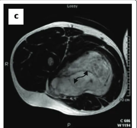
Figure 5Axial T2WSE The mass contains ill-defined areas of lower intensity relative to subcutaneous fat on T2WSE.Internal septations are evident (curved double arrow).
that are usually painless and relative mobile. Owing to the tumor’s hypervascularity, localized warmth can be depicted over the lesion at palpation [4,6,7,14,15]. The lesions can become symptomatic when compression of nearby structures occurs [6,15]. No evidence of a
Figure 7Partial loss of fat signal intensity is depicted on STIR images.
Figure 8 Sagital reformatted image clearly exhibits large intratumoral vessels (black arrows).
Figure 9Axial reformatted image.
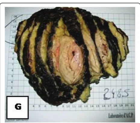
malignant form of hibernoma has been reported in the English literature, except for the case published as an abstract by Teplitz et al. [24] that involved a sarcoma with hibernoma-like features. Incomplete excision results in local recurrence of the tumor; therefore mar-ginal but complete resection is considered as the treat-ment of choice for these lesions [14,24]. Even though core needle biopsy is not recommended in cases of sus-pected hibernoma due to the tumor’s hypervascularity [9,14,25] all of the presented cases were preoperatively biopsied without any complications. From a macro-scopic aspect, hibernomas are well-defined, encapsulated soft, lobulated masses and the color ranges from tan to red brown [15] (Figure 4G.). They usually measure from 5 to 10 cm in diameter, but they may reach up to 20 cm [4,15]. Microscopically, the tumor is characterized by multivacuolated cells with eccentric nuclei and gran-ular eosinophilic cytoplasm, univacuolated cells with peripheral nuclei, and smaller round cells with granular cytoplasm. The hypervascularity and the presence of cells with eosinophilic granular cytoplasm full of
mitochondria give hibernomas their brown color [4,6,18]. From an histological point of view this entity must be distinguished from granular cell tumor, that is a benign peripheral nerve derived tumor composed of granular cells rich in mitochondria. In this regard immunohistochemistry does not help, because both tumors intensely stain for S-100 protein. The main his-tological difference is that hibernoma shows much more pleomorphism and focally show typical mature adipo-cytes, in between the granular cells. The diagnosis of lipomatoustumors is often very difficult. Molecular pathology can better classify these lesions and made past classifications out of date. But cytogenetics studies do not help in the diagnosis of hibernoma [26].
According to the 2002 WHO classification there are six histologic subtypes of hibernomas [27]. These are only of diagnostic relevance and not of prognostic value. Histopathologic evaluation of hibernomas, as previously described, is well-established and pathognomonic. On
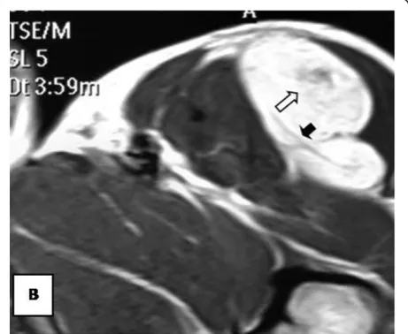
Figure 11Homogeneous enhancement is observed (arrow).
the contrary, CT and MRI features are not specific and vary with the nature and amount of lipid component [4,12,18,19,22,23]. Non contrast CT usually demon-strates a well-demarcated soft tissue mass of predomi-nantly low attenuation which is close but not identical to subcutaneous fat. On the other hand, more heteroge-neous patterns can be encountered as well, as in this
series. Internal linear, curvilinear or branching septa-tions-like densities may be contained [7,9,10]. On post contrast scans, enhancement of the septa as well as more diffuse uptake, usually occurs [7, 9, and 23]. Dif-fuse enhancement was depicted in all the present cases whereas internal enhancing linear or curvilinear densi-ties were shown in four out of eight cases, indicating
Figure 13Axial T1WSE before injection.
Figure 14Axial T1WSE after injection enhanced (white arrows) and unenhanced (black arrows) thin curvilinear structures corresponding to fibrovascular and fibrous tissue, respectively.
Figure 15Axial T1WSE before injection at another level.
thus internal vasculature. Even though vessels were shown in the remaining four cases on post contrast images; the absence of septations in these lesions prior to contrast infusion was attributed to the fact that these lesions had attenuations closer to muscle than fat. On MR images, as in previously published data [6-16,18], five out of six lesions presented, on T1WSE sequences, slightly to moderately decreased signal intensity rela-tively to subcutaneous fat and only one showed a het-erogeneous-mixed signal intensity including areas of increased and decreased intensity but on the whole slightly lower than subcutaneous fat, probably due to a greater “hibernoma” component. Three lesions on T2WSE images demonstrated slightly lower intensities than subcutaneous fat; although most authors report signal intensities closer to fat [5,7,9,11-14]. The
heterogeneous lesion on T1WSE remained heteroge-neously hyperintense on T2WSE images as well. Finally, like in most cases [5-7,13-17], STIR and T2 fat sat sequences failed to achieve full suppression of the
Figure 17On T2 GRE sequence, internal thin vessels are also seen (thin black arrows).
Figure 18Ultrasonography exhibits a mild heterogeneous hyperechoic mass.
Figure 19It contains prominent vessels with Doppler.
examined hibernomas and displayed the most heteroge-neous patterns. Gadolinium enhancement, either hetero-geneous or homohetero-geneous, is usually present in hibernomas [5,7,11-18]; even though Cook M et al [8] and Lee J [6] et al did not report any significant gadoli-nium uptake in their cases. Although, internal curvi-linear structures of low signal intensity were observed on T1WSE and T2WSE sequences in all lesions, they didn’t exhibit the same degree of enhancement most likely corresponding to hypocellular fibrous and fibro-vascular tissue interspersed with the fatty and non fatty portions of the tumor [6,14]. Little is known regarding the imaging of hibernomas on18F FDG-PET scans. The reported high FDG accumulation in these fat-containing tumors may be attributed to the metabolically active cel-lular elements rather than reflect their malignant or not potential [28,29].
Various differential considerations, based on imaging, can be suggested when a complex fatty mass is encoun-tered, including benign entities like lipoma, angiolipoma and hemangioma as well as malignant tumors like lipo-sarcoma. Lipomas present as homogeneous fatty masses with few scattered internal septa and no signs of enhancement [6]. Angiolipomas and hemangiomas can
be distinguished in terms of different morphology of internal vasculature [13,16,17]. Several studies [4,16-18] stress the importance of large branching intratumoral vessels with early contrast enhancement and AV shunt-ing in the differential diagnosis of hibernomas. However these features are not always present, although fine enhancing strands may be seen [6]. In the present series, internal vessels were apparent in six MRI exams; while in total three lesions contained vessels of larger caliber as well. So, vascularity either in the form of thin enhan-cing septa or in the form of vessels is primarily antici-pated in hibernomas. On the other hand, absence of large intratumoral vessels should not exclude hiberno-mas from the differential diagnosis. Well-differentiated liposarcomas are characterized by the presence of irre-gularly thick (>2 mm) and/or nodular septa, foci of high T2 and prominent areas of enhancement [6,15]. More-over, the fatty component of a well-differentiated lipo-sarcoma appears isointense to subcutaneous fat, on T1WSE; distinguishing them from hibernomas [6]. Other lesions like myxoid liposarcoma and clear cell
Figure 21T1WSE FAT SAT: the lesion is poorly pre saturated.
sarcoma could be similar to brown fat tumors but the former displays intense heterogeneity on T2 sequences and the latter primarily involves a tendon, ligament or aponeurosis [13].
This study has limitations, such as limited number of cases, and examinations performed with different techni-ques. None the less, this study comprises the largest number of cases of this rare tumor published thus far and elaborates effectively on its various imaging appear-ances. Conclusively, even if CT and MRI features are not specific, hibernoma should be strongly suggested if a soft tissue mass, exhibits higher attenuation than subcuta-neous fat on CT, slightly lower signal intensity relative to subcutaneous fat on T1WSE, marked enhancement and partial fat suppression on STIR and fat-saturated sequences. These differences compared to subcutaneous
Figure 23 On18 F FDG-PET scan, the lesion has shown
increased FDG accumulation.
Figure 24Gross surgical specimen reveals an encapsulated, lobular mass with yellow-tan to dark brown cut surfaces.
Figure 25Hematoxylin-Eosin stain: Multiple multivacuolated cells are identified with some scattered white adipocytes.
fat, especially on MRI, reflect the different nature of lipid component of hibernomas and comprise the cornerstone in differentiating them from malignant lipomatous tumors. However, as in this study, other atypical findings such as more heterogeneous patterns of mixed fatty and non fatty components on unenhanced CT and MR T1W may be encountered. Furthermore internal septations, regardless of enhancement, and thin vessels contribute in establishing the diagnosis. The role of large intratumoral vessels remains questionable in characterizing hiberno-mas. While complete surgical resection is curative for hibernomas, knowledge of its MRI and CT features can help narrow the field of differential diagnosis and modify adequately the pre-operative planning of complex lipo-matous tumors.
Author details
1
Research, The Rizzoli Institute, Via del Barbiano 1/10, 40106, Bologna, Italy.
2Pathology C, The Rizzoli Institute, Via del Barbiano 1/10, 40106, Bologna,
Italy.3Centre Oscar Lambret, Lille, France.
Authors’contributions
All authors have read and approved the final manuscript.
ZP looked at the cases and wrote the article, MA checked the histology, and the text, ST gave one case and checked the text, CE checked the surgical part, PP checked the research part, DV proposed the article, reviewed the cases and checked the text.
Competing interests
The authors declare that they have no competing interests.
Received: 17 January 2011 Accepted: 25 July 2011 Published: 25 July 2011
References
1. Merkel H:On a pseudolipoma of the breast.Beitr Pathol Anat1906, 39:152-57.
2. Gery L:Discussions.Bull Mem Soc Anat (Paris)1914,89:111.
3. Furlong MA, Fanburg-Smith JC, Miettinen M:The morphologic spectrum of hibernoma: a clinicopathologic study of 170 cases.Am J Surg Pathol
2001,25(6):809-14.
4. Murphey MD, Carroll JF, Flemming DJ, Pope TL, Gannon FH, Kransdorf MJ: From the archives of the AFIP: benign musculoskeletal lipomatous lesions.Radiographics2004,24(5):1433-66.
5. Ritchie DA, Aniq H, Davies AM, Mangham DC, Helliwell TR:Hibernoma– correlation of histopathology and magnetic-resonance-imaging features in 10 cases.Skeletal Radiol2006,35(8):579-89.
6. Lee JC, Gupta A, Saifuddin A, Flanagan A, Skinner JA, Briggs TW, Cannon SR: Hibernoma: MRI features in eight consecutive cases.Clin Radiol2006, 61(12):1029-34.
7. Dursun M, Agayev A, Bakir B, Ozger H, Eralp L, Sirvanci M, Guven K, Tunaci M:CT and MR characteristics of hibernoma: six cases.Clin Imaging
2008,32(1):42-7.
8. Cook MA, Stern M, de Silva RD:MRI of a hibernoma.J Comput Assist Tomogr1996,20(2):333-5.
9. Alvine G, Rosenthal H, Murphey M, Huntrakoon M:Hibernoma.Skeletal Radiol1996,25(5):493-6.
10. Lewandowski PJ, Weiner SD:Hibernoma of the medial thigh. Case report and literature review.Clin Orthop Relat Res1996, ,330:198-201. 11. Peer S, Kühberger R, Dessl A, Judmaier W:MR imaging findings in
hibernoma.Skeletal Radiol1997,26(8):507.
12. Mugel T, Ghossain MA, Guinet C, Buy J, Bethoux J, Texier P, Vadrot D:MR and CT findings in a case of hibernoma of the thigh extending into the pelvis.Eur Radiol1998,8(3):476-8.
13. Anderson SE, Schwab C, Stauffer E, Banic A, Steinbach LS:Hibernoma: imaging characteristics of a rare benign soft tissue tumor.Skeletal Radiol
2001,30(10):590-5.
14. Kallas KM, Vaughan L, Haghighi P, Resnick D:Hibernoma of the left axilla; a case report and review of MR imaging.Skeletal Radiol2003,32(5):290-4. 15. da Motta AC, Tunkel DE, Westra WH, Yousem DM:Imaging findings of a
hibernoma of the neck.AJNR Am J Neuroradiol2006,27(8):1658-9. 16. Colville J, Feigin K, Antonescu CR, Panicek DM:Hibernoma: Report
emphasizing large intratumoral vessels and high T1 signal.Skeletal Radiol
2006,35(7):547-50.
17. Tomihama RT, Lindskog DM, Ahrens W, Haims AH:Hibernoma: a case report demonstrating usefulness of MR angiography in characterizing the tumor.Skeletal Radiol2007,36(6):541-5.
18. Kumazoe H, Nagamatsu Y, Nishi T, Kimura YN, Nakazono T, Kudo S: Dumbbell-shaped thoracic hibernoma: computed tomography and magnetic resonance imaging findings.Jpn J Radiol2009,27(1):37-40. 19. Kransdorf M, Murphey M:Lipomatous tumors.Imaging of soft tissue tumors
Philadelphia, Pa: Saunders; 1997, 57-101.
20. Rasmussen A:The so-called hibernating gland.J Morphol1923, 147-50. 21. Heaton JM:The distribution of brown adipose tissue in the human.Anat
1972,112(Pt 1):35-9.
22. Drevelegas A, Pilavaki M, Chourmouzi D:Lipomatous tumors of soft tissue: MR appearance with histological correlation.Eur J Radiol2004, 50(3):257-67.
23. Bancroft LW, Kransdorf MJ, Peterson JJ, O’Connor MI:Benign fatty tumors: classification clinical course imaging appearance and treatment.Skeletal Radiol2006,35(10):719-33.
24. Lele SM, Chundru S, Chaljub G, Adegboyega P, Haque AK:Hibernoma: a report of 2 unusual cases with a review of the literature.Arch Pathol Lab Med2002,126(8):975-8.
25. Lung RJ, Lapidus S, Miller SH, Graham WP:Hibernoma: report of two cases.J Surg Oncol1977,9(6):563-6.
26. Weiss SW, Goldblum JR:Benign lipomatous tumors.InSoft tissue tumors..5 edition. Edited by: Weiss SW, Goldblum JR. St. Louis: Mosby; 2008:466. 27. Miettinen MM, Fanburg-Smith JC, Mandhl N:Hibernoma.InWorld Health
Organization classification of tumours. Pathology and genetics of tumours of soft tissue and bone.Edited by: Fletcher CDM, Unni KK, Mertens F. Lyon: IARC Press; 2002:33-35.
28. Burdick MJ, Jolles PR, Grimes MM, Henry DA:Mediastinal hibernoma simulates a malignant lesion on dual time point FDG imaging.Lung Cancer2008,59(3):391-4.
29. Subramaniam RM, Clayton AC, Karantanis D, Collins DA:Hibernoma: 18F FDG PET/CT imaging.J Thorac Oncol2007,2(6):569-70.
doi:10.1186/2045-3329-1-3
Cite this article as:Papathanassiouet al.:Imaging of hibernomas: A retrospective study on twelve cases.Clinical Sarcoma Research20111:3.
Submit your next manuscript to BioMed Central and take full advantage of:
• Convenient online submission
• Thorough peer review
• No space constraints or color figure charges
• Immediate publication on acceptance
• Inclusion in PubMed, CAS, Scopus and Google Scholar
• Research which is freely available for redistribution