S T U D Y P R O T O C O L
Open Access
The brain slice method for studying drug
distribution in the CNS
Irena Loryan
1, Markus Fridén
2and Margareta Hammarlund-Udenaes
1*Abstract
The high-throughput brain slice method is a precise and robust technique for estimating the overall uptake of drugs into brain tissue through determination of the unbound volume of distribution in the brain (Vu,brain; ml·g brain
-1 ). Vu,braindescribes the relationship between the total drug concentration in the brain and the concentration of unbound drug in the brain interstitial fluid, regardless of blood–brain barrier function. The brain slice method is more physiologically based than the brain homogenate method with respect to the assessment of drug distribution in the brain because the cell-cell interactions, pH gradients and active transport systems are all conserved. The method provides information that is directly relevant to issues such as nonspecific binding to brain tissue, lysosomal trapping, and active uptake into the cells. For these reasons, the brain slice method is recommended for estimation of
target-site pharmacokinetics in the early drug discovery process and fundamental pharmacological studies. This article provides a detailed protocol for the rat and mouse brain slice methods, with the aim of enabling simple, cost-effective profiling of compounds with diverse physicochemical properties. The procedure for assessing the viability of the brain slices after the 5 h incubation period is also described. The results are interpreted for a set of compounds covering a wide range of physicochemical properties and various pharmacological targets. Application of the method for evaluating the unbound intracellular-to-extracellular concentration ratio (Kp,uu,cell) and the unbound brain-to-plasma concentration ratio (Kp,uu,brain) is discussed.
Keywords:Brain slice method, Unbound volume of distribution in the brain, Neuropharmacokinetics, Drug
discovery, High-throughput screening
Background
It is generally accepted that the cerebral concentration of unbound drug is the main pharmacokinetic determinant of CNS activity for neurotherapeutics [1-3]. Preliminary assessment of the clinically relevant pharmacokinetic parameters required for approximation of the unbound-drug concentration in the brain interstitial fluid is thus pivotal in guiding early drug discovery research [4]. Be-cause of the cost and complexity of the methodology, many of the available “gold standard” pharmacokinetic methods are not appropriate for use in the early stages of drug discovery. Consequently, there is an urgent need for adequate high-throughput in vitro systems and methods for CNS drug development programs.
The implementation of the high-throughput equilib-rium dialysis-based assay for estimation of the fraction of unbound drug in the brain tissue (fu,brain), combined
with measurement of whole brain concentrations
in vivo, was groundbreaking for the field [5]. However, homogenization of the brain as used in this method changes the brain tissue binding properties, leading to implicit errors in the readouts [6].
In contrast, the brain slice method has a more physio-logical basis and has several benefits over the brain hom-ogenate method. The brain slice preparation method was implemented by Henry McIlwain and is now exten-sively used in neurobiology, biophysics and quantitative pharmacology [7-9]. It has the advantage of offering a strongly regulated in vitro environment, while preserving much of the complex cellular integrity, including cellular barriers and intact circuitry, and as a result conserving functionality – resulting in an in vitro environment * Correspondence:[email protected]
1Department of Pharmaceutical Biosciences, Translational PKPD Research Group, Uppsala University, Associate member of SciLife Lab, Box 591, SE-75124, Uppsala, Sweden
Full list of author information is available at the end of the article
more comparable to the in vivo brain than seen in the homogenate method.
Several research groups have used the method to esti-mate the uptake of exogenous compounds into the brain [10-15]. Moreover, studies have investigated mechanistic pharmacokinetic/pharmacodynamic links using brain slice methodology [13,16].
Measurements obtained from in vivo microdialysis have also been compared to those from the in vitro brain slice and homogenate methods [17]. The reasonable cor-respondence (within a 3-fold range) between the cere-bral microdialysis and brain slice method results in this study indicates that the brain slice method is the choice of preference [17].
The brain slice method has recently been further developed for high-throughput, making it more access-ible for use by pharmaceutical companies [18]. It is now a precise, robust technique for estimating the overall uptake of drugs into brain tissue through deter-mination of the unbound volume of distribution in the brain (Vu,brain; ml·g brain-1). Vu,brain describes the rela-tionship between the total drug concentration in the brain and the unbound-drug concentration in the brain interstitial fluid, regardless of blood–brain barrier function. The key assumption of the experiment is that, at equilib-rium, the unbound-drug concentration in the brain slice interstitial fluid (ISF) or extracellular fluid (ECF) is equal to the drug concentration in the buffer in the beaker.
This article provides a detailed protocol for the rat and mouse brain slice methods, with the aim of encouraging simple, cost-effective profiling of compounds with diverse physicochemical properties and unifying procedures among laboratories in order to aid the achievement of comparable results.
Methods and Design Animals
The protocols presented below are based on animal experiments approved by the Animal Ethics Committee of Uppsala, Sweden (C21/9 and C351/11). Drug-naïve male Sprague–Dawley 250–300 g rats and Naval Medical Re-search Institute (NMRI) 25–30 g mice were used (Taconic, Lille Skensved, Denmark). The fresh brain slices can be prepared from various strains of wild type and genetically modified mice and rats, depending on the purpose of the study and the respective laboratory traditions. The brain slices could as well be genetically manipulated using meth-ods such as viral infection [19], biolistics [20], etc.
Preparatory steps
Artificial extracellular fluid
To ensure maintenance of the brain slices in a healthy state, the artificial settings should mimic the in vivo cellular environment. The composition of artificial cerebrospinal
fluid or extracellular fluid (aECF) is crucial. A large number of formulations for these artificial fluids can be found in the literature. In the experimental settings used in the stud-ies underlying this paper, the HEPES-buffered aECF con-tained 129 mM NaCl, 3 mM KCl, 1.4 mM CaCl2, 1.2 mM MgSO4, 0.4 mM K2HPO4, 25 mM HEPES, 10 mM glucose and 0.4 mM ascorbic acid [18]. Ascorbic acid is used as a natural free-radical scavenger to protect the cell mem-branes from lipid peroxidation and the brain slices from swelling [21].
Prior to starting an experiment, a stock solution of
aECF (1290 mM NaCl, 30 mM KCl, 12 mM MgSO4,
4 mM K2HPO4, 250 mM HEPES) is prepared and stored at room temperature. The 400 mM stock solution of as-corbic acid should be stored at +4°C.
On the day before the experiment, 1 L of Milli-Q water should be dispensed. On the day of experiment, this is used to prepare the working aECF solution according to the for-mulation (Table 1). The solution is then equilibrated with 100% oxygen for 15 minutes in an ice-water bath. The pH of the aECF should be 7.6 at 23°C at the beginning of the experiment and about 7.3 at 37°C directly after the 5 h in-cubation. See Table 2 for a summary of the critical steps in the brain slice experiment protocol.
Preparation of cassettes
This protocol allows simultaneous investigation of a selection of up to ten compounds within the same experiment, allowing coverage of a wide range of physicochemical properties and various pharmaco-logical targets in the same cassette.
When deciding on the compounds of each cassette, the pKa values of the compounds should be taken into account. Because high concentrations of weak bases can increase the pH of the acidic intracellular compartments, the extent of lysosomal trapping of a weak base could be affected by the existence of another weak base. The inter-action between two weak bases is mainly regulated by the concentrations of the free compounds in the cassette and their potency in increasing intralysosomal pH [22]. Conse-quently, it is recommended that the final aECF concentra-tion of each studied compound in the cassette should be
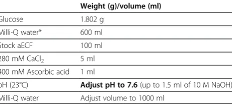
Table 1 Composition of the working aECF solution
Weight (g)/volume (ml)
Glucose 1.802 g
Milli-Q water* 600 ml
Stock aECF 100 ml
280 mM CaCl2 5 ml
400 mM Ascorbic acid 1 ml
pH (23°C) Adjust pH to 7.6(up to 1.5 ml of 10 M NaOH)
100–200 nM and the total concentration of the studied compounds should not exceed 1μM [18].
Each cassette of compounds is prepared individually, ex tempore, in scintillation vials (20 ml glass vials with screw lids; one vial per rat or mouse brain). Initially, the required volume of stock drug solution is added to an empty scintillation vial. To reduce possible toxic effects of the solvents (methanol, acetonitrile, etc.) on the brain slices, the solvents are evaporated under a gentle stream of nitrogen before diluting the sample with aECF. When using dimethyl sulfoxide (DMSO) to dissolve the com-pounds, it is strongly recommended that the final con-centration of DMSO is kept as low as possible (no higher than 1%). Subsequently, 20 ml of ice-cold aECF, pre-equilibrated with 100% oxygen, is added to each scintilla-tion vial and ultrasonicated for 10 minutes to facilitate the dissolution of the compounds. These ready-to-use solutions are maintained at 37°C until incubation.
Preparation of slices
The glassware and tools are set up for the dissection prior to preparing the brain slices. The vibrating blade microtome (e.g. Leica VT1200 (Leica Microsystems AB, Sweden)) is prepared for slicing and the chambers are chilled.
Drug-naïve rats/mice are anesthetized with inhalation anesthesia using 5% isoflurane. When deep anesthesia is reached, up to 10 ml of blood (rats) is collected intracar-dially. The animal is then decapitated and the skull quickly opened. The isolated brain is immediately placed into blank ice-cold aECF saturated with oxygen. In our experi-ence, the brains should be sliced within 15–20 minutes of harvesting to retain their viability. Slices from three rat/ mouse brains are generally equilibrated during the same day with one cassette of drugs.
The pre-chilled chamber of the vibratome is filled with ice-cold oxygenated aECF just before use and is then placed in the ice-filled tray of the vibratome.
It is advantageous to put one or two drops of cyano-acrylate glue on the cutting platform one minute before mounting the brain, to allow the glue to dry slightly.
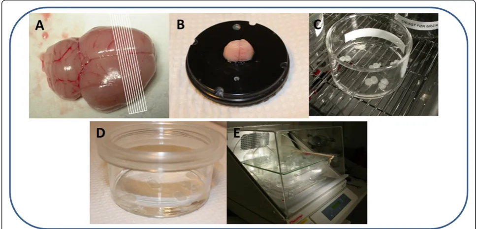
Working quickly, the brain is placed on a chilled Petri dish covered with filter paper. Using a #22 surgical blade, a 3 mm piece is cut from the rostral area on a coronal plane, leaving a piece of about 10 mm for subse-quent slicing. A caudal cut is then made (Figure 1A). The 10 mm piece of the brain is glued to the slicing platform in a coronal position (Figure 1B), and the plat-form is then positioned in the slicing chamber filled with the blank ice-cold aECF. The razor blade (Gillette, super-stainless) is then mounted and the clearance angle is fixed at 21°. We use a motorized blade holder section-ing speed of 0.8 mm/s with a 1 mm amplitude, in 0.05 mm steps.
After discarding the first one or two brain slices, 6 (rat brain) or 10 (mouse brain) consecutive 300 μm brain slices are cut on a coronal plane, starting approximately 1.7 mm anterior to the bregma (rostral striatum).
The 300μm thickness provides good cell preservation without compromising the diffusion of oxygen into the core of the slice. The equilibration time during incuba-tion is inversely related to the square of the brain slice thickness [16].
The slices are moved, using a micro spatula, to the brain slice storage beaker filled with oxygenated blank aECF which is kept in an ice bucket before the incu-bation. It is recommended that only brain slices with intact edges be used for the experiment, to reduce the amount of debris detaching from the brain tissue dur-ing incubation.
The slicing platform should be scrubbed to remove the brain and glue remnants, and the chamber refilled with fresh ice-cold oxygenated aECF, before proceeding with the next brain. The vibratome chambers should be cleaned, disinfected and dried at the end of each experi-mental day.
Incubation
The incubation-equilibration process is started by gently transferring the 6 (rat) or 10 (mouse) brain slices from the storage beaker (using a micro double-ended spatula) into one 45 mm high, 80 mm diameter, flat-bottomed glass bea-ker (Duran Group, VWR, Sweden) containing 15 ml (rat) or 10 ml (mouse) of the aECF containing the selection of drugs to be investigated (Figure 1C). The beaker is then filled with humidified 100% oxygen over the aECF and covered with a custom-fabricated lid (Figure 1D) composed of a Teflon fluorinated ethylene-propylene (FEP) film (50 Å, 12.7 μm thick; DuPont, Katco Ltd,
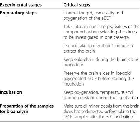
Table 2 Critical steps in the brain slice experiment
Experimental stages Critical steps
Preparatory steps Control the pH, osmolarity and oxygenation of the aECF
Take into account the pKavalues of the compounds when selecting the drugs to be investigated in one cassette
Do not take longer than 1 minute to extract the brain
Keep cold-chain during the brain slicing procedure
Preserve the brain slices in ice-cold oxygenated aECF before starting the incubation
Incubation Keep oxygenation, temperature and
stirring constant during the incubation
Preparation of the samples for bioanalysis
UK) as designed by Potter and DeMarse with minor modifications [23]. A “blank” beaker is also incubated in parallel to check stability of the compounds added into the buffer.
The transparent Teflon FEP film is used in preference to a glass lid because it is selectively permeable to gases (e.g. oxygen) while remaining relatively impermeable to water vapor. This significantly decreases evaporation (allowing improved control of osmolarity and pH), thus permitting the use of a non-humidified incubator.
Finally, the beaker is placed inside the small plastic box in the incubated shaker (e.g. MaxQ4450 Thermo Fisher Scientific, NinoLab, Sweden) for 5 h at 37°C (Figure 1E). Control of the temperature by an external thermometer is recommended. A rotation speed of 45 rpm and oxygen flow of about 75–80 ml per mi-nute through a glass frit apparatus appear sufficient.
The pH of the aECF should be measured at 37°C im-mediately after the 5 h incubation. A reduction of more than 0.15 pH units over the 5 hours indicates more than acceptable acidification of the buffer.
Preparation of the samples for bioanalysis
It is necessary to take several samples for bioanalysis during the experiment:
I. aECF samples
a. for thermostability testing of the compounds studied (sampled before and after incubation in aECF without the brain slices)
b. for measurement of Cbuffer–the final concentrations
of the unbound compounds in aECF (sampled at the end of the 5 h incubation with the brain slices) II.Brain slices samples
a. for measurement of Abrain–the amount of drug in
t-he brain slices (sampled at tt-he end of tt-he 5 h incubation)
Procedural details for preparation of the aECF and brain slice samples are given below.
I. aECF samples
a. Assessment of the thermostability of the compounds provides valuable information and is recommended f-or each drug selection tested. A 200μl sample of aE-CF is taken directly from the beaker at the beginning of the study and at the end of 5 h incubation without brain slices for analysis of initial and final concentra-tions of the drugs in aECF. The aECF sample is
trans-ferred into an Eppendorf tube containing 200 μl
blank brain homogenate that has been prepared be-forehand with 4 volumes of blank aECF. The brain h-omogenate is included to prevent nonspecific binding of drugs to the plastic as well as to match the matrix of the slice homogenates, as required for the follow-ing LC-MS-MS analysis.
b. Since the unbound drug concentration in the brain slice interstitial fluid at equilibrium is taken to be equal to the drug concentration in the aECF in the beaker, the presence of any minor debris from the
brain tissue should be avoided in the sampled aECF after the 5 h incubation. To achieve this, the beaker needs to sit still for 5 minutes after finishing the i-ncubation before sampling the aECF. To sample,
2-00 μl of the aECF is aspirated from just below the
surface (without wetting the tip before sampling) a-nd dispensed into an Eppea-ndorf tube containing the
200 μl of blank brain homogenate, as described in
Ia. It is recommended that duplicate samples of the aECF be taken at this stage. Filtration of aECF, wh-ich is recommended by several authors, is not suit-able for drug distribution studies because of the possible loss of the compounds in the filter.
II.Brain slice samples
a. After sampling the aECF, the brain slices in the aECF a-re individually a-removed, dried on filter paper (about 30 s), weighed (~33 mg per rat brain slice and ~13 mg per mouse brain slice) and homogenized separately in 9 vo-lumes (w/v) of aECF with an ultrasonic processor (e.g. VCX-130; Sonics, Chemical Instruments AB, Sweden).
The samples are then stored at−20°C pending bioana-lysis (e.g. LC-MS-MS).
Assessment of viability of brain slices
The brain slices must remain viable during the experiment. There are several methods of assessing viability based on biochemical and/or electrophysiological parameters. In our laboratory, the viability of the brain slices is assessed by measuring the relative activity of released lactate dehydro-genase (LDH) using a cytotoxicity detection kit [24] according to the manufacturer's instructions (Roche Diagnostics GmbH, Germany), with some modifications.
A brief description of the preparation of the main con-trols and samples required for analysis is given in Tables 3 and 4.
To calculate the viability of the brain slices (as a percent-age) after the 5 h incubation-equilibration period, the fol-lowing steps are taken for each experimental setup:
Background absorbance control - provides
informa-tion about the background absorbance of the assay
medium (aECF). For this purpose, 200μl sample of
aECF is taken at the beginning of the experiment fro-m the scintillation vial and fro-mixed with 200μl blank aECF. The obtained absorbance value is then sub-tracted from all other values.
Low control - provides information about the
activ-ity of LDH released from the brain slices as a result of damage to the membranes caused by slicing the brain. A 200μl aECF sample is taken 5 minutes after transferring the brain slices into the beaker for incu-bation and is then mixed with 200μl blank aECF.
High control - provides information about the
max-imum possible activity of releasable LDH in the brain slice. To achieve this, one rat or three mouse brain s-lices are used after the 5 h incubation for each ex-perimental setup. After drying the brain slice(s) on filter paper and individually weighing them, 9 volum-es (w/v) of 2% Triton X-100 solution in aECF is add-ed. To facilitate the release of LDH from the brain slice(s), the Eppendorf tube is placed in an ultra-sound bath for one hour at +4°C followed by 30 min-utes of incubation in a water bath at 37°C. The supernatant obtained after centrifugation of the tube for 5 minutes at 10,000 rpm and +4°C is stored at +-4°C pending analysis (no longer than 5 days).
For preparation of the experimental samples (experi-mental value), 200μl of aECF sample is taken after the
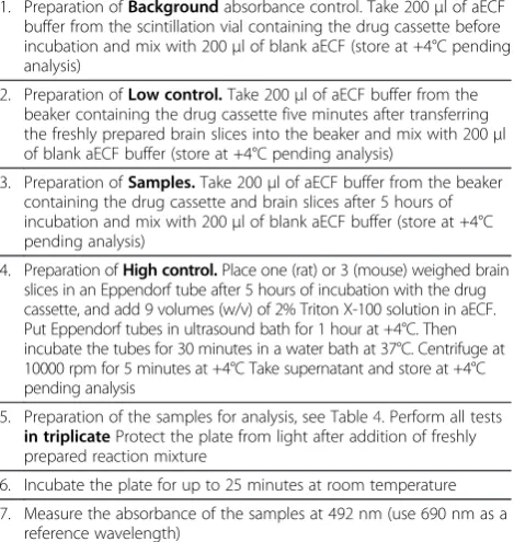
Table 3 Brief description of assessment of viability of brain slices based on the activity of released lactate dehydrogenase
1. Preparation ofBackgroundabsorbance control. Take 200μl of aECF buffer from the scintillation vial containing the drug cassette before incubation and mix with 200μl of blank aECF (store at +4°C pending analysis)
2. Preparation ofLow control.Take 200μl of aECF buffer from the beaker containing the drug cassette five minutes after transferring the freshly prepared brain slices into the beaker and mix with 200μl of blank aECF buffer (store at +4°C pending analysis)
3. Preparation ofSamples.Take 200μl of aECF buffer from the beaker containing the drug cassette and brain slices after 5 hours of incubation and mix with 200μl of blank aECF buffer (store at +4°C pending analysis)
4. Preparation ofHigh control.Place one (rat) or 3 (mouse) weighed brain slices in an Eppendorf tube after 5 hours of incubation with the drug cassette, and add 9 volumes (w/v) of 2% Triton X-100 solution in aECF. Put Eppendorf tubes in ultrasound bath for 1 hour at +4°C. Then incubate the tubes for 30 minutes in a water bath at 37°C. Centrifuge at 10000 rpm for 5 minutes at +4°C Take supernatant and store at +4°C pending analysis
5. Preparation of the samples for analysis, see Table4. Perform all tests in triplicateProtect the plate from light after addition of freshly prepared reaction mixture
6. Incubate the plate for up to 25 minutes at room temperature
7. Measure the absorbance of the samples at 492 nm (use 690 nm as a reference wavelength)
Table 4 Preparation of samples for assessment of viability of brain slices based on the activity of released lactate dehydrogenase
aECF buffer
Low control
Sample High
control
Reaction mixture*
Background 100μl 100μl
Low control 100μl 100μl
Sample 100μl 100μl
High control 100μl 100μl
5 h incubation-equilibration period and mixed with 200 μl blank aECF. For evaluation of the effects of changes in viability of the brain slices during the incuba-tion, aECF samples can be taken at different time points (after 1, 2, 3, etc. hours).
Once the absorbance of the control and experimental samples has been obtained (Table 3) the relevant viabil-ity (%) of the brain slices can be calculated according to Equations 1 and 2:
Cytotoxicityð Þ ¼% experimental valuelow control high controllow control 100
ð1Þ
Viabilityð Þ ¼% 100%Cytotoxicityð Þ% ð2Þ
In practice, it is recommended that 85-90% viability be aimed for; however, a viability of around 60% provides similar results according to our experience (data not shown). Viability values lower than 50% after the 5 h incu-bation period are associated with dramatic changes in the estimation of Vu,brain, especially for weak bases and results from the experiments should be discarded.
Estimation of Vu,brain
Bioanalytically determined drug concentrations in the brain slices and the 5 h aECF samples are used to estimate Vu,brain. It is critical to remember to scale back the obtained concentrations (or areas under the concentration-time peaks) to the undiluted buffer and brain slice concentra-tions by multiplying by the dilution factors as appropriate. The concentration in each brain slice sample is multiplied by 10 to account for the dilution during preparation of the homogenate. The concentration in aECF is multiplied by 2 to account for the dilution during 1:1 mixing of the aECF sample with blank brain homogenate (in 4 volumes (w/v) of aECF). The dilutions associated with protein precipita-tion are not accounted for because they are the same for all samples.
Vu,brain (ml · g brain-1), as defined in Equation 3, is equivalent to the ratio of the amount of drug in the brain or brain slice (Abrain, nanomoles · gram brain-1) to the measured final aECF concentration (Cbuffer, micromoles per liter) or unbound brain interstitial fluid concentra-tion (Cu,brainISF) measured using cerebral microdialysis technique:
Vu;brain¼CAbrain
u;brainISF¼ Abrain
Cbuffer ð
3Þ
Because of incomplete absorption of aECF by the filter paper, the brain slices have a surrounding layer of aECF, and it is important to measure the volume of this layer (Vi, milliliters per gram of slice) and compensate for this aECF buffer layer, i.e. (1-Vi) in
Equation 4. Vi should be measured in a separate ex-periment using [14C] inulin as described in Fridén et al. [18]. Vi was reported to be 0.094 ml ∙g slice-1 [18]. In view of this, Equation 3 can be reorganized to obtain Vu,brain corrected for the remaining aECF volume on the brain slice:
Vu;brain¼
AbrainVi_Cbuffer
Cbuffer_ð1ViÞ ð4Þ
High-throughput screening capacity
Once the brain slice methodology is established in the laboratory (Table 5), it can be used in a high-throughput manner. One trained individual can perform up to four experiments per day (using rats or mice). The method allows up to 10 compounds to be tested simultaneously (consultation with an analytical chemist is required). A series of three experiments is enough to obtain consist-ent results for one cassette.
Table 5 Checklist before starting the experiments
aECF Stock aECF
280 mM CaCl2
400 mM ascorbic acid
Dispensed the day before 1 L MQ water
Oxygen supply
pH meter
Apparatus Orbital shaking incubator
Vibratome
Nitrogen evaporator
Ultrasonic bath
Ultrasonic processor
Centrifuge
Water bath
ELISA reader
Glass- and lab-ware Petri dishes
45 mm high, 80 mm diameter, flat-bottomed glass beakers
Custom-fabricated lids of Teflon FEP film for beakers
Scintillation vials
Surgical instruments for dissection of brain
Ice buckets
Pre-labeled Eppendorf tubes
Nunc 96-well plates
Blank brain homogenate (in 4 volumes (w/v) of aECF)
Miscellaneous Cyanoacrylate glue
Discussion
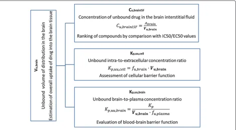
The high-throughput rat or mouse fresh brain slice method is a powerful tool for estimating the intracereb-ral distribution of diverse compounds in an in vitro setup with preserved cellular barrier functionality. The method allows the estimation not only of nonspecific binding of compounds to the brain tissue but also of the cellular accumulation of compounds through uptake transporters, trapping in acidic intracellular compart-ments (i.e. lysosomes), and active efflux from the cellular membrane [6]. Consequently, the determination of more physiological Vu,brain values using fresh brain slices in-stead of brain homogenates permits more precise assess-ment of Cu,brainISF (Figure 2) with reduced risk of misrepresentation during subsequent evaluation of exposure-target engagement relationships.
The estimated Vu,brain value can be put into context by comparison with the physiological volume of brain fluids, e.g. Vu,brain values higher than 0.8 ml · g brain-1 (the volume of total brain fluids is 0.8 ml · g brain-1) are interpreted as intracellular distribution of the drug in question [4,18]. Values higher than this indicate that proportionally more of the drug is intracellularly dis-tributed, e.g. into lysosomes, and/or bound to parenchy-mal components. A value lower than 0.8 ml · g brain-1 shows restricted distribution. The lowest volume pos-sible in vivo is the brain interstitial fluid volume of 0.2 ml · g brain-1; however, in the slices, this is somewhat higher because of damage to the surface layer of cells.
Table 6 shows Vu,brain values of nine drugs covering a wide range of physicochemical properties and pharma-cological targets. Vu,brain has extensive variability with a range from 3.75 to 2650 ml · g brain-1.
An additional very important aspect of drug discov-ery is the ability to extrapolate the results to other species. Recently, it has been shown that unbound fraction of drug in brain homogenate value obtained
from the Wistar Han rat brain homogenate can be
used as a representative value for any preclinical spe-cies and also humans [25]. The results in Table 6 Figure 2A number of applications of Vu,brainfor integrative pharmacology.
Table 6 Unbound volume of distribution in the brain (Vu,brain) determined using Sprague–Dawley (SD) rat and
Naval Medical Research Institute (NMRI) mouse brain slices
Ion class Vu,brainml·g brain-1
SD rat NMRI mouse
Verapamil Base 46.6 (1.8) 47.3 (4.4)
Docetaxel Base 777 (217) 796 (177)
Oxycodone Base 4.20 (0.13)* 3.75 (0.22)
Digoxin Base 33.1 (6.0) 44.9 (5.7)
Gabapentin Zwitterion 4.49 (0.29) 4.22 (0.93)
Indomethacin Acid 14.1 (1.8) 12.0 (1.8)
Paroxetine Base 714 (72) 596 (97)
Thioridazine Base 2650 (232) 1930 (170)
Diazepam Neutral 17.8 (1.1) 17.1 (2.3)
* Data from Fridén et al., 2011 [6].
indicate an absence of any significant dissimilarity in
Vu,brain values between Sprague–Dawley rats and
NMRI mice. However, a more systematic investiga-tion is desirable before the possibility of interchange-able use of Vu,brain measurements can be supported for translational studies.
It has been proposed by Fridén et al. [17] that in vitro determination of Vu,brain in combination with in vivo determination of the total brain-to-plasma con-centration ratio (Kp,brain) and in vitro determination of the fraction of unbound drug in plasma (fu,plasma) would allow rapid evaluation of unbound brain-to-plasma concentration ratio (Kp,uu,brain) (Figure 2). Moreover, combining Vu,brainwith fu,brainwould allow es-timation of the unbound intracellular-to-extracellular concentration ratio (Kp,uu,cell). It is essential to emphasize that, in terms of predicting Cu,brainISF or Kp,uu,brain, rank ordering of compounds with respect to Vu,brain is futile, since there is no causal relationship [4].
Complex evaluation of the abovementioned neurophar-macokinetic parameters gives insight to drug distribution in the brain. For instance, analogue of γ-aminobutyric acid gabapentin has Kp,brain equal to 0.64 [26]. How-ever, after correction of Kp,brain for brain tissue uptake (using Vu,brain derived from the brain slice method) and plasma protein binding (using fu,plasma) the BBB net flux was estimated as a 0.14 meaning that only 14% of unbound drug in plasma is passing the BBB. Moreover, after passing BBB gabapentin (substrate to the large neutral amino acid transporter) tends to accu-mulate in the cells as it could be judged from Kp,uu,cell equal to 4.55 [6].
The brain slice method can also be used to identify suitable positron emission tomography (PET) tracers, which should have a low degree of nonspecific binding (i.e. a low Vu,brain value) to obtain higher specificity for their targets.
In summary, the brain slice method, used for assess-ment of the volume of distribution of unbound drug in the brain, is a useful tool for both drug discovery and fundamental pharmacology research.
Abbreviations
aECF: Artificial extracellular fluid; Abrain: Amount of drug in brain tissue; BBB: Blood–brain barrier; Cbuffer: Final drug concentration in aECF; Cu,
brainISF: Concentration of unbound drug in the brain interstitial fluid; CNS: Central nervous system; DMSO: Dimethyl sulfoxide; ECF: Extracellular fluid; HTS: High-throughput screening; fu,brain: Unbound fraction of drug in brain homogenate; fu,plasma: Unbound fraction of drug in plasma; Kp,
brain: Total brain-to-plasma concentration ratio; Kp,uu,brain: Unbound brain-to-plasma concentration ratio; Kp,uu,cell: Unbound intracellular-to-extracellular concentration ratio; LDH: Lactate dehydrogenase; Vu,brain: Unbound volume of distribution in brain.
Competing interests
Irena Loryan is funded by a post-doc stipend from Johnson&Johnson. Markus Fridén is an employee of AstraZeneca R&D. Margareta Hammarlund-Udenaes: no conflicts of interest or other issues.
Authors’contributions
I L: Writing of manuscript, development of slice method and viability test. M F: Feedback on manuscript, proofreading. Development of slice method for high throughput studies, development of pH partitioning method etc. M H-U: Feedback on manuscript, proofreading. All authors have read and approved the final version of the manuscript.
Authors’information
You may choose to use this section to include any relevant information about the author(s) that may aid the reader's interpretation of the article, and understand the standpoint of the author(s). This may include details about the authors' qualifications, current positions they hold at institutions or societies, or any other relevant background information. Please refer to authors using their initials. Note this section should not be used to describe any competing interests.
Acknowledgements
We thank Britt Jansson and Waqas Sadiq for excellent assistance with LC-MS-MS analysis of verapamil, oxycodone, docetaxel and digoxin; the results were used for estimation of the unbound volume of distribution in the brain.
Author details
1Department of Pharmaceutical Biosciences, Translational PKPD Research Group, Uppsala University, Associate member of SciLife Lab, Box 591, SE-75124, Uppsala, Sweden.2AstraZeneca Research and Development, Respiratory and Inflammatory Innovative Medicines, Department of Drug Metabolism and Pharmacokinetics, Mölndal, Sweden.
Received: 7 November 2012 Accepted: 2 January 2013 Published: 21 January 2013
References
1. Hammarlund-Udenaes M, Friden M, Syvanen S, Gupta A:On the rate and extent of drug delivery to the brain.Pharm Res2008,25:1737–1750. 2. Hammarlund-Udenaes M:Active-site concentrations of chemicals - are
they a better predictor of effect than plasma/organ/tissue concentrations?Basic Clin Pharmacol Toxicol2010,106:215–220. 3. Watson J, Wright S, Lucas A, Clarke KL, Viggers J, Cheetham S, Jeffrey P,
Porter R, Read KD:Receptor occupancy and brain free fraction. Drug Metab Dispos2009,37:753–760.
4. Hammarlund-Udenaes M, Bredberg U, Friden M:Methodologies to assess brain drug delivery in lead optimization.Curr Top Med Chem2009,9:148–162. 5. Kalvass JC, Maurer TS:Influence of nonspecific brain and plasma binding on CNS exposure: implications for rational drug discovery.Biopharm Drug Dispos2002,23:327–338.
6. Friden M, Bergstrom F, Wan H, Rehngren M, Ahlin G, Hammarlund-Udenaes M, Bredberg U:Measurement of unbound drug exposure in brain: modeling of pH partitioning explains diverging results between the brain slice and brain homogenate methods.Drug Metab Dispos2011,39:353–362.
7. McIlwain H:Metabolic response in vitro to electrical stimulation of sections of mammalian brain.Biochem J1951,48.
8. McIlwain H, Buchel L, Cheshire JD:The inorganic phosphate and phosphocreatine of Brain especially during metabolism in vitro. Biochem J1951,48:12–20.
9. Li CL, McIlwain H:Maintenance of resting membrane potentials in slices of mammalian cerebral cortex and other tissues in vitro.J Physiol1957,
139:178–190.
10. Patlak CS, Hospod FE, Trowbridge SD, Newman GC:Diffusion of
radiotracers in normal and ischemic brain slices.J Cereb Blood Flow Metab 1998,18:776–802.
11. Becker S, Liu X:Evaluation of the utility of brain slice methods to study brain penetration.Drug Metab Dispos2006,34:855–861.
12. Kakee A, Terasaki T, Sugiyama Y:Brain efflux index as a novel method of analyzing efflux transport at the blood–brain barrier.J Pharmacol Exp Ther1996,277:1550–1559.
13. Gredell JA, Turnquist PA, Maciver MB, Pearce RA:Determination of diffusion and partition coefficients of propofol in rat brain tissue: implications for studies of drug action in vitro.Br J Anaesth2004,
14. Blasberg R, Levi G, Lajtha A:A comparison of inhibition of steady state, new transport, and exchange fluxes of amino acids in brain slices. Biochim Biophys Acta1970,203:464–483.
15. Newman GC, Hospod FE, Wu P:Glucose utilization of ischemic hippocampal slices.J Neurosci Methods1989,28:23–34. 16. Benkwitz C, Liao M, Laster MJ, Sonner JM, Eger EI:2nd, Pearce RA:
Determination of the EC50 amnesic concentration of etomidate and its diffusion profile in brain tissue: implications for in vitro studies. Anesthesiology2007,106:114–123.
17. Friden M, Gupta A, Antonsson M, Bredberg U, Hammarlund-Udenaes M:In vitro methods for estimating unbound drug concentrations in the brain interstitial and intracellular fluids.Drug Metab Dispos2007,35:1711–1719. 18. Friden M, Ducrozet F, Middleton B, Antonsson M, Bredberg U,
Hammarlund-Udenaes M:Development of a high-throughput brain slice method for studying drug distribution in the central nervous system.Drug Metab Dispos2009,37:1226–1233.
19. Stokes CE, Murphy D, Paton JF, Kasparov S:Dynamics of a transgene expression in acute rat brain slices transfected with adenoviral vectors. Exp Physiol2003,88:459–466.
20. Wellmann H, Kaltschmidt B, Kaltschmidt C:Optimized protocol for biolistic transfection of brain slices and dissociated cultured neurons with a hand-held gene gun.J Neurosci Methods1999,92:55–64.
21. Rice ME:Use of ascorbate in the preparation and maintenance of brain slices.Methods1999,18:144–149.
22. MacIntyre AC, Cutler DJ:The potential role of lysosomes in tissue distribution of weak bases.Biopharm Drug Dispos1988,9:513–526. 23. Potter SM, DeMarse TB:A new approach to neural cell culture for
long-term studies.J Neurosci Methods2001,110:17–24.
24. Dos-Anjos S, Martinez-Villayandre B, Montori S, Salas A, Perez-Garcia CC, Fernandez-Lopez A:Quantitative gene expression analysis in a brain slice model: influence of temperature and incubation media.Anal Biochem 2008,378:99–101.
25. Di L, Umland JP, Chang G, Huang Y, Lin Z, Scott DO, Troutman MD, Liston TE:Species independence in brain tissue binding using brain homogenates.Drug Metab Dispos2011,39:1270–1277.
26. Friden M, Winiwarter S, Jerndal G, Bengtsson O, Wan H, Bredberg U, Hammarlund-Udenaes M, Antonsson M:Structure-brain exposure relationships in rat and human using a novel data set of unbound drug concentrations in brain interstitial and cerebrospinal fluids.J Med Chem 2009,52:6233–6243.
doi:10.1186/2045-8118-10-6
Cite this article as:Loryanet al.:The brain slice method for studying drug distribution in the CNS.Fluids and Barriers of the CNS201310:6.
Submit your next manuscript to BioMed Central and take full advantage of:
• Convenient online submission • Thorough peer review
• No space constraints or color figure charges • Immediate publication on acceptance
• Inclusion in PubMed, CAS, Scopus and Google Scholar
• Research which is freely available for redistribution