Open Access
Research
Normal mitochondrial respiratory function is essential for spatial
remote memory in mice
Daisuke Tanaka
†1, Kazuto Nakada
†1, Keizo Takao
2,3, Emi Ogasawara
1,
Atsuko Kasahara
1,4, Akitsugu Sato
1, Hiromichi Yonekawa
1,5,
Tsuyoshi Miyakawa
2,3and Jun-Ichi Hayashi*
1Address: 1Graduate School of Life and Environmental Sciences, University of Tsukuba, Tsukuba, Ibaraki 305-8572, Japan, 2Division of Systems Medical Science, Institute for Comprehensive Medical Science, Fujita Health University, 1-98 Dengakugakubo, Kutsukake-cho, Toyoake, Aichi 470-1192, Japan, 3Genetic Engineering and Functional Genomics Group, Frontier Technology Center, Graduate School of Medicine, Kyoto University, Yoshida-Konoe-cho, Sakyo-ku, Kyoto 606-8501, Japan, 4Department of Cell Physiology and Metabolism, University of Geneva Medical School, Rue Michel Servet 1, 1211 Geneva 4-CH, Switzerland and 5Department of Laboratory Animal Science, The Tokyo Metropolitan Institute of Medical Science, 3-18-22 Honkomagome, Bunkyo-ku, Tokyo 113-8613, Japan
Email: Daisuke Tanaka - [email protected]; Kazuto Nakada - [email protected]; Keizo Takao - [email protected]; Emi Ogasawara - [email protected]; Atsuko Kasahara - [email protected];
Akitsugu Sato - [email protected]; Hiromichi Yonekawa - [email protected]; Tsuyoshi Miyakawa - [email protected]; Jun-Ichi Hayashi* - [email protected]
* Corresponding author †Equal contributors
Abstract
Background: Mitochondrial DNA (mtDNA) with pathogenic mutations has been found in patients with cognitive disorders. However, little is known about whether pathogenic mtDNA mutations and the resultant mitochondrial respiration deficiencies contribute to the expression of cognitive alterations, such as impairments of learning and memory. To address this point, we used two groups of trans-mitochondrial mice (mito-mice) with heteroplasmy for wild-type and pathogenically deleted (Δ) mtDNA; the "low" group carried 50% or less ΔmtDNA, and the "high" group carried more than 50% ΔmtDNA.
Results: Both groups had normal phenotypes for not only spatial learning, but also memory at short retention delays, indicating that ΔmtDNA load did not affect learning and temporal memory. The high group, however, showed severe impairment of memory at long retention delays. In the visual cortex and dentate gyrus of these mice, we observed mitochondrial respiration deficiencies, and reduced Ca2+/calmodulin-dependent kinase II-α (α-CaMKII), a protein important for the
establishment of spatial remote memory.
Conclusion: Our results indicated that normal mitochondrial respiratory function is necessary for retention and consolidation of memory trace; deficiencies in this function due to high loads of pathogenically mutated mtDNA are responsible for the preferential impairment of spatial remote memory.
Published: 16 December 2008
Molecular Brain 2008, 1:21 doi:10.1186/1756-6606-1-21
Received: 25 September 2008 Accepted: 16 December 2008 This article is available from: http://www.molecularbrain.com/content/1/1/21
© 2008 Tanaka et al; licensee BioMed Central Ltd.
Background
Mitochondria are intracellular organelles containing their own genomes (mtDNA), and playing a crucial role in ATP production through oxidative phosphorylation. Mamma-lian mitochondria have multiple copies of mtDNA (103 ~
104 copies/cell) that is replicated and expressed within the
organellar system [1,2]. Mammalian mtDNA encodes 13 polypeptides, which are essential subunits of complexes I, III, IV, and V for oxidative phosphorylation on the inner mitochondrial membrane, and 22 tRNAs and 2 rRNAs, which are necessary for the translation of these 13 polypeptides. The remaining mitochondrial proteins for oxidative phosphorylation, metabolic enzymes, DNA and RNA polymerases, and ribosomal proteins are all encoded by the nuclear genome [1].
The accumulation of pathogenic mtDNAs with large-scale deletion or point mutations, and the resultant mitochon-drial respiration deficiencies are associated with a wide variety of disorders, such as mitochondrial diseases, neu-rodegenerative diseases, diabetes, and aging [1]. Moreo-ver, dementia and ataxia are found in patients with traditional mitochondrial diseases caused by accumula-tion of mutated mtDNAs, such as MELAS (mitochondrial encephalopathy, lactic acidosis, and stroke-like episodes), MERRF (myoclonic epilepsy and ragged red fibers), KSS (Kearns-Sayre Syndrome), CPEO (chronic progressive external ophthalmoplegia), and NARP (neurogenic mus-cle weakness, ataxia, and retinitis pigmentosa) [3-8], and the same mutated mtDNAs have been identified in patients with dementia, ataxia, and Alzheimer's disease [9,10]. Besides, there appears to be a relationship between mtDNA polymorphisms and cognitive function in humans [11]. These findings suggest that mtDNAs with both pathogenic mutations and polymorphisms contrib-ute to various cognitive disorders, thus leading to demen-tia, and ataxia. It has been demonstrated that polymorphisms, at least, in mtDNAs are responsible for changes in mammalian cognitive function, since the exchange of mtDNAs between NZB/BINJ and CBA/H mice affected their learning and exploration processes [12]. However, there is no direct experimental evidence that mitochondrial dysfunction induced by pathogenic mtDNAs results in cognitive disorders, because no proce-dures are available for the direct introduction of muta-genized mammalian whole mtDNA into the mitochondria of living cells, or even into isolated mito-chondria.
Trans-mitochondrial mice carrying pathogenic mtDNAs are very useful for addressing whether mitochondrial res-piration deficiencies induced by the mtDNA mutations are responsible for cognitive alterations, and, if so, how they affect brain function. Mito-mice are a type of trans -mitochondrial mice generated by the direct introduction
of mitochondria carrying ΔmtDNA isolated from cultured cells into normal pronuclear embryos by a cytoplast fusion technique [13]. The ΔmtDNA has an expanded deletion of 4696 bp, from nucleotide position 7,759 in the tRNALys gene to position 12,454 in the ND5 gene [13],
and is similar to the "common deletion" found in KSS, and CPEO patients [14]. The great advantages of mito-mice are that they all share exactly the same nuclear genomic background (C57BL6/J; B6), and their genetic variation is restricted to the proportions of introduced pathogenic ΔmtDNA. Therefore, mito-mice could provide direct evidence that mitochondrial respiration deficien-cies induced by mtDNA accumulation are sufficient in themselves for expression of the clinical phenotypes observed in patients with mutated mtDNA.
We used these mito-mice to examine the direct relation-ship between mutated mtDNA and cognitive alteration, and we succeeded in showing that mutated mtDNA and the resultant mitochondrial respiration deficiencies were responsible for impairments of spatial remote memory. Furthermore, our study demonstrated that mitochondrial respiration deficiencies gave rise to downregulation of α -CaMKII, which is important for the establishment of spa-tial remote memory in the hippocampus and cortex. These findings suggested that mito-mice with impaired spatial remote memory would be valuable models for understanding the molecular mechanisms of cognitive alteration of mitochondrial origin.
Results
Behavioral and physical features of mito-mice
Mito-mice that are models for mitochondrial diseases carry both wild-type mtDNA and ΔmtDNA (Figure 1A), and the proportions of ΔmtDNA differ among individuals [13]. Not all mito-mice are useful for examining cognitive alteration, because in mice with severe mitochondrial dis-ease phenotypes due to high loads of ΔmtDNA (approxi-mately more than 80%), the abnormal cognitive function can be overshadowed by other abnormalities in the phe-notype. In fact, a high load of ΔmtDNA induces locomo-tor defects in male mito-mice, leading to impaired mating activity [15]. Furthermore, the proportion of ΔmtDNA in various tissues of mito-mice increases progressively with aging, and the mice begin to show the onset of mitochon-dria-driven diseases other than those manifested cogni-tively [16]; we therefore had to finish all of our behavioral analyses before ΔmtDNA accumulated to more than approximately 80%.
the mito-mice, because the proportions are very similar throughout all the tissues of individual mito-mice at age 4 weeks [13,17]. We then classified the mito-mice into two groups: low (n = 13), carrying 50% or less ΔmtDNA in their tails, and high (n = 17), carrying more than 50% (Figure 1B). From our previous studies, we expected that the mito-mice in the low group would maintain normal mitochondrial respiration activity, and would thus be quite healthy during the behavioral analyses, whereas those in the high group would show slight mitochondrial respiration abnormalities, and resultant lactic acidosis,
one of the markers for the onset of mitochondrial dis-eases, at about the time the study ended [13,15]. Since the genetic variation in the mito-mouse population was lim-ited to the proportions of exogenously introduced
ΔmtDNA, the low group was used as experimental normal controls.
Before we focused on cognitive function in the mito-mice, we examined the locomotor activity and physical per-formance of the two groups (n = 10 as a low group, and n = 14 as a high group), because the results of spatial learn-Genetic characterization and locomotor activity of mito-mice before and after cognitive analyses
Figure 1
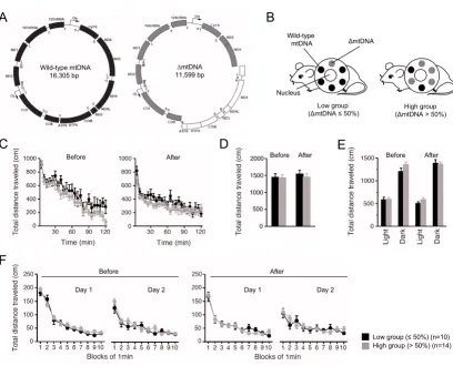
Genetic characterization and locomotor activity of mito-mice before and after cognitive analyses. (A) Gene map of mouse wild-type mtDNA (black) and ΔmtDNA (gray). The white arc in the ΔmtDNA indicates the deleted region expanded from the tRNALys to ND5 genes. (B) Two groups of mito-mice were used in the experiment. Mito-mouse population (n = 24)
was divided into two groups, low (n = 10), carrying 50% or less ΔmtDNA in their tails (at age 4 weeks), and high (n = 14), car-rying more than 50%. The two groups shared the same nuclear genome background (C57BL/6). (C-F) To examine locomotor activity in the low group (black), and high (gray) group, four kinds of traditional behavioral test, the open field test (C) (P = 0.1638 in before, P = 0.1502 in after), elevated plus maze test (D) (P = 0.9589 in before, P = 0.5703 in after), light-dark transi-tion test (E) (before: P = 0.8434 in light side, P = 0.1114 in dark side; after: P = 0.1379 in light side, P = 0.8239 in dark side), and Porsolt forced-swim test (F) (before: P = 0.3685 in day 1, P = 0.2449 in day 2; after: P = 0.7663 in day 1, P = 0.7735 in day 2), were performed before and after cognitive analyses. Results of total distance analyses indicated that there were no differences in locomotor activity between the low and high groups. All values are means ± SE.
E
F
C
D
A
mtDNA Wild-type
mtDNA
Nucleus
Low group (mtDNA 50%)
High group (mtDNA > 50%)
B
Wild-type mtDNA 16,305 bp
mtDNA 11,599 bp
Before After Before After Before After
After Before
Day 1 Day 2 Day 1 Day 2
ing and memory tests for the study of cognitive function are easily affected by locomotor and physical abnormali-ties. We therefore performed an open field test (Figure 1C), elevated plus maze test (Figure 1D and 2A), light-dark transition test (Figure 1E and 2B), and Porsolt forced-swim test (Figure 1F and 2C), before and after the
cogni-tive analyses. There were no significant differences between the low and high groups in terms of locomotor activity and physical performance, including anxiety-, and depression-related behaviors, at the two time points (Fig-ure 1C–1F and 2). From these results, we concluded that the two groups of mito-mice could be used as an in vivo
Behavioral analyses of mito-mice before and after cognitive analyses Figure 2
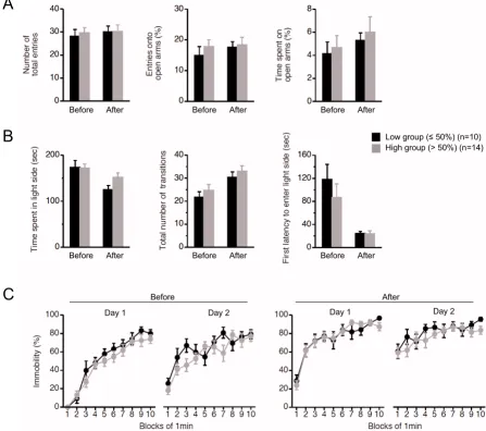
Behavioral analyses of mito-mice before and after cognitive analyses. To examine behavioral performances in the low group (black), and high (gray) group, three kinds of traditional behavioral tests were performed before and after cognitive analyses. (A) Elevated plus maze test. There was no difference in phenotypes of anxiety-related behavior between the low and high groups at the two time points (number of total entries: P = 0.6823 in before, P = 0.9775 in after; entries onto open arms:
P = 0.4360 in before, P = 0.8226 in after; time spent on open arms: P = 0.7157 in before, P = 0.6756 in after). (B) Light-dark transition test. There was no difference in phenotypes of anxiety-related behavior between the low and high groups at the two time points (time spent in light side: P = 0.9125 in before, P = 0.0625 in after; total number of transitions: P = 0.4083 in before,
P = 0.4569 in after; first latency to enter light side: P = 0.3812 in before, P = 0.9798 in after). (C) Porsolt forced-swim test. There was no difference in phenotypes of depressant-related behaviors between the low and high groups at the two time points (before: P = 0.1479 in day 1, P = 0.0526 in day 2; after: P = 0.9900 in day 1, P = 0.3132 in day 2). All values are means ± SE.
A
B
C
Before After Before After Before After
Before After Before After Before After
After Before
Day 1 Day 2 Day 1 Day 2
model to confirm the direct relationship between mutated mtDNA and cognitive alteration.
Mito-mice showed abnormalities at long retention delays in cognitive analyses
To examine cognitive function in mito-mice (n = 10 as a low group, and n = 14 as a high group), we performed Barnes circular maze tests (see Methods), which can reveal spatial learning and memory ability without the effects by
difference in motor coordination or emotional aspects. During the course of training (21 trials for 7 days), two groups of mito-mice learned to locate a single target among 12 holes. Underneath the target hole was an escape box containing shredded paper. We measured three parameters, latency, number of errors, and distance traveled until they located the target hole. In both groups, all parameters were progressively reduced with training, indicating acquisition of spatial learning (Figure 3A).
Spatial learning and memory analyses using Barnes circular maze test in mito-mice Figure 3
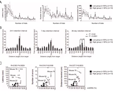
Spatial learning and memory analyses using Barnes circular maze test in mito-mice. (A) Training course. Latency (P = 0.7381), number of errors (P = 0.5051), and distance traveled (P = 0.7885) to target hole for low (black) and high (gray) groups were plotted. (B) Probe tests at 24-h, 7-day, and 36-day after the last training. There was no significant difference in the probe test at 24-h (between the target hole and right side hole, P < 0.005 in the low and P < 0.001 in the high groups; between the target hole and left side hole, P < 0.05 in the low and P < 0.002 in the high groups) and 7-day retention intervals (between the target and right side holes, P < 0.001 in the low and P < 0.001 in the high groups; between the target and left side holes, P
< 0.05 in the low and P < 0.002 in the high groups). In the probe test at the 36-day retention interval, single and double aster-isks indicate significant differences between the target and right side holes in the low group (P < 0.02 in the low and P = 0.9804 in the high groups) and between the target and left side holes in the low group (P < 0.0001 in the low and P = 0.2106 in the high groups), respectively. (C) A single retraining after the probe test at the 36-day retention interval. Each score of individual mito-mice in the low (black) and high (gray) groups were plotted against the proportion of ΔmtDNA in the tails at age 4 weeks. Mice 52, 64, and 76 carried 52%, 64%, and 76% ΔmtDNA, respectively. Pearson’s product-moment correlation coefficients and the associated probabilities are indicated as R and P, respectively. All values are means ± SE.
** *
A
: P < 0.02 : P < 0.0001
* **
B
C
Number of trials Number of trials Number of trials
Mouse 76
Mouse 64
Mouse 52
Mouse 76
Mouse 64
Mouse 52
Mouse 76
Mouse 64
Mouse 52
mtDNA (%)
Low group ( 50%) (n=10) High group (> 50%) (n=14)
Low group ( 50%) (n=10) High group (> 50%) (n=14)
Through the training trials, there were no statistical differ-ences between the two groups in latency, number of errors, and distance traveled to the target hole (Figure 3A). These results indicated that a high load of ΔmtDNA was not required for acquisition of spatial memory, although we could not rule out the possibility that a load of more than 80% – the pathogenic threshold for expression of systemic mitochondrial disease phenotypes – would induce impairment of spatial learning.
To assess the spatial memory of mito-mice at long reten-tion delays, we performed probe tests at 24-h, 7-day, and 36-day retention intervals after the last training trial (see Methods). Since the probe tests were conducted without an escape box, there was no box for them to escape into under the target hole. If the mouse had memorized a tar-get, it would visit the hole where the escape box had pre-viously been located and spend a long time around it. In the probe tests at the 24-h and 7-day retention intervals, which we used to examine temporal memory ability, there were no statistical differences in the times spent around the target hole by the two groups (Figure 3B, left and center panels). However, in the probe test at the 36-day retention interval, which was used to examine remote memory ability, the high group spent significantly less time around the target hole than did the low group (Fig-ure 3B, right panel). These results indicated that abnor-malities in spatial remote memory were induced by a high load of ΔmtDNA.
Phenotypic expression of mitochondrial diseases is regu-lated by a threshold effect of the mutated mtDNAs: that is, most phenotypes of mitochondrial disease appear only when mutated mtDNAs have accumulated to a certain level [17]. If the ΔmtDNA load were responsible for abnormalities in spatial remote memory, then only mito-mice with a high load of ΔmtDNA – beyond a certain threshold – would be likely not to memorize the target hole. To confirm this point, we performed a single retrain-ing of each mito-mouse after the probe test at the 36-day retention interval, and we measured three parameters dur-ing the traindur-ing session: latency, number of errors, and distance traveled to the target hole with the escape box under it (see Methods). Increases in the three parameters were observed in each mito-mouse when the ΔmtDNA load was more than approximately 60% (Figure 3C). The results showed that a high load of ΔmtDNA was definitely responsible for the impairment of spatial remote mem-ory; they also suggested that the pathological threshold for expressing the memory impairment phenotype was approximately 60%.
Accumulation of ΔmtDNA and resultant mitochondrial respiration deficiencies in brain tissues of mito-mice
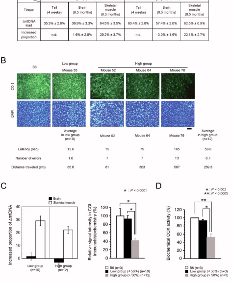
It was not clear whether the impaired spatial remote memory in the high group was induced by mitochondrial respiration deficiencies due to high load of ΔmtDNA in the brain tissues. To address this point, we first estimated the proportion of ΔmtDNA in brain tissues from the two groups. We previously reported that ΔmtDNA can accu-mulate over time in cultured cells and various tissues because it has replication advantages [13,16]. In consider-ation of this feature of ΔmtDNA, we selected a high group of mito-mice in which ΔmtDNA would accumulate only to less than 80%, even when all the analyses in the study were finished. The proportion of ΔmtDNA in the skeletal muscle tissues at age 8.5 months was higher in both groups than in the tail samples at age 4 weeks. The increase in the proportion of ΔmtDNA in the skeletal muscle was 29.2% ± 3.7% in the low group and 22.1% ± 2.7% in the high group (Figure 4A and 4C). The propor-tion of ΔmtDNA in the brain tissues, however, increased very little or decreased; at age 8.5 months the proportion of ΔmtDNA in the brain ranged from 25% to 53% in the low group (increase compared with tail samples at 4 weeks, 1.6% ± 2.8%) and from 49% to 71% in the high group (increase compared with tail samples at 4 weeks, -3.0% ± 1.6%) (Figure 4A and 4C).
COX activity between normal B6 mice and low group mito-mice (Figure 4D). However, we observed a half of the COX activity in whole brain tissues from the high group (Figure 4D). From these results, together with the results of retraining after the last probe test (Figure 3C), we confirmed that the mitochondrial respiration deficien-cies due to high load of ΔmtDNA were consistent with the phenotypic expression of impairments of spatial remote memory. The three parameters – latency, number of errors, and distance traveled to the target hole – increased gradually in the order of Mice 52, 64, and 76 (Figure 4B), reflecting the hypothesis that phenotypic expression and severity of impairment of remote memory depended upon the degree of mitochondrial respiration deficiency.
Expression of α-CaMKII protein in mito-mice
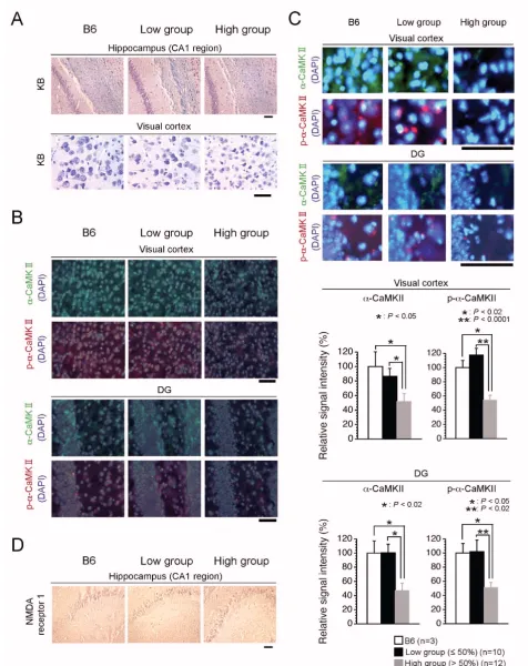
Since it has been suggested that temporal and remote memory is regulated, respectively, as hippocampal and cortex-hippocampal tasks, the results of the Barnes circu-lar maze tests suggested that a high load of ΔmtDNA affected cortex-hippocampal tasks, rather than hippocam-pal ones. However, the mechanisms by which mitochon-drial respiration deficiencies of the brain tissues induced abnormalities selectively in cortex-hippocampal tasks were unclear. We therefore compared histological changes in the hippocampus and visual cortex between the two groups. No marked histological abnormalities in cell number and death were seen in either group, with the exception of the fact that some of the neuronal cells in the visual cortical sections from high group mice were slightly smaller than those from low group mice (Figure 5A). These observations indicated that mitochondrial respira-tion deficiencies, at least in the high group expressing impairment of spatial remote memory, did not affect cell quantity and formation in the visual cortex and hippoc-ampus, although a very high load of ΔmtDNA (more than 80%) might have induced some histological abnormali-ties.
α-CaMKII is important for the establishment of remote memory, including for cortical plasticity and consolida-tion of memory traces in cortical networks [18]. A part of
α-CaMKII mRNAs is targeted dendritically, then trans-lated at the site, and then finally matured into constitu-tively active forms [19]. Since the occurrence of impairment of spatial remote memory was correlated with mitochondrial respiration deficiency due to high load of ΔmtDNA, there was a possibility that mito-mice in the high group possessed some abnormalities in expres-sion and/or dendritic distribution of α-CaMKII protein. To test this, we examined expression of α-CaMKII and phosphorylated-α-CaMKII (p-α-CaMKII) as the translated and constitutively active forms, respectively. In visual cor-tical sections from the low group, both α-CaMKII and
p-α-CaMKII proteins were expressed well and distributed in dendrites and cytoplasm as well as those in the B6 mouse (Figure 5B, 5C and 6A). In contrast, we observed a notable general reduction in both α-CaMKII and p-α-CaMKII pro-tein expression in the visual cortical sections from the high group (Figure 5B, 5C and 6A). However, we did not observed significant difference in the expression levels of
α-CaMKII mRNA among the three groups (Figure 6B), although the high group mice possessed a slight increase of the α-CaMKII mRNA level (Figure 6B), probably due to the compensation of decreased α-CaMKII protein (see Fig-ure 6A). These results indicated that mitochondrial respi-ration deficiency affected translation machinery and/or dendritically-targeting process of α-CaMKII mRNA, rather than phosphorylation of the protein and transcription of the mRNA, thus leading to impairment of spatial remote memory. Moreover, we performed immunohistochemical analyses to visualize α-CaMKII and p-α-CaMKII proteins in section of the dentate gyrus (DG), because the DG is considered to be important in spatial pattern separation [20], and the specific occurrence of adult neurogenesis [21]. Consistent with the visual cortical sections, the levels of expression of both α-CaMKII and p-α-CaMKII proteins ΔmtDNA load and mitochondrial respiration in brain tissues
Figure 4 (see previous page)
ΔmtDNA load and mitochondrial respiration in brain tissues. (A) Accumulation of ΔmtDNA. Because ΔmtDNA is dis-tributed nearly uniformly throughout all the tissues of a mito-mouse at age 4 weeks, we estimated the increased proportion of
in DG sections from the low group were similar to those in the B6 mouse, whereas these expressions in DG sec-tions from the high group were notably reduced (Figure 5B and 5C). These immunohistochemical results sug-gested that a notable reduction in α-CaMKII and p-α -CaMKII protein production in the visual cortex and DG may have caused the impairment of spatial remote mem-ory. The N-methyl-D-aspartate (NMDA) receptor, a major molecule upstream of α-CaMKII, plays an essential role in learning and memory in the hippocampus [22], and deg-radation of NMDA receptor subunits in the hippocampus contributes to learning and memory defects [23]. We also performed immunohistochemical analyses with anti-NMDA receptor 1, but there was no difference in the expression of NMDA receptor 1 protein between the two groups (Figure 5D).
Discussion
In healthy mouse models (NZB/BINJ and CBA/H mice) it has been well documented that mtDNA polymorphisms are among the genetic candidates for the control of learn-ing ability [12]. However, there was no direct experimen-tal evidence as to whether mtDNA with pathogenic mutations and the resultant mitochondrial respiration deficiencies are responsible for cognitive alterations. Since mito-mice share the same nuclear genome background, and their genetic variations are restricted to the propor-tions of pathogenic ΔmtDNA – similar to the case with common deletion types in human diseases [13] – their use could provide unambiguous evidence that pathologi-cal phenotypes exclusively observed in mito-mice carrying high loads of ΔmtDNA are caused by ΔmtDNA-induced mitochondrial respiration deficiencies. Our experiments clearly showed that loads of 50% or less ΔmtDNA did not induce mitochondrial respiration deficiencies in the brain tissues, and were also not associated with behavioral abnormalities (Figure 1C–1F, 2, 3, 4B and 4C), whereas loads of more than 50% were able to induce
mitochon-drial respiration deficiencies, and downregulation of α -CaMKII protein in brain tissues, leading to impairment of spatial remote memory (Figure 3B, 4B, 4C, 4D, 5B, 5C and 6A). We therefore succeeded for the first time in showing experimental evidence that a high load of patho-genically mutated mtDNA and the resultant mitochon-drial respiration deficiencies, in the absence of severe mitochondrial disease phenotypes, are responsible for the impairment of spatial remote memory.
It has been considered that remote memory traces are established by the acquired formation of neuronal circuits between the hippocampus and cortex and by the resultant cortical plasticity, whereas learning and temporal memory are regulated by the existing neuronal circuits in the hip-pocampus [24]. Frankland et al. [18] have been reported the possibility that the α-CaMKII modulates the synaptic events required for the consolidation of memory trace in cortical networks. Heterozygous mice for a null mutation of α-CaMKII showed abnormal cortical plasticity and DG maturation, resulting in impairment of remote memory [18,25]. These mice showed normal hippocampal plastic-ity, resulting in no impairment of learning, and temporal memory [18,25]. Consistent with these reports, expres-sion levels of α-CaMKII protein were reduced in the corti-ces and DGs of mito-mice from the high group, and these mice showed impairment of spatial remote memory (Fig-ure 3B, 5B and 5C). In contrast, hippocampal plasticity, which is regulated mainly by existing neuronal circuits in the hippocampus, is required for initial learning, and tem-poral memory formation [24]. Genetic disruption experi-ments in mouse models have indicated that NMDA receptors are important not only in the formation of neu-ronal circuits during brain development, but also in the learning and hippocampus-dependent temporal memory that use these circuits [23,26]. In mito-mice, irrespective of the ΔmtDNA load, the level of expression of NMDA receptor 1 was not altered (Figure 5D); this probably Histological and immunohistochemical analyses of brain tissues
Figure 5 (see previous page)
explains why all the mice had normal spatial learning and temporal memory (Figure 3A and 3B). Therefore, the occurrence of mito-mouse phenotypes in which spatial remote memory was impaired but spatial learning and temporal memory were not indicated that mitochondrial respiratory function plays an integral role in the forma-tion of acquired neuronal circuits between the cortex and hippocampus, and/or neuronal plasticity in the cortex, rather than in the use of existent neuronal circuits in the hippocampus.
The pathophysiological mechanisms of mitochondria-related impairment of spatial remote memory can be con-sidered in the following way. When more than 60%
ΔmtDNA accumulates in the brain, mitochondrial respi-ration deficiencies are induced. Reduction and depletion of the mitochondrial energy supply would affect the proc-esses of translation and/or targeting of α-CaMKII mRNA. The reduced α-CaMKII protein would cause impairment of spatial remote memory. There are several possible rea-sons why α-CaMKII protein was downregulated in the brain tissues of mito-mice with high ΔmtDNA loads. The Quantification of α-CaMKII protein and mRNA in visual cortex
Figure 6
Quantification of α-CaMKII protein and mRNA in visual cortex. (A) Quantification of α-CaMKII protein in the visual cortex by Western blotting analysis. Extracted protein of the visual cortex from B6, low group, and high group were blotted with an anti-α-CaMKII antibody. Bars indicate values of α-CaMKII protein expression in the visual cortex from the B6 (white bar), low group (black bar), and high group (gray bar). In the high group, remarkable downregulation of α-CaMKII proteins (P = 0.5148 B6 vs low group, P < 0.005 B6 vs high group, P < 0.005 low group vs high group) was observed. The β-actin was used as a loading control in the Western blotting. Values are the means ± SE. Asterisks indicate significant differences. (B) Quantifica-tion of α-CaMKII mRNA in the visual cortex. Total mRNAs were extracted from frozen samples of the visual cortex from B6, low group and high group, and cDNAs were obtained by a reverse transcription method. Relative expression levels of α -CaM-KII mRNA were determined by a real-time monitoring PCR technique. Bars indicate values of relative expression levels of α -CaMKII mRNA in the B6 (white bar), low group (black bar), and high group (gray bar). The β-actin was used as a loading con-trol in the real-time monitoring PCR technique. There was no significant difference among the three groups (P = 0.9972 B6 vs low group, P = 0.6214 B6 vs high group, P = 0.6238 low group vs high group). Values are means ± SE.
A
B
*
*
*
: P < 0.005Relative
-C
aMKII p
rotein
level (%)
Relative
-C
aMKII m
RNA
level (%)
first is that mitochondrial energy supply is necessary for the dendritically targeted processes of α-CaMKII mRNA. It is well known that the mRNAs of α-CaMKII are targeted to dendrites, and that additional transcription and matura-tion events occur after this targeting [19]. Many proteins, including cytoskeletal components and ion-motive ATPases, that are critical for the dendritically targeted processes of α-CaMKII mRNA require ATP [27,28]. Thus, one possible reason is that mitochondrial respiration defi-ciencies could induce abnormalities in the localization of
α-CaMKII mRNAs, and the resultant downregulation of its protein form. This possibility was supported by normal level of α-CaMKII mRNA in the high group (Figure 6B). However, the reduction of α-CaMKII protein in high group brain did not always occur specially in dendrites (Figure 5C). The second possibility is that exogenous sig-nals derived from other tissues carrying ΔmtDNA at high levels contribute to the reduction of α-CaMKII protein production in the brain tissues of the mito-mice. Taking this study together with our previous one [16], most tis-sues in the high group were allowed to accumulate
ΔmtDNA with aging, and these tissues fell into mitochon-drial respiration deficiency, even though the proportion of ΔmtDNA in the brain tissues did not increase (Figure 4A and 4C). Systemic mitochondrial respiration deficien-cies give rise to lactic acidosis, which is one of the diagnos-tic markers of mitochondrial diseases [1], and the lactate can bind Ca2+ [29]. The onset of lactic acidosis, therefore,
could disturb calcium homeostasis and signaling in nerve cells, probably leading to the general reduction of α -CaM-KII protein (Figure 5C), although all vital functions that occur via calcium signaling should in fact be affected by the lactic acidosis.
In the brain tissues of the mito-mice, a ΔmtDNA load of more than 60% could induce mitochondrial respiration deficiencies (Figure 4B). The threshold ΔmtDNA load leading to mitochondrial respiration deficiencies in the brain tissues was clearly lower than that in other tissues; a
ΔmtDNA load of more than approximately 80% is needed for the occurrence of mitochondrial respiration deficien-cies in skeletal and cardiac muscle, and in renal, pancre-atic, and testicular tissues [13,15,17,30]. Considering that the brain is a high-energy-demand tissue [1], the lower load of ΔmtDNA was sufficient to induce mitochondrial respiration deficiencies in the brain tissues. In addition, it has been reported that a calcium-regulated signal pathway controls mitochondrial biogenesis, since transgenic mice with overexpression of CaMKIV in their skeletal muscles show enhanced mitochondrial biogenesis, including enhanced mtDNA replication, mitochondrial respiration, and fatty acid metabolism [31]. If mitochondrial biogen-esis in the brain is also controlled by a calcium-regulated signal pathway, then a reduction in CaMKII production might participate in mitochondrial respiration
deficien-cies in brain tissues. The potential involvement of CaMKII in mitochondrial respiration requires further investiga-tion. Moreover, the brain tissues of mito-mice showed lit-tle or negative accumulation of ΔmtDNA without neuronal cell death (Figure 4A and 4C) [16], even when the proportions of ΔmtDNA in other tissues increased (Figure 4A and 4C). This phenomenon suggested that the replication frequency of mtDNA molecules differs in each tissue, and that the frequency in the brain is the lowest among various tissues [16]. In human brains, however, it has been reported that deleted mtDNAs accumulate clon-ally in single cells during aging [32]. Therefore, it remains to be answered why the mouse brain was sensitive to a low load of ΔmtDNA, more than 60%, and why ΔmtDNA did not accumulate in the mouse brain.
The disease phenotypes caused by pathogenic mtDNAs differ among the types of mutation. For instance, muta-tions in particular tRNA genes are responsible for MELAS, MERRF, and cardiomyopathy, whereas ones in structural genes induce Leigh syndrome, and Leber's disease [33]. We previously generated a novel mouse model (mito-mouse COI) carrying homoplasmic mtDNA with a path-ogenic point mutation in the COI gene (COImut-mtDNA) [34]. The mito-mice COI looked healthy throughout life, because of the lower pathogenicity of COImut-mtDNA compared with that of ΔmtDNA, but they showed slight mitochondrial respiration deficiencies in all tissues examined [34]. We have obtained prelimi-nary data that mito-mice COI do not have impairment of spatial remote memory, but they have other cognitive function phenotypes (unpublished data). These findings suggest that the occurrence of clinical phenotypes in cog-nitive function also differs among different types of muta-tion, and is directly regulated by the intensity of the pathology of mitochondrial respiration. Experimental support for this possibility will require further investiga-tion.
Conclusion
resultant mitochondrial respiration deficiencies. We also suggest that improving mitochondrial respiration might be an effective strategy for treating memory disorders, although there are biological differences between such disorders in humans and mice.
Methods
MiceThirty male mito-mice carrying various proportions of
ΔmtDNA were used for this study. The proportion of
ΔmtDNA in mito-mice was deduced from tail DNA sam-ples, because it was very similar in all the tissues of the same individual mouse [13,17]. Age-matched male B6 mice (n = 6) were used as normal controls in histological, immunohistochemical, biochemical, Western blotting, and real-time monitoring PCR analyses.
Estimation of ΔmtDNA proportions by real-time monitoring PCR
Real-time monitoring PCR was used to estimate the pro-portions of ΔmtDNA in tails mito-mice at age 4 weeks and in brain and skeletal muscle tissues of mito-mice after all behavioral analyses (at age 8.5 months). It was performed with a TaqMan PCR reagent kit and an ABI PRISM 7900HT sequence detection system (Applied Biosystems, Foster City, CA). To estimate the absolute copy number of wild-type mtDNA and ΔmtDNA, we used the standard curve method. The standard curve for the assay was calcu-lated using a series of 10-fold dilution of titrated synthetic standard DNA. Each measurement was repeated three times, and the proportions of ΔmtDNA and total mtDNA were calculated. The primer set specific for ΔmtDNA was TTTCACTATGAAGCTAAGAGCGTTAACCT and GGT-GGAATCGGACCAGTAGGA. The reporter dye 6-carboxy-fluorescein (FAM)-labeled TaqMan minorgroove-binder probe specific for ΔmtDNA was AACTGGTGTATGGA-GATTT. The primer set specific for wild-type mtDNA was AACCTGGCACTGAGTCACCA and GGGTCTGAGTG-TATATATCATGAAGAGAAT. The reporter dye FAM and the quencher dye 6-carboxy-tetramethyl-rhodamine (TAMRA)-labeled probe was TCTGTAGCCCTTTTTGTCA-CATGATC.
Behavioral analysis
Animals and experimental design
Mice were housed four per cage in a room with a 12-hr light-dark cycle (lights on at 7:00 a.m.) with access to food and water ad libitum. Behavioral testing was performed between 9:00 a.m. and 6:30 p.m. After the tests, the appa-ratus were cleaned with super hypochlorous water to pre-vent a bias due to olfactory cues. All behavioral tests were conducted in a manner similar to those described previ-ously [35], and performed with male mice between 5 and 8 months after birth. All behavioral testing procedures
were approved by the Animal Care and Use Committee of Kyoto University Graduate School of Medicine.
Open field test
Open field test was performed with male mice before (at age 5 months) and after (at age 8 months) cognitive anal-yses, respectively. Each mouse was placed in the center of the open field apparatus (40 × 40 × 30 cm; Accuscan Instruments, Columbus, OH). Total distance traveled (in cm), and number of fecal boli were recorded. Data were collected for 120 min.
Elevated plus maze test
Elevated plus maze test was performed with male mice before (at age 5 months) and after (at age 8 months) cog-nitive analyses, respectively. The elevated plus maze (O'Hara & Co., Tokyo, Japan) consisted of two open arms (25 × 5 cm) and two enclosed arms of the same size, with 15-cm high transparent walls. The arms and central square were made of white plastic plates, and were elevated to a height of 55 cm above the floor. To minimize the likeli-hood of animals falling from the apparatus, 3-mm high plastic ledges were provided for the open arms. Arms of the same type were arranged at opposite sides to each other. Each mouse was placed in the central square of the maze (5 × 5 cm), facing one of the closed arms. Mouse behavior was recorded during a 10-min test period. The number of entries onto, and the time spent on open and enclosed arms, were recorded. For data analysis, we used the following four measures: total distance traveled (cm), the number of total entries, the percentage of entries onto the open arms, and the time spent on the open arms (s). Data acquisition and analysis were performed automati-cally using Image EP software (see 'Image analyses').
Light-dark transition test
Light-dark transition test was performed with male mice before (at age 5 months) and after (at age 8 months) cog-nitive analyses, respectively. The apparatus used for the light/dark transition test consisted of a cage (21 × 42 × 25 cm) divided into two sections of equal size by a partition containing a door (O'Hara & Co., Tokyo, Japan). One chamber was brightly illuminated (390 lux), whereas the other chamber was dark (2 lux). Mice were placed into the dark side, and allowed to move freely between the two chambers with the door open for 10 min. Total distance traveled, time spent in each side, total number of transi-tions between chambers, and first latency to enter the light side were recorded automatically.
Porsolt forced-swim test
The cylinders were filled with water (23°C) up to a height of 7.5 cm. Mice were placed into the cylinders, and their behavior recorded over a 10-min test period for 2 days. Data acquisition and analysis were performed automati-cally, using Image PS software (see 'Image analyses'). The total distance traveled, and the percentage of immobility were measured by Image OF software (see 'Image analy-ses') using stored image files.
Barnes circular maze test
The Barnes circular maze test was conducted on "dry land", a white circular surface, 1.0 m in diameter, with 12 holes equally spaced around the perimeter (O'Hara & Co., Tokyo, Japan). The circular open field was elevated 75 cm from the floor, and evenly illuminated by overhead fluo-rescent white room lighting (1000 lux). A black Plexiglas escape box (17 × 13 × 7 cm) containing shredded paper was located under one of the holes. The hole above the escape box represented the target, analogous to the hid-den platform in the Morris task. The location of the target was consistent for a given mouse, but was randomized across mice. The maze was rotated daily, with the spatial location of the target unchanged with respect to the distal visual room cues, to prevent a bias based on olfactory or proximal cues within the maze. Three trials per day were conducted for 7 successive days. On day 8, a probe test was conducted without the escape box, to confirm that this spatial task was acquired based on navigation using distal environment room cues. Three trials were con-ducted immediately after the probe test, and 7 and 36 days later additional probe tests were conducted again. A single retraining was conducted 7 days later after the probe test at the 36-day retention interval. Latency, number of errors, and distance traveled until they located to the target hole, and time spent around each hole were recorded by Image BM software (see 'Image analyses').
Image analyses
The applications used for the behavioral studies (Image EP, Image PS, Image OF, and Image BM) were based on the public domain NIH Image program (developed at the U.S. National Institutes of Health, and available on the Internet at http://rsb.info.nih.gov/nih-image/ and ImageJ program http://rsb.info.nih.gov/ij/, which were modified for each test by Tsuyoshi Miyakawa (available through O'Hara & Co., Tokyo, Japan).
Histological and immunohistochemical procedures
Brain samples fixed with 4% paraformaldehyde were used for immunohistochemistry with anti-α-CaMKII (BD Bio-sciences, San Jose, CA), anti-p-α-CaMKII (Promega, Mad-ison, WI), and anti-COI (Molecular Probes, Inc., Eugene, OR) antibodies. Frozen sections of the fixed samples were reacted to the primary antibodies, and then visualized with secondary antibodies Alexa Fluor 488 (for anti-α
-CaMKII and anti-COI antibodies) and 594 (for anti-p-α -CaMKII) conjugated goat anti-IgGs (Molecular Probes, Inc., Eugene, OR). The sections were stained with DAPI to visualize all nuclei. For Kluver-Barrera (KB) staining and immunohistochemistry with an anti-NMDA receptor 1 antibody, brain tissues fixed in 4% formaldehyde were used. Paraffin sections (10 μm thick) of the fixed samples were stained with KB method. The sections were also reacted to an anti-NMDA receptor 1 antibody (Acris Anti-bodies GmbH, Hiddenhausen, Germany) followed by a secondary antibody, biotinylated goat anti-IgGs, and VECTASTAIN ABC kit (Vector Laboratories, Inc., Burlin-game, CA). The sections stained with anti-NMDA receptor 1 antibody were counterstained with hematoxylin to visu-alize all nuclei. Quantification of COI-, α-CaMKII-, and
p-α-CaMKII-positive signals were performed by ImageJ pro-gram http://rsb.info.nih.gov/ij/.
Analysis of complex IV (COX) activity
Estimation of COX activity was carried out by examining the rate of cyanide-sensitive oxidation of reduced cyto-chrome c [36] with modifications. Biochemical analysis was based on the procedure described before [37].
Western blotting
To detect α-CaMKII protein, frozen visual cortex regions were lysed on ice in 2% SDS (Wako, Osaka, Japan), 50 mM Tris-HCl (pH 6.8), 10% Glycerol (Wako, Osaka, Japan), and 5% 2-Mercaptoethanol (Wako, Osaka, Japan). After centrifugation, the supernatant was used as a sample. Proteins were resolved by SDS-PAGE under reducing conditions. The resolved proteins were trans-ferred electrophoretically to a nitrocellulose membrane (GE Healthcare Bio-Sciences KK, Tokyo, Japan). After incubation with phosphate-buffered saline (PBS, pH 7.4) in 1% Bovine serum albumin (BSA; Sigma-Aldrich, St. Louis, MO) for at least 1 h at room temperature, the mem-brane was incubated with an anti-α-CaMKII antibody (BD Biosciences, San Jose, CA) for 1 h at room temperature, washed extensively with PBS in 0.1% Tween-20 (ICN Bio-medicals Inc., Aurora, OH), and then incubated with biotinylated goat anti-IgGs, and VECTASTAIN ABC kit (Vector Laboratories, Inc., Burlingame, CA). Proteins were detected using Immunostaining HRP-1000 (Konica Minolta, Tokyo, Japan). For loading controls, incubated with a monoclonal anti-β-actin antibody (Sigma-Aldrich, St. Louis, MO) followed by incubation with a horseradish peroxidase-conjugated goat anti-mouse IgG. Each meas-urement was repeated three times, and the expression of
Real-time monitoring RT-PCR
Total RNA was extracted by ISOGEN (Nippon Gene, Tokyo, Japan) from frozen organs in mouse brain visual cortex. RNA samples were subjected to DNase I treatment (Invitrogen, Carlsbad, CA) to eliminate DNA contami-nants, and reverse transcribed using Oligo (dT)12–18 Primer (Invitrogen, Carlsbad, CA), 10 mM dNTP Mix (Invitrogen, Carlsbad, CA), 0.1 M DTT (Invitrogen, Carlsbad, CA), RNase Out Recombinant Ribonuclease Inhibiter (Invitrogen, Carlsbad, CA), and SuperScript II RNase H-Reverse Transcriptase (Invitrogen, Carlsbad, CA). cDNA samples were subjected to RNaseH treatment (Invitrogen, Carlsbad, CA). Real-time monitoring PCR was performed with SYBR Green PCR Master Mix and an ABI PRISM 7900HT sequence detection system (Applied Biosystems, Foster City, CA). The primer set specific for α -CaMKII was GTCGGAATTCCATCCTCACCACTATGCTG and AAGGATCCATCGATGAAAGTCCAGGCCC. Quanti-tatve-PCR for β-actin mRNA was also performed on the same samples, to correct for any residual differences in the initial level of RNA in the specimens. Results were then normalized using β-actin mRNA levels in the same sam-ples. The primer set specific for β-actin was GGTCATCAC-TATTGGCAACGAG and GTCAGCAATGCCTGGGTACA. Each measurement was repeated three times, and the expression levels of α-CaMKII mRNA were calculated.
Statistical analyses
Statistical analyses were performed by using a commer-cially available software package (STATVIEW, SAS Insti-tute, Cary, NC) or the data analyses add-in for Microsoft Excel. One-way ANOVA followed by Student's t-tests were used for Figures 1D, 1E, 2A, 2B, 4C, 4D, 5C, 6A, and 6B. Two-way repeated-measures ANOVA followed by the two-tailed Student's t-tests were used for Figures 1C, 1F, 2C, and 3A. Paired comparisons t-tests were used for Figure 3B. The relationship between latency, number of errors, or distance and proportion of ΔmtDNA in tails followed by calculating the Pearson's correlation coefficient were used for Figure 3C. Values with P < 0.05 were considered signif-icant.
Competing interests
The authors declare that they have no competing interests.
Authors' contributions
JIH is responsible for the original concept of this study. KN, TM, HY, and JIH designed the study. AS, KN, and HY generated and provided the mito-mouse. DT, KN, KT, AK, and AS performed the behavioral experiments. DT, KN, and EO carried out histological, immunohistochemical, and biochemical experiments. KN, TM, DT, KT, and JIH wrote the manuscript. All authors read and approved the final manuscript.
Acknowledgements
This work was supported by Grants-in-Aid for Scientific Research (S) from the Japan Society for Promotion of Science (JSPS) to J.-I.H. and by the Research Grant (20–13) for Nervous and Mental Disorders from the Min-istry of Health, Labor and Welfare to K.N.
References
1. Wallace DC: Mitochondrial diseases in man and mouse. Science
1999, 283:1482-1488.
2. Clayton DA: Replication of animal mitochondrial DNA. Cell
1982, 28:693-705.
3. Dubeau F, De Stefano N, Zifkin BG, Arnold DL, Shoubridge EA:
Oxi-dative phosphorylation defect in the brains of carriers of the tRNAleu(UUR) A3243G mutation in a MELAS pedigree. Ann Neurol 2000, 47:179-185.
4. Melone MA, Tessa A, Petrini S, Lus G, Sampaolo S, di Fede G,
San-torelli FM, Cotrufo R: Revelation of a new mitochondrial DNA
mutation (G12147A) in a MELAS/MERFF phenotype. Arch Neurol 2004, 61:269-272.
5. Austin SA, Vriesendorp FJ, Thandroyen FT, Hecht JT, Jones OT, Johns
DR: Expanding the phenotype of the 8344 transfer RNAlysine mitochondrial DNA mutation. Neurology 1998, 51:1447-1450.
6. Nakai A, Goto Yi, Fujisawa K, Shigematsu Y, Kikawa Y, Konishi Y,
Nonaka I, Sudo M: Diffuse leukodystrophy with a large-scale
mitochondrial DNA deletion. Lancet 1994, 343:1397-1398.
7. Uusimaa J, Moilanen JS, Vainionpää L, Tapanainen P, Lindholm P,
Nuu-tinen M, Löppönen T, Mäki-Torkko E, Rantala H, Majamaa K:
Preva-lence, segregation, and phenotype of the mitochondrial DNA 3243A>G mutation in children. Ann Neurol 2007,
62:278-287.
8. Holt IJ, Harding AE, Petty RK, Morgan-Hughes JA: A new
mito-chondrial disease associated with mitomito-chondrial DNA heter-oplasmy. Am J Hum Genet 1990, 46:428-433.
9. Nelson I, Hanna MG, Alsanjari N, Scaravilli F, Morgan-Hughes JA,
Harding AE: A new mitochondrial DNA mutation associated
with progressive dementia and chorea: a clinical, pathologi-cal, and molecular genetic study. Ann Neurol 1995, 37:400-403.
10. Tysoe C, Robinson D, Brayne C, Dening T, Paykel ES, Huppert FA,
Rubinsztein DC: The tRNA(Gln) 4336 mitochondrial DNA
var-iant is not a high penetrance mutation which predisposes to dementia before the age of 75 years. J Med Genet 1996,
33:1002-1006.
11. Skuder P, Plomin R, McClearn GE, Smith DL, Vignetti S, Chorney MJ,
Chorney K, Kasarda S, Thompson LA, Detterman DK, Petrill SA,
Daniels J, Owen MJ, McGuffin P: A polymorphism in
mitochon-drial DNA associated with IQ? Intelligence 1995, 21:1-11.
12. Roubertoux PL, Sluyter F, Carlier M, Marcet B, Maarouf-Veray F,
Chérif C, Marican C, Arrechi P, Godin F, Jamon M, Verrier B,
Cohen-Salmon C: Mitochondrial DNA modifies cognition in
interac-tion with the nuclear genome and age in mice. Nat Genet 2003,
35:65-69.
13. Inoue K, Nakada K, Ogura A, Isobe K, Goto Yi, Nonaka I, Hayashi JI:
Generation of mice with mitochondrial dysfunction by intro-ducing mouse mtDNA carrying a deletion into zygotes. Nat Genet 2000, 26:176-181.
14. Hayashi JI, Ohta S, Kikuchi A, Takemitsu M, Goto Yi, Nonaka I:
Intro-duction of disease-related mitochondrial DNA deletions into HeLa cells lacking mitochondrial DNA results in mitochon-drial dysfunction. Proc Natl Acad Sci USA 1991, 88:10614-10618.
15. Nakada K, Sato A, Yoshida K, Morita T, Tanaka H, Inoue S, Yonekawa
H, Hayashi JI: Mitochondria-related male infertility. Proc Natl
Acad Sci USA 2006, 103:15148-15153.
16. Sato A, Nakada K, Shitara H, Kasahara A, Yonekawa H, Hayashi JI:
Deletion-mutant mtDNA increases in somatic tissues but decreases in female germ cells with age. Genetics 2007,
177:2031-2037.
17. Nakada K, Inoue K, Ono T, Isobe K, Ogura A, Goto Yi, Nonaka I,
Hayashi JI: Inter-mitochondrial complementation:
Mitochon-dria-specific system preventing mice from expression of dis-ease phenotypes by mutant mtDNA. Nat Med 2001, 7:934-940.
18. Frankland PW, O'Brien C, Ohno M, Kirkwood A, Silva AJ:
Alpha-CaMKII-dependent plasticity in the cortex is required for permanent memory. Nature 2001, 411:309-313.
Publish with BioMed Central and every scientist can read your work free of charge
"BioMed Central will be the most significant development for disseminating the results of biomedical researc h in our lifetime."
Sir Paul Nurse, Cancer Research UK
Your research papers will be:
available free of charge to the entire biomedical community
peer reviewed and published immediately upon acceptance
cited in PubMed and archived on PubMed Central
yours — you keep the copyright
Submit your manuscript here:
http://www.biomedcentral.com/info/publishing_adv.asp
BioMedcentral
20. Kesner RP, Gilbert PE, Wallenstein GV: Testing neural network
models of memory with behavioral experiments. Curr Opin Neurobiol 2000, 10:260-265.
21. Eriksson PS, Perfilieva E, Björk-Eriksson T, Alborn AM, Nordborg C,
Peterson DA, Gage FH: Neurogenesis in the adult human
hip-pocampus. Nat Med 1998, 4:1313-1317.
22. Tang YP, Shimizu E, Dube GR, Rampon C, Kerchner GA, Zhuo M, Liu
G, Tsien JZ: Genetic enhancement of learning and memory in
mice. Nature 1999, 401:63-69.
23. Tsien JZ, Huerta PT, Tonegawa S: The essential role of
hippoc-ampal CA1 NMDA receptor-dependent synaptic plasticity in spatial memory. Cell 1996, 87:1327-1338.
24. Lisman J, Morris RG: Memory. Why is the cortex a slow
learner? Nature 2001, 411:248-249.
25. Yamasaki N, Maekawa M, Kobayashi K, Kajii Y, Maeda J, Soma M,
Takao K, Tanda K, Ohira K, Toyama K, Kanzaki K, Fukunaga K, Sudo Y, Ichinose H, Ikeda M, Iwata N, Ozaki N, Suzuki H, Higuchi M, Suhara
T, Yuasa S, Miyakawa T: Alpha-CaMKII deficiency causes
imma-ture dentate gyrus, a novel candidate endophenotype of psy-chiatric disorders. Mol Brain 2008, 1:6.
26. Kutsuwada T, Sakimura K, Manabe T, Takayama C, Katakura N,
Kush-iya E, Natsume R, Watanabe M, Inoue Y, Yagi T, Aizawa S, Arakawa
M, Takahashi T, Nakamura Y, Mori H, Mishina M: Impairment of
suckling response, trigeminal neuronal pattern formation, and hippocampal LTD in NMDA receptor epsilon 2 subunit mutant mice. Neuron 1996, 16:333-344.
27. Howard J: Molecular motors: structural adaptations to
cellu-lar functions. Nature 1997, 389:561-567.
28. Wang F, Chen L, Arcucci O, Harvey EV, Bowers B, Xu Y, Hammer JA
3rd, Sellers JR: Effect of ADP and ionic strength on the kinetic
and motile properties of recombinant mouse myosin V. J Biol
Chem 2000, 275:4329-4335.
29. Jackson DC: Surviving extreme lactic acidosis: the role of
cal-cium lactate formation in the anoxic turtle. Respir Physiol Neu-robiol 2004, 144:173-178.
30. Nakada K, Sato A, Sone H, Kasahara A, Ikeda K, Kagawa Y, Yonekawa
H, Hayashi JI: Accumulation of pathogenic ΔmtDNA induced
deafness but not diabetic phenotypes in mito-mice. Biochem Biophys Res Commun 2004, 323:175-184.
31. Wu H, Kanatous SB, Thurmond FA, Gallardo T, Isotani E,
Bassel-Duby R, Williams RS: Regulation of mitochondrial biogenesis in
skeletal muscle by CaMK. Science 2002, 296:349-352.
32. Kraytsberg Y, Kudryavtseva E, McKee AC, Geula C, Kowall NW,
Khrapko K: Mitochondrial DNA deletions are abundant and
cause functional impairment in aged human substantia nigra neurons. Nat Genet 2006, 38:518-520.
33. Schapira AH: Mitochondrial disease. Lancet 2006, 368:70-82.
34. Kasahara A, Ishikawa K, Yamaoka M, Ito M, Watanabe N, Akimoto M,
Sato A, Nakada K, Endo H, Suda Y, Aizawa S, Hayashi JI: Generation
of trans-mitochondrial mice carrying homoplasmic mtDNAs with a missense mutation in a structural gene using ES cells.
Hum Mol Genet 2006, 15:871-881.
35. Miyakawa T, Yamada M, Duttaroy A, Wess J: Hyperactivity and
intact hippocampus-dependent learning in mice lacking the M1 muscarinic acetylcholine receptor. J Neurosci 2001,
21:5239-5250.
36. Seligman AM, Karnovsky MJ, Wasserkrug HL, Hanker JS:
Nondrop-let ultrastructural demonstration of cytochrome oxidase activity with a polymerizing osmiophilic reagent, diami-nobenzidine (DAB). J Cell Biol 1968, 38:1-14.
37. Miyabayashi S, Narisawa K, Iinuma K, Tada K, Sakai K, Kobayashi K,
Kobayashi Y, Morinaga S: Cytochrome C oxidase deficiency in
two siblings with Leigh encephalomyelopathy. Brain Dev 1984,