resulting from homozygous or com pound heterozygous SLC26A4 sequence variations and characterized by the combination of sensorineural hearing loss and a partial iodide organification defect, clinically revealed by a positive perchlorate discharge test, with or with out overt goiter or hypothyroidism (4). In addition, monoallelic (5–7) or bial lelic (7) sequence variations in the pen drin gene are found in nonsyndromic EVA (nsEVA), characterized by deafness without thyroid involve ment. On the other hand, monoallelic SLC26A4 sequence variations can be found in deafness not associated with EVA (8). However, whether monoallelic SLC26A4 sequence variations can be regarded as the unique determinant of deafness is currently uncertain.
According to the Human Gene Mutation Database (http://www.hgmd. cf.ac.uk/ac/index.php) (9), close to Sequence variations in the pendrin
gene (SLC26A4) are found in individ uals with syndromic (Pendred syn drome) or nonsyndromic hearing loss, both associated with inner ear malfor mations such as an enlarged vestibular aqueduct (EVA). Pendred syndrome is an autosomal recessive disease INTrODUCTION
Pendrin (SLC26A4, PDS), first described as the protein encoded by the gene linked to Pendred syndrome (OMIM ID: 274600) (1), is a multifunc tional anion exchanger expressed in the inner ear (2) and thyroid (3), among other tissues.
of Functionally affected Pendrin (sLC26a4) Protein Variants
Vanessa C S de Moraes,
1*Emanuele Bernardinelli,
2*Nathalia Zocal,
1Jhonathan A Fernandez,
1Charity Nofziger,
2Arthur M Castilho,
3Edi L Sartorato,
1Markus Paulmichl,
2and Silvia Dossena
21Center of Molecular Biology and Genetic Engineering (CBMEG), Molecular Biology Laboratory, State University of Campinas, UNICAMP, Campinas/São Paulo, Brazil; 2Institute of Pharmacology and Toxicology, Paracelsus Medical University, Salzburg, Austria; and 3Otology, Audiology and Implantable Ear Prostheses, State University of Campinas, UNICAMP, Campinas/São Paulo, Brazil
Sequence alterations in the pendrin gene (SLC26A4)leading to functionally affected protein variants are frequently involved in the pathogenesis of syndromic and nonsyndromic deafness. Considering the high number of SLC26A4 sequence alterations reported to date, discriminating between functionally affected and unaffected pendrin protein variants is essential in contrib-uting to determine the genetic cause of deafness in a given patient. In addition, identifying molecular features common to the functionally affected protein variants can be extremely useful to design future molecule-directed therapeutic approaches. Here we show the functional and molecular characterization of six previously uncharacterized pendrin protein variants found in a cohort of 58 Brazilian deaf patients. Two variants (p.T193I and p.L445W) were undetectable in the plasma membrane, completely retained in the endoplasmic reticulum and showed no transport function; four (p.P142L, p.G149R, p.C282Y and p.Q413R) showed reduced function and significant, although heterogeneous, expression levels in the plasma membrane. Importantly, total expres-sion levels of all of the functionally affected protein variants were significantly reduced with respect to the wild-type and a fully functional variant (p.R776C), regardless of their subcellular localization. Interestingly, reduction of expression may also reduce the transport activity of variants with an intrinsic gain of function (p.Q413R). As reduction of overall cellular abundance was identified as a common molecular feature of pendrin variants with affected function, the identification of strategies to prevent reduction in expression levels may represent a crucial step of potential future therapeutic interventions aimed at restoring the transport activity of dysfunctional pendrin variants.
Online address: http://www.molmed.org doi: 10.2119/molmed.2015.00226
*VCSdM and EBcontributed equally to this work.
Address correspondence to Silvia Dossena, Institute of Pharmacology and Toxicology, Paracelsus Medical University, Strubergasse 21, Haus C, A-5020, Salzburg, Austria. Phone: +43-(0)662-2420-80564; Fax: +43-(0)662-2420-80569; E-mail: silvia.dossena@pmu. ac.at; or Markus Paulmichl, Institute of Pharmacology and Toxicology, Paracelsus Medical University, Strubergasse 21, Haus C, A-5020, Salzburg, Austria. Phone: +43-(0)662-2420-80560; Fax: +43-(0)662-2420-80569; E-mail: [email protected].
results, were characterized in the present study, with the aim of identifying pen drin as the genetic cause of deafness in a given patient by discriminating between functionally affected and unaffected vari ants and assessing possible correla tions between (a) genotype and presence of EVA in these cohorts and (b) function and molecular features of the protein variants.
MaTErIaLs aND METHODs
Patients
A total of 58 Brazilian individuals of undetermined ethnicity (30 females and 28 males aged between 4 and 55 years) diagnosed with profound sensorineu ral hearing loss were included in the study. These patients were divided in two groups on the basis of the radio logical findings, as follows: group I, 32 deaf individuals with no EVA; group II, 26 deaf individuals with EVA (Table 1). In this last group, we included 23 patients from our former study for which we characterized the genotype (18); however, no functional shtm, table 1.3.1), and representing
the fifth largest population in the world (http://unstats.un.org/unsd/ demographic/products/socind/default. htm), former reports described dele tions (c.279delT and c.1197delT) found in homozygo sity in the pendrin gene in large inbred families, leading to likely nonfunc tional proteins caused by frameshift, pre mature stop codon and truncation (15–17). More recently, we reported the occurrence of 11 known and 2 novel (p.P142L and p.G149R) variants of pendrin in a cohort of 23 unrelated Brazilian patients with non syndromic hearing loss and EVA (18,19).
Genetic screening of our cohort of 58 Brazilian patients with a diagnosis of profound deafness (32 without and 26 with EVA) (Table 1) led to identifi cation of SLC26A4 sequence alterations encoding 12 distinct pendrin proteins variants, of which one was novel (p.C282Y) and five were uncharacterized (p.P142L, p.G149R, p.T193I, p.Q413R, p.L445W). These variants, together with a variant (p.R776C) for which previous functional studies led to contradictory 500 different sequence alterations of the
pendrin gene have been reported to date, but most of the corresponding protein variants miss a precise functional and molecular characterization (10). In the absence of careful genetic and functional assessments, two factors, i.e., the clinical condition of pseudoPendred syndromes (the association of deafness and goiter with no pendrin mutations [11–15]) and the existence of pendrin variants with no functional impairment (10), could lead to an incorrect assignment of the genetic cause of the disease.
Notable effort was devoted to assess the prevalence of pendrin mutations within specific cohorts and the respective molec ular defects. Specifically, for the Brazilian population, accounting for 199 million people (according to the World Health Organization estimate for 2012, http:// www.who.int/countries/bra/en/), with more than 9 million having hearing problems (http://www.ibge.gov.br/ home/estatistica/populacao/censo2010/ caracteristicas_religiao_deficiencia/ caracteristicas_religiao_deficiencia_tab_xls.
Table 1. Genotype and phenotype of patients without (group I) and with (group II) EVA carrying variations in the SLC26A4 sequence.a
Genotype
Group Patient ID hearing lossOnset of b
Nucleotide change Amino acid change Phenotype
Allele 1 Allele 2 Allele 1 Allele 2 EVA Deafness Goiter
I C15 Undetermined c.412G>T WT p.V138F WT No Progressive;
profound at 53 years of age
No
C26c 3 c.845G>A WT p.C282Y WT No Profound No
C01 2 c.1826T>G WT p.V609G WT No Profound No
C04 40 c.1826T>G WT p.V609G WT No Profound No
C09 Prelingual c.1826T>G WT p.V609G WT No Profound No
II 21 1 month c.425C>T c.279delT p.P142L p.S93Rfs3* Bilateral Profound No
16 Prelingual c.445G>A WT p.G149R WT Unilateral Profound No
18 5 c.578C>T WT p.T193I WT Bilateral Profound No
22 Prelingual c.1226G>A c.1226G>A p.R409H p.R409H Bilateral Profound No 02 Prelingual c.1229C>T c.1707+5G>A p.T410M SS Bilateral Profound No 06 Prelingual c.1238A>G c.412G>T p.Q413R p.V138F Bilateral Profound No 23 Prelingual c.1334T>G c.1001+1G>A p.L445W SS Bilateral Profound Yes
L1c 1 c.1826T>G WT p.V609G WT Bilateral Profound No
15 5 c.2326C>T WT p.R776C WT Bilateral Profound No
aThe newly identified pendrin sequence variant is indicated in bold. Light gray cells denote the pendrin variants characterized in the
present study; dark gray cells indicate pendrin variants for which the functional test described here could not be applied. SS, splicing site variant; *, stop codon.
bAge of onset of hearing loss in years, except when otherwise specified.
transfected by the calcium phosphate coprecipitation method or with Metafec tene Pro (Biontex), following the manu facturer’s instructions. Further details are available in the Supplementary Materials and Methods.
Pendrin Functional Test
For testing the function of pendrin variants, the influx of iodide was measured in wildtype and mutated pendrinoverexpressing and control cells. Because a high transfection ef ficiency is an essential prerequisite, HEK 293 Phoenix cells, in which transfection efficiencies of ~90% are obtained, are particularly suitable for this approach. Cells were cotrans fected with a plasmid encoding for EYFP p.H148Q;I152L (an EYFP variant with substantially improved sensitiv ity for iodide [24]) and the pTARGET plasmid bearing the cDNA of wild type or mutated pendrin. Control cells were cotransfected with EYFP p.H148Q;I152L and the empty pTAR GET vectors. The functional test was performed as already described (8), with adaptations allowing for the use of a multiplate reader (further details are given in the Supplementary Ma terials and Methods). Experiments were performed at room temperature. Data are expressed as % fluorescence variations (ΔF%), and a negative ΔF% indicates a flux of iodide from the extracellular to the intracellular milieu.
Colocalization Experiments
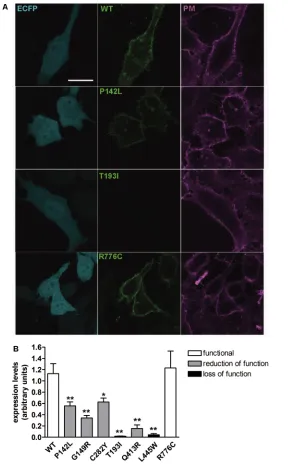
Subcellular localization of pendrin variants was determined by colocal ization between wildtype or mutant PDSEYFP and markers of the plasma membrane (PM) (CellMask™ Deep Red plasma membrane stain, C10046; Invi trogen Molecular Probes) or endoplas mic reticulum (ER) (ERTracker™ Red, glibenclamide BODIPY®TR, E34250; Invitrogen Molecular Probes). Being that HEK 293 Phoenix cell morphology is scarcely suitable for colocalization experiments, HeLa cells were used for this purpose.
a monoallelic SLC26A4 variant were submitted to the genetic analysis for FOXI1 (OMIM ID: 601093, GenBank ID: NG_012068.1) and KCNJ10 (OMIM ID: 602208, GenBank ID: NG_016411.1). The primer pairs used to amplify the coding exons of FOXI1 and KCNJ10 are reported in the Supplementary Table S1. The same primers were also used for sequencing. The newly identified SLC26A4 sequence variants were submitted to the Leiden Open Variation Database (https://grenada. lumc.nl/LOVD2/Usher_ montpellier/ home.php).
Plasmid Constructs
The pTARGET (Promega Corpora tion) vector, containing the open read ing frame (ORF) of wildtype human pendrin cloned from normal thyroid tissue, was originally provided by P BeckPeccoz, University of Milan (Italy), and was used for functional tests and Western blot.
For colocalization and determination of pendrin expression levels via imaging, the ORF of wildtype pendrin was sub cloned into the pEYFPN1 vector, in frame with the ORF of the enhanced yellow fluorescent protein (EYFP). After trans fection of this construct in cells, pendrin is produced with the EYFP fused to its C terminus (PDSEYFP). Wildtype PDSEYFP showed better plasma mem brane trafficking than EYFPPDS ( pendrin with the EYFP fused to its Nterminus; data not shown), in agreement with previous observations (5).
The pendrin mutants were made using the QuikChange® sitedirected mutagenesis kit (Stratagene) according to the manufacturer’s protocol, using the primers listed in Supplementary Table S2. All plasmid inserts were sequenced before use in experiments (Microsynth AG, Switzerland) with the primers listed in Supplementary Table S2.
Cell Lines and Transient Transfection Human embryonic kidney (HEK) 293 Phoenix (23) and HeLa ( cervical adeno carcinoma, American Type Cell Culture Collection ATCCCCL2) cells were test of the identified pendrin variants
was performed. DNA samples of all individuals were obtained from the Clinic of Otorhinolaryngology (State University of Campinas, Brazil). The research was prospectively reviewed and approved by a duly constituted ethics committee ( project: numbers 633/2003 and 396/2006). Written informed consent was obtained from all subjects or their legal representatives before blood sampling and genetic testing. For all patients, imaging studies of the inner ear by computer tomography (CT) and magnetic resonance imaging (MRI) of the temporal bones were performed. EVA was defined as an enlargement of the vestibular aqueduct >1.5 mm midway between the endolymphatic sac and the vestibule. The thyroid function of patients was not analyzed (the perchlo rate discharge test was not performed); however, it was noticeable that one patient (patient ID 23, group II) presented overt goiter.
Genomic DNa analysis
Determination of Wild-type and Mutant Pendrin Expression Levels in the Plasma Membrane region
HeLa cells expressing wildtype or mutant PDSEYFP and the enhanced cyan fluorescent protein (ECFP)
(pECFPC1 vector, Clontech) were stained with CellMask™ Deep Red plasma membrane stain, thoroughly washed and imaged in HBSS. EYFP fluorescence intensity of three regions of interest of the plasma membrane of a single cell was subtracted for the background flu orescence, averaged and normalized for the backgroundsubtracted ECFP fluores cence intensity measured in the cytosol of the same cell, to obtain wildtype or mutant PDSEYFP expression levels nor malized for the transfection efficiency of the single cell. Imaging was performed by confocal microscopy as described above. Further details are available in the Supplementary Materials and Methods.
salts and Chemicals
All salts and chemicals used were per analysis grade.
statistical analysis
All data are expressed as arithmetic means ± standard error of the mean (SEM). For statistical analysis, GraphPad Prism software (version 4.00 for Win dows, GraphPad Software) was used. Significant differences between datasets were tested by Fisher exact test or one way analysis of variance (ANOVA) with Bonferroni or Dunnett posttests, as appropriate. Statistically significant differences were assumed at p < 0.05; (n) corresponds to the number of indepen dent measurements.
All supplementary materials are available online at www.molmed.org.
rEsULTs
Detection of sequence Variations in the
GJB2,GJB6 and MT-RNR1 Genes All patients were negative for two common deletions [del(GJB6D13S1830) and del(GJB6D13S1854)] affecting for 10 min, washed three times and
imaged in Hanks balanced salt solution (HBSS). The nuclear staining with DAPI gives a signal that, expressed as average levels of gray, correlates to the number of cells in the imaged field, therefore giving an indication of cell density. Fluorescence intensity of the whole imaging field ( average levels of gray) in the EYFP emission channel was subtracted for the background fluorescence and normalized for the backgroundsubtracted fluorescence intensity in the DAPI emission channel, to obtain wildtype or mutant PDSEYFP expression levels normalized for the cell density. Imaging was performed by confocal microscopy as described above. Further details are available in the Sup plementary Materials and Methods.
Determination of Wild-type and Mutant Pendrin Expression Levels in Total Cell Membranes Lysates by Western Blot
HEK 293 Phoenix cells expressing wildtype or mutant pendrin were collected and centrifuged at 216g for 15 min at 4°C. The extraction of the total membrane protein fraction, including both plasma membrane proteins and membrane proteins from cellular or ganelles, was obtained with the Plasma Membrane Extraction Kit (MBL Inter national Corporation), according to the manufacturer’s instructions. Sodium dodecyl sulfate– polyacrylamide gel elec trophoresis (SDSPAGE) and Western blot were performed following standard procedures (see Supplementary Materi als and Methods). The primary antibod ies were rabbit anti pendrin, 1:10,000, provided by D Eladari (25), or rabbit antipendrin, polyclonal, raised against amino acids 586–780 of human pendrin (sc50346, Santa Cruz Biotechnology) 1:500 and rabbit anti calreticulin (ab4, Abcam) 1:1,000. Detection of the signal of immunocomplexes was performed with the ODYSSEY infrared imaging system (LICOR). Blot images were den sitometrically analyzed with the ImageJ 1.46r software.
Imaging was performed by sequential acquisition with a Leica TCS SP5II AOBS confocal microscope (Leica Microsystems) equipped with a HCX PL APO 63×/1.20 Lambda blue water immersion ob jective and controlled by the LAS AF SP5 software (Leica Microsystems). Further details are given in the Supplementary Materials and Methods.
semiquantitative reverse
Transcription–PCr for Verification of Equal Transfection Efficacy of the Different Plasmid Constructs
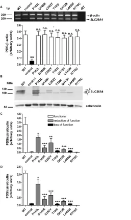
Extraction of total RNA from HEK 293 Phoenix cells transfected with the pTARGET constructs coding for wildtype or mutated pendrin or with the pTARGET empty vector (control) was performed with the All Prep DNA/RNA mini kit (Qiagen). At total of 1 μg total RNA was used for the reverse transcription reaction with the QuantiTect® reverse tran scription kit for cDNA synthesis with integrated removal of genomic DNA contamination (Qiagen). For detecting the SLC26A4 transcript, the following primers were used: forward, 5′TTCCA GCAAC AGCAC GAG3′, and reverse, 5′GCCAC TAGCC CAGTA CTAAC TC3′. The SLC26A4 signal was normal ized to the βactin signal, detected by using the following primers: forward, 5′GGCAT GGGTC AGAAG GATTC3′, and reverse, 5′AGAGG CGTAC AGGGA TAGCA C3′. These primers span an in tronexon boundary and would disclose an eventual contamination from ge nomic DNA as a band at 740 bp, which was not detected ( Figure 4A). Densi tometric analysis was done with the ImageJ 1.46r software (Wayne Rasband, National Institutes of Health, USA).
Determination of Wild-Type and Mutant Pendrin Total Expression Levels by Imaging
Function of Pendrin Variants The impact of the amino acid sub stitutions p.V138F, p.V609G, p.R409H and p.T410M and the deletion c.279delT (p.S93Rfs3*) on pendrin functionality were previously described by us or others (10; Table 2) and were not characterized further. For the splicing site mutations c.1707+5G>A and c.1001+1G>A (also called IVS15+5G>A and IVS8+1G>A, respectively; evidenced in dark gray in Table 1) most likely leading to an aberrant protein product (26) or no protein prod uct at all (27), the functional test described here could not be applied. Therefore, five previously uncharacterized pendrin vari ants (p.P142L, p.G149R, p.T193I, p.Q413R, p.L445W), the novel variant p.C282Y was detected in this cohort (patient
ID C26). All the other patients were homo zygous wildtype for the pendrin gene.
In the cohort of deaf patients with EVA (group II, Table 1), as previously described (18), sequence alterations in the pendrin gene were found in 9 of 26 subjects. Of these patients, five carried biallelic and four monoallelic sequence alterations, respectively. All the other subjects were homozygous wildtype for the pendrin gene.
sequencing of FOXI1 and KCNJ10
Coding regions
Sequence alterations in the FOXI1 or KCNJ10 coding regions were not detec ted in these patients.
GJB6 (encoding connexin 30) and for the m.1555A>G mutation in the mito chondrially encoded 12S rRNA gene MT-RNR1. Two patients (patient ID C26, group I, and L1, group II; Table 1) showed a monoallelic mutation (c.35delG) in the GJB2 gene encoding Connexin 26. All the other patients were homozygous wildtype for GJB2.
Pendrin allelic Variants in Deaf Brazilian Patients with and without EVa
In the cohort of deaf patients without EVA (group I, Table 1), monoallelic alterations in the sequence of the pen drin gene were found in 5 of 32 subjects. A novel (previously unknown and uncharacterized) pendrin variant (p.C282Y)
Table 2. Functional and molecular features of pendrin variants identified patients without (group I) and with (group II) EVA.a
Group Patient ID
Protein variants: Allele 1 Allele 2
Function: Allele 1 Allele 2
Localization: Allele 1 Allele 2
Expression: Allele 1 Allele 2
Pendrin-related deafness
Reference: Allele 1 Allele 2
I C15 p.V138F Lost ER ? Probably not (44)
WT WT PM WT —
C26b p.C282Y Reduced (41%) PM Reduced Probably not Present work
WT WT PM WT —
C01 p.V609G Reduced (45%) ? ? Probably not (45)
WT WT PM WT —
C04 p.V609G Reduced (45%) ? ? Probably not (45)
WT WT PM WT —
C09 p.V609G Reduced (45%) ? ? Probably not (45)
WT WT PM WT —
II 21 p.P142L Reduced (76%) PM Reduced Yes Present work
p.S93Rfs3* Lost ? ? (46)
16 p.G149R Reduced (55%) PM/ER Reduced ? Present work
WT WT PM WT —
18 p.T193I Lost ER Greatly reduced ? Present work
WT WT PM WT —
22 p.R409H Lost or reduced (33%) PM/ER Reduced Yes (47) and data not shown p.R409H Lost or reduced (33%) PM/ER Reduced (47) and data not shown
02 p.T410M Lost ER ? yes (44)
SS (c.1707+5G>A) Most likely lost — Lost (27)
06 p.Q413R Reduced (60%) ER Reduced Yes Present work
p.V138F Lost ER ? (44)
23 L445W Lost ER Greatly reduced Yes Present work
SS (c.1001+1G>A) Most likely lost ? (26)
L1b p.V609G Reduced (45%) ? ? ? (45)
WT WT PM WT —
15 p.R776C Unaffected PM Unaffected No Present work
WT WT PM WT —
aThe magnitude of reduction in pendrin transport activity is indicated in %. Question mark (?) denotes an undetermined feature. SS,
splicing site variant; *, stop codon.
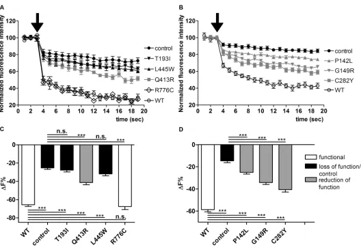
subcellular Localization of Pendrin Variants
To define their subcellular localization, wildtype or mutated PDSEYFP were expressed in HeLa cells (Figure 2), and colocalization with the PM or ER was determined. As shown in Figures 2A and D, wildtype pendrin is preferentially targeted to the PM, with poor colocal ization with the ER, thereby providing evidence that the Cterminal EYFP tag does not alter proper pendrin trafficking. Similarly, pendrin p.P142L, p.G149R, p.C282Y and p.R776C (Figure 2A) were efficiently transposed to the PM. On the (Figures 1A, C) was not significantly
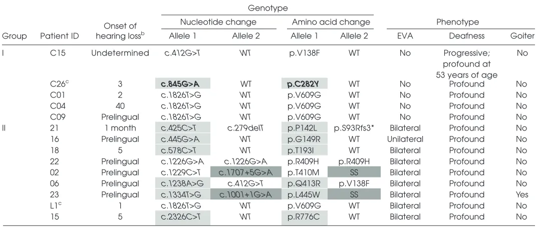
different from that measured in con trol cells, indicating a complete loss of function of these variants. In contrast, iodide influx measured in cells express ing pendrin p.Q413R (Figures 1A, C), p.P142L, p.G149R and p.C282Y (Figures 1B, D) was significantly different from that measured in wildtype and control cells (p < 0.001), indicating that these variants retain residual transport activity. The transport activity of pendrin p.R776C (Figures 1A, C) was indistinguishable from wildtype. The results of the func tional test are summarized in Table 2. and one variant (p.R776C) for which
functionality was ambiguously defined (5,14,28) were subjected to a functional test (Figure 1; the pendrin variants were subdivided into two groups for technical reasons).
Iodide flux in cells expressing wild type pendrin (Figure 1) was significantly higher (p < 0.001) compared with that measured in control cells and confirmed that pendrin is an iodide transporter (29,30), most likely acting as a Cl–/I– anion exchanger in this system (31).
Iodide influx measured in cells expres sing pendrin p.T193I and p.L445W
fully functional variant p.R776C, were substantially reduced with respect to wildtype. A dramatic reduction in the expression levels was observed, especially for those mutants (p.T193I, p.Q413R and p.L445W) entrapped in the ER, whereas the expression levels of those variants (p.P142L and p.C282Y) preferentially targeted to the PM were significantly higher with respect to those of the variants prefer entially retained in the ER (p < 0.001). (Table 2). Pendrin p.G149R represents an
intermediate situation, since this variant was found to colocalize with both the PM (Figure 2A) and ER (Figure 2D).
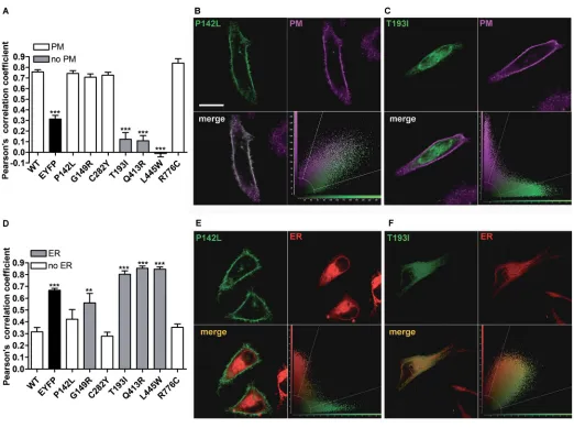
Total Expression Levels of Pendrin Variants
Total expression levels of pendrin variants were evaluated by confocal imaging (Figure 3). Interestingly, the total expression levels of all of the pen drin variants, with the exception of the other hand, pendrin p.T193I, p.Q413R
and p.L445W failed to reach the PM. Figure 2D shows that pendrin p.G149R, p.T193I, p.Q413R and p.L445W are pref erentially located in the ER, whereas pendrin p.P142L, p.C282Y and p.R776C are not preferentially found in this compartment. To conclude, wildtype, p.P142L, p.C282Y and p.R776C pendrin variants show preferential trafficking to the PM, whereas p.T193I, p.Q413R and p.L445W are largely retained in the ER
nonglycosylated, partially glycosylated and maturely glycosylated forms of pendrin (Figure 4B) and correspond to what was previously reported (32,33). untagged (that is, with no EYFP fused
to the C terminus) wildtype and mutated pendrin ( Figures 4B–D). Bands between 100 and 130 kDa represent the Expression levels of pendrin vari
ants in total cellular membrane pro tein extracts were also evaluated by Western blot after transfection of
A signal corresponding to pendrin p.R776C was not detected, most likely due to conformational disruption of the epitope recognized by the antibody used in these experiments (25). Densitometry showed that all functionally impaired pendrin variants exhibit cellular expres sion levels significantly reduced with respect to the wildtype (Figure 4C) and confirmed the results obtained by imag ing, thereby showing evidence that the presence of a Cterminal tag does not significantly influence protein stability. Notably, the pendrin variants with loss of function (p.T193I and p.L445W) only show the nonglycosylated form (Figures 4B, D). Furthermore, the pres ence of the higher band, corresponding to the maturely glycosylated protein (Figure 4D), correlates with function (Figure 1) and with expression in the plasma membrane region (Figure 5). In these experiments, an antibody directed against the C terminal 29 amino acids of rat pendrin was used (25). Similar results were obtained with a commercial antibody directed against the amino acids 586–780 of human pendrin (data not shown).
Verification of the Transfection Efficacy of the Different Plasmid Constructs
As differences in the protein abun dance of the different pendrin variants may potentially arise from differences in the transfection efficacy of the corre sponding plasmid constructs, the latter was verified by semiquantitative reverse transcription PCR. Significant differences between the abundance of cDNA (and hence, mRNA) encoding the various pendrin variants in the corresponding batches of transfected cells were not detected (Figure 4A).
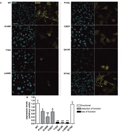
Expression Levels of Pendrin Variants in the Plasma Membrane region
Whereas in Figure 2A we showed the ability of the investigated pendrin variants to reach the PM, the technique used did not allow quantification and comparison of the amount of the different protein vari ants within this compartment. As shown Figure 4. RNA and protein levels of wild-type pendrin and pendrin variants. (A) Top: The
expression levels of p.Q413R are very low (Figure 3), a residual amount of this variant within the plasma membrane was detected (Figure 5).
DIsCUssION
Genotype–phenotype correlations are difficult to assess for SLC26A4. A recent analysis of six studies with a total enrollment of 769 hearingimpaired probands with EVA revealed that, on average, 45–50% of patients carry at least one SLC26A4 sequence variant. Of these, only 25% carry biallelic SLC26A4 sequence variants, whereas for a nota ble proportion of deaf individuals with EVA (~55%), SLC26A4 sequence variants were not detected (34). Deafness can unequivocally be assigned to pendrin dysfunction only when both SLC26A4 alleles encode for hypo or nonfun ctional pendrin protein variants. Before
the com pletion of the present study, deafness and EVA could unequivocally be linked to pendrin malfunction in only 2 of 26 individuals in our cohort of deaf patients with EVA (patient IDs 22 and 02, Group II, Table 2), bearing previ ously characterized pendrin sequence alterations affecting function. The func tional assessment of p.P142L, p.Q413R and p.L445W pendrin protein variants presented here allowed for the conclu sion that deafness and EVA are most likely due to pendrin dysfunction also for patients 21, 06 and 23 (Table 2). The lack of functional impact of the amino acid substitution p.R776C allowed for the exclusion of pendrin as the genetic cause of deafness and EVA for patient 15 (Table 2).
The functional test used in the pres ent work evaluates the chloride/iodide exchange activity of pendrin (8,31,35,36). Despite that iodide transport may be of no physiological relevance in the inner ear, the chloride/iodide exchange activity generally reflects the chloride/ bicarbonate exchange activity (10) and can therefore be used as a tool to discrim inate between variants with and without pathogenic potential. Accordingly, when differences between the chloride/iodide Expression levels of the different pen
drin variants in the PM region mirrored total expression levels (Figures 3, 4). It is important to note that, although the total in Figure 5, the amount of pendrin and
its variants in the PM region was evalu ated by confocal imaging with a different approach (see Materials and Methods).
of the protein in the PM region that could not be quantified in colocaliza tion experiments, as mentioned earlier. The low expression levels of pendrin p.Q413R, however, seems to be insuffi cient to justify its significant transport function. It is possible that the amino acid substitution Q413R leads to protein misfolding and consequent degradation and simultaneously confers an intrinsic gain of transport function, therefore leading to significant iodide intake (Fig ure 1C) despite dramatically reduced expression levels (Figures 3–5). Expres sion levels of the variant p.R776C were not reduced with respect to the wild type, therefore confirming its nature of a benign polymorphism. We conclude that functionally impaired pendrin vari ants show cellular expression levels re duced with respect to the wildtype and functionally unaffected variants, with mutants with reduced function showing significant, although heterogeneous, expression levels at the PM and mutants with null function being undetectable in the PM and completely retained in the ER.
Misfolded proteins are degraded via a series of pathways collectively denoted as ERassociated degradation (ERAD), requiring ubiquitination by different E3 ubiquitin ligases as an essential step (38). Single amino acid substitutions may impair the ability of the affected polypeptide to reach the functional conformation; consequently, prolonged interaction with molecular chaperones, enhanced proteolytic degradation and reduced cellular halflife of mutant pro teins are events occurring in a number of genetic diseases (39). A previous report gathered evidence that wildtype pendrinGFP colocalizes with ubiqui tin in the cell, therefore suggesting a role for polyubiquitination in pendrin degradation (40). Accordingly, ubiq uitination prediction programs (http:// www.ubpred.org/) identify pendrin lysine residues at positions 632, 647, 734 and 753 as putative ubiquitination sites ( Supplementary Figure S1). Recently, Lee et al. (33) identified the specific for three cases, the assignment of the
genetic cause of deafness and EVA was not conclusive (Table 2).
The identification of pendrin variants with different degrees or no functional impairment led us to attempt correlat ing transport function and molecular features such as cellular distribution and expression levels of pendrin vari ants. Pendrin variants showing good colocalization with the PM (p.P142L, p.G149R, p.C282Y; Figure 2A) retain a significant transport activity (Figure 1D), whereas poor colocalization with the PM and retention in the ER (as is the case of variants p.T193I and p.L445W [ Figures 2A, D]; the localization of pendrin p.L445W is in agreement with previous findings [5]) correlates with loss of function ( Figure 1C).
Retention in the subcellular compart ments is an indication of protein misfold ing, often leading to degradation (37). This observation prompted us to quan tify the cellular expression levels of the different pendrin protein variants, on the basis of the hypothesis that abundance of variants with significant retention in the ER should be reduced with respect to variants with targeting to the PM. In terestingly, however, total (Figures 3 and 4) and PM (Figure 5) expression levels of all of the functionally impaired pendrin variants were significantly reduced with respect to the wildtype, regardless of their cellular localization. Abundance of all protein variants with significant residual activity (p.P142L, p.G149R, p.C282Y and p.Q413R), evaluated in the PM region by means of quantitative imaging (Figure 5), was significantly higher than that of variants with total loss of function (p.T193I and p.L445W). Differences in expression levels of pen drin protein variants did not arise from differences in the transfection efficiency of the corresponding plasmid construct or mRNA abundance ( Figure 4A, Sup plementary Materials and Methods and Supplementary Table S3).
The significant residual activity of pendrin p.Q413R appears to be substan tiated by detection of a small amount and chloride/bicarbonate exchange
efficiencies were observed, they were con sidered to be “moderate” and most likely of no pathophysiological relevance (5).
For patients bearing monoallelic pen drin mutations, interpretation of results is less straightforward. Variants with reduction or loss of function are found both in the group of deaf patients with EVA (p.G149R, p.T193I and p.V609G) and in the group of deaf patients with no EVA (p.V138F, p.C282Y and p.V609G), with no apparent correlation between occurrence of monoallelic, functionally impaired pendrin variants and presence of EVA in these cohorts. In addition, a same variant (that is, p.V609G) could be found in both cohorts with and without EVA. On the basis of these observations, it seems that some defined monoallelic pendrin sequence alterations, although encoding hypofunctional protein vari ants, as it is the case of p.V609G, are per se not sufficient to cause deafness and EVA, and the association with other genetic, epigenetic and/or envi ronmental factors should be taken into account when attempting to identify the determinant for the deafness phenotype of a given patient. Noteworthy in the present study, the p.V609G amino acid substitution was found in association with a monoallelic GJB2 pathogenic variant in patient L1, possibly leading to EVA. However, the association between monoallelic GJB2 and SLC26A4 sequence alterations affecting functionality does not invariably lead to EVA, since this genetic configuration is also found in patient C26(carrying the newly identi fied monoallelic p.C282Y pendrin variant and the c.35delG GJB2 sequence alteration and not showing EVA).
6. Pryor SP, et al. (2005) SLC26A4/PDS genotype phenotype correlation in hearing loss with enlargement of the vestibular aqueduct (EVA): evidence that Pendred syndrome and non syndromic EVA are distinct clinical and genetic entities. J. Med. Genet. 42:159–65.
7. Albert S, et al. (2006) SLC26A4 gene is frequently involved in nonsyndromic hearing impairment with enlarged vestibular aqueduct in Caucasian populations. Eur. J. Hum. Genet. 14:773–9. 8. Pera A, et al. (2008) Functional assessment of
allelic variants in the SLC26A4 gene involved in Pendred syndrome and nonsyndromic EVA.
Proc. Natl. Acad. Sci. U. S. A. 105:18608–13. 9. Stenson PD, et al. (2014) The Human Gene
Mutation Database: building a comprehensive mutation repository for clinical and molecular genetics, diagnostic testing and personalized genomic medicine. Hum Genet. 133:1–9. 10. Dossena S, et al. (2011) Molecular and functional
characterization of human pendrin and its allelic variants. Cell Physiol. Biochem. 28:451–66. 11. Fugazzola L, et al. (2002) Differential diagnosis
between Pendred and pseudoPendred syn dromes: clinical, radiologic, and molecular studies. Pediatr. Res. 51:479–84.
12. Kara C, Kilic M, Ucakturk A, Aydin M. (2010) Congenital goitrous hypothyroidism, deafness and iodide organification defect in four siblings: Pendred or pseudoPendred syndrome? J. Clin.
Res. Pediatr. Endocrinol. 2:81–4.
13. Davis N, Lunardi C, Shield JP. (2006) Sensorineural deafness and hypothyroidism: autoimmunity causing ‘pseudoPendred syndrome.’ Horm. Res. 65:267–8.
14. Pfarr N, et al. (2006) Goitrous congenital hypo thyroidism and hearing impairment associated with mutations in the TPO and SLC26A4/PDS genes. J. Clin. Endocrinol. Metab. 91:2678–81. 15. Kopp P, et al. (1999) Phenocopies for deafness
and goiter development in a large inbred Brazil ian kindred with Pendred’s syndrome associated with a novel mutation in the PDS gene. J. Clin.
Endocrinol. Metab. 84:336–41.
16. Camargo R, et al. (2001) Aggressive metastatic follicular thyroid carcinoma with anaplastic transformation arising from a longstanding goiter in a patient with Pendred’s syndrome.
Thyroid. 11:981–8.
17. LofranoPorto A, et al. (2008) Pendred syn drome in a large consanguineous Brazilian family caused by a homozygous mutation in the
SLC26A4 gene. Arq. Bras. Endocrinol. Metabol. 52:1296–303.
18. de Moraes VC, et al. (2013) Molecular analysis of SLC26A4 gene in patients with nonsyndromic hearing loss and EVA: identification of two novel mutations in Brazilian patients. Int. J. Pediatr.
Otorhinolaryngol. 77:410–3.
19. Ramos PZ, et al. (2013) Etiologic and diagnostic evaluation: algorithm for severe to profound sensorineural hearing loss in Brazil. Int. J. Audiol. 52:746–52.
of subcellular localization. Therefore, a molecular feature common to patho genic pendrin protein variants has been identified and could represent the target of future moleculedirected therapies aimed at restoring pendrin function.
aCKNOWLEDGMENTs
This work was supported by the Seventh Framework Programme (grant PIRSESGA2008230661) and Fonds zur Förderung der wissenschaftlichen Forschung (FWF) (grant P18608) to M Paulmichl and Fundação de Amparo à Pesquisa do Estado de São Paulo and Coordenação de Aperfeiçoamento de Pessoal de Nível Superior to EL Sar torato. C Nofziger was supported by the Roche Postdoc Fellowship Program (grant 231). The antipendrin antibody was a gift from D Eladari, Institut National de la Santé et de la Recherche Médicale, Paris, France. We sincerely thank Elisabeth Mooslechner for her expert secretarial assistance.
DIsCLOsUrE
The authors declare that they have no competing interests as defined by Molecular Medicine, or other interests that might be perceived to influence the results and discussion reported in this paper.
rEFErENCEs
1. Everett LA, et al. (1997) Pendred syndrome is caused by mutations in a putative sulphate trans porter gene (PDS). Nat. Genet. 17:411–22. 2. Royaux IE, et al. (2003) Localization and func
tional studies of pendrin in the mouse inner ear provide insight about the etiology of deafness in Pendred syndrome. J. Assoc. Res. Otolaryngol. 4:394–404.
3. Royaux IE, et al. (2000) Pendrin, the protein encoded by the Pendred syndrome gene (PDS), is an apical porter of iodide in the thyroid and is regulated by thyroglobulin in FRTL5 cells. Endocrinology. 141:839–45.
4. Bizhanova A, Kopp P. (2010) Genetics and phenomics of Pendred syndrome. Mol. Cell.
Endocrinol. 322:83–90.
5. Choi BY, et al. (2009) Hypofunctional SLC26A4 variants associated with nonsyndromic hearing loss and enlargement of the vestibular aqueduct: genotypephenotype correlation or coincidental polymorphisms? Hum. Mutat. 30:599–608.
ER resident E3 ubiquitin ligase involved in the degradation of wildtype and mutated pendrin. It is therefore likely to hypothesize that the reduced global and PM expression levels of function ally impaired pendrin variants are the consequence of an enhanced degrada tion by the ubiquitin proteasome system.
The mutations analyzed in the pres ent work do not lie in close proximity of a glycosylation site (Supplemen tary Figure S1) and most likely do not directly impede glycosylation. Some amino acid substitutions, however, as in the case of p.T193I and p.L445W, may cause protein misfolding and premature degradation, therefore in directly interfering with or preventing glycosylation (Figure 4D).
Studies on mouse models lacking pen drin expression selectively in the endo lymphatic sac (41), or in the cochlea and the vestibular labyrinth (42), showed that pendrin activity is only required during a critical time period of embryonic devel opment and suggested that a temporally and spatially limited therapy directed to the endolymphatic sac and focused on the prenatal phase could restore normal hearing in patients with deafness linked to pendrin mutations (43). On the basis of these considerations and on the find ings presented here, we suggest that pre vention of reduction of expression levels, together with the assistance of proper protein folding, should be regarded as an essential component of potential ther apeutic approaches aimed at restoring or increasing the transport activity of dysfunctional pendrin variants. Further studies are needed to elucidate the mo lecular mechanism leading to the reduc tion of cellular abundance of pathogenic pendrin protein variants.
CONCLUsION
36. Fugazzola L, et al. (2007) High phenotypic intrafamilial variability in patients with Pen dred syndrome and a novel duplication in the
SLC26A4 gene: clinical characterization and func
tional studies of the mutated SLC26A4 protein.
Eur. J. Endocrinol. 157:331–8.
37. Tamura T, Sunryd JC, Hebert DN. (2010) Sorting things out through endoplasmic reticulum qual ity control. Mol. Membr. Biol. 27:412–27.
38. Smith MH, Ploegh HL, Weissman JS. (2011) Road to ruin: targeting proteins for degradation in the endoplasmic reticulum. Science. 334:1086–90. 39. Bross P, et al. (1999) Protein misfolding and
degradation in genetic diseases. Hum. Mutat. 14:186–98.
40. Shepshelovich J, et al. (2005) Protein synthesis inhibitors and the chemical chaperone TMAO reverse endoplasmic reticulum perturbation induced by overexpression of the iodide trans porter pendrin. J. Cell Sci. 118:1577–86. 41. Hulander M, et al. (2003) Lack of pendrin
expression leads to deafness and expansion of the endolymphatic compartment in inner ears of
Foxi1 null mutant mice. Development. 130:2013–25.
42. Li X, et al. (2013) SLC26A4 targeted to the endolymphatic sac rescues hearing and balance in Slc26a4 mutant mice. PLoS. Genet. 9:e1003641. 43. Wangemann P. (2013) Mouse models for pen
drinassociated loss of cochlear and vestibular function. Cell. Physiol. Biochem. 32:157–65. 44. Taylor JP, Metcalfe RA, Watson PF, Weetman
AP, Trembath RC. (2002) Mutations of the PDS gene, encoding pendrin, are associated with protein mislocalization and loss of iodide efflux: implications for thyroid dysfunction in Pendred syndrome. J. Clin. Endocrinol. Metab. 87:1778–84. 45. Dossena S, et al. (2011) Identification of allelic
variants of pendrin (SLC26A4) with loss and gain of function. Cell. Physiol. Biochem. 28:467–76. 46. Palos F, et al. (2008) Pendred syndrome in two
Galician families: insights into clinical pheno types through cellular, genetic, and molecular studies. J. Clin. Endocrinol. Metab. 93:267–77. 47. Gillam MP, Bartolone L, Kopp P, Benvenga S.
(2005) Molecular analysis of the PDS gene in a nonconsanguineous Sicilian family with Pendred’s syndrome. Thyroid. 15:734–41.
Cite this article as: de Moraes VCS, et al. (2016) Reduction of cellular expression levels is a common feature of functionally affected pendrin (SLC26A4) protein variants. Mol. Med. 22:41–53.
20. del Castillo FJ, et al. (2005) A novel deletion involving the connexin30 gene, del( GJB6 d13s1854), found in trans with mutations in the GJB2 gene (connexin26) in subjects with DFNB1 nonsyndromic hearing impairment.
J. Med. Genet. 42:588–94.
21. Friedman RA, et al. (1999) Maternally inherited non syndromic hearing loss. Am. J. Med. Genet. 84:369–72. 22. Iwasaki S, Tamagawa Y, Ocho S, Hoshino T,
Kitamura K. (2000) Hereditary sensori neural hearing loss of unknown cause involving mitochondrial DNA 1555 mutation.
ORL J. Otorhinolaryngol. Relat. Spec. 62:100–3. 23. DiCiommo DP, Duckett A, Burcescu I, Bremner R,
Gallie BL. (2004) Retinoblastoma protein purification and transduction of retina and retinoblastoma cells using improved alphavirus vectors. Invest.
Ophthal-mol. Vis. Sci. 45:3320–9.
24. Galietta LJ, Haggie PM, Verkman AS. (2001) Green fluorescent proteinbased halide indicators with improved chloride and iodide affinities.
FEBS Lett. 499:220–4.
25. Knauf F, et al. (2001) Identification of a chlo rideformate exchanger expressed on the brush border membrane of renal proximal tubule cells.
Proc. Natl. Acad. Sci. U. S. A. 98:9425–30. 26. Bogazzi F, et al. (2000) A novel mutation in the
pendrin gene associated with Pendred’s syn drome. Clin. Endocrinol. (Oxf). 52:279–85. 27. Ganaha A, et al. (2013) Pathogenic substitution
of IVS15 + 5G>A in SLC26A4 in patients of Okinawa Islands with enlarged vestibular aque duct syndrome or Pendred syndrome. BMC Med.
Genet. 14:56.
28. Yuan Y, et al. (2012) Molecular epidemiology and functional assessment of novel allelic variants of
SLC26A4 in nonsyndromic hearing loss patients
with enlarged vestibular aqueduct in China.
PLoS One. 7:e49984.
29. Scott DA, Wang R, Kreman TM, Sheffield VC, Karniski LP. (1999) The Pendred syndrome gene encodes a chlorideiodide transport protein. Nat.
Genet. 21:440–3.
30. Gillam MP, et al. (2004) Functional characteriza tion of pendrin in a polarized cell system: evi dence for pendrinmediated apical iodide efflux.
J. Biol. Chem. 279:13004–10.
31. Dossena S, et al. (2006) Fast fluorometric method for measuring pendrin (SLC26A4) Cl–/I– trans
port activity. Cell Physiol. Biochem. 18:67–74. 32. Yoon JS, et al. (2008) Heterogeneity in the pro
cessing defect of SLC26A4 mutants. J. Med. Genet. 45:411–9.
33. Lee K, Hong TJ, Hahn JS. (2012) Roles of 17AAGinduced molecular chaperones and Rma1 E3 ubiquitin ligase in folding and degrada tion of Pendrin. FEBS Lett. 586:2535–41. 34. Pique LM, et al. (2014) Mutation analysis of the
SLC26A4, FOXI1 and KCNJ10 genes in individu
als with congenital hearing loss. PeerJ. 2:e384. 35. Dror AA, et al. (2010) Calcium oxalate stone for