Studies on indomethacin intraocular implants using different in vitro release methods
6
0
0
Full text
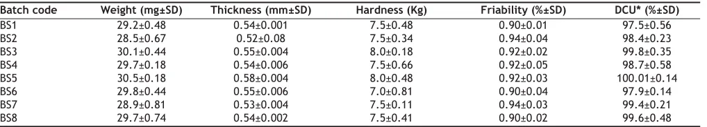
(2) www.ijpsonline.com. Fabrication of implants: Indomethacin and sodium alginate alone and in combinations with HPMC, with or without calcium chloride were mixed together well in a mortar and slugged on a Manesty E2 (Manesty, U.K) single punch machine using 12 mm diameter punches. The resultant slugs were broken and sieved through sieve #60 and #100. The granules passing through #60 and retained over #100 were compressed directly using 2×7.5mm punches on Manesty E2 single punch machine to yield track-Þeld type implants.. Evaluation of implants: For uniformity of weight, ten implants from each batch were weighed individually and their average determined. Friability and hardness were tested using Roche’s friabilator (n=6) and Monsanto hardness tester (n=10). For determination of uniformity of drug content, 6 implants from each batch were weighed individually and dissolved in 50 ml of phosphate buffer (pH 7.4). The resultant solution was filtered through G2 glass Þlter. An aliquot of the Þltrate was diluted suitably and analyzed for indomethacin content at 319.5 nm (Shimadzu, UV-1601, Japan). Dissolution studies by agar diffusion method: Ten millilitres of 1 and 2% w/v sterilized agar solution was poured on Petri dishes (90 mm diameter), aseptically and allowed to set. A circular hole was made in the center of the agar plates and the implant was placed at the center of the bore and covered with the agar plug. The implants were removed at pre-. determined time intervals and dissolved in phosphate buffer (pH 7.4), filtered, diluted and residual drug content was measured spectrophotometrically at 319.5 nm. The agar gel was dissolved in hot phosphate buffer (pH 7.4) and analyzed for indomethacin content using an appropriate blank. Dissolution studies using USP apparatus: USP apparatus-I (Campbell Electronics, Mumbai) was used with 500 ml of phosphate buffer (pH 7.4) at 37±1° as the dissolution medium with the basket speed maintained at 50 rpm. At pre-determined time intervals aliquots were withdrawn and replaced with equal volume of pre-warmed buffer. The samples were analyzed for indomethacin content at 319.5 nm, spectrophotometrically. Dissolution studies by static method: Individually weighed implants were placed in stainless steel mesh holder of dimensions 2×4×6 mm and suspended in amber coloured vials containing 10 ml of phosphate buffer (pH 7.4) and placed in a water bath thermostated at 37±1°. At pre-determined time intervals the dissolution medium was completely withdrawn and replaced with pre-warmed buffer to ensure sink conditions. The withdrawn samples were analyzed for indomethacin content as described earlier. Dissolution studies on flow-through apparatus: The studies were conducted using an in house fabricated dissolution cell, described elsewhere 5.. TABLE 1: COMPOSITION OF SODIUM ALGINATE IMPLANTS (FORMULA/ IMPLANT) Batch code BS1 BS2 BS3 BS4 BS5 BS6 BS7 BS8. Sodium alginate (mg) 25 25 12.5 10 20 15 17.5 15. Calcium chloride (mg) 5 7.5 10. HPMC K-100 (mg) 12.5 15 10 -. Indomethacin (mg) 5 5 5 5 5 5 5 5. TABLE 2: PHYSICO-CHEMICAL PROPERTIES OF THE PREPARED IMPLANTS Batch code BS1 BS2 BS3 BS4 BS5 BS6 BS7 BS8. Weight (mg±SD) 29.2±0.48 28.5±0.67 30.1±0.44 29.7±0.18 30.5±0.18 29.8±0.44 28.9±0.81 29.7±0.74. Thickness (mm±SD) 0.54±0.001 0.52±0.08 0.55±0.004 0.54±0.006 0.58±0.004 0.55±0.006 0.53±0.004 0.54±0.002. Hardness (Kg) 7.5±0.48 7.5±0.34 8.0±0.18 7.5±0.66 8.0±0.48 7.0±0.81 7.5±0.11 7.5±0.41. Friability (%±SD) 0.90±0.01 0.94±0.04 0.92±0.02 0.92±0.05 0.92±0.03 0.90±0.04 0.94±0.03 0.90±0.02. DCU* (%±SD) 97.5±0.56 98.4±0.23 99.8±0.35 98.7±0.58 100.01±0.14 97.9±0.14 99.4±0.21 99.6±0.48. *DCU– Drug content uniformity. March - April 2008. Indian Journal of Pharmaceutical Sciences. 217.
(3) www.ijpsonline.com. 350. 350 B. 300. Mean amount / unit area (mcg /mm) 2. Mean amount / unit area (mcg / mm)2. A. 250. 200. 150. 100. 50. 300. 250. 200. 150. 100. 50. 0. 0 0. 1. 2. 3. 4. 5. 0.0. 6. 0.5. 1.0. 2.0. 2.5. 3.0. 1/2. 300. 350 C. Mean amount / unit area (mcg / mm)2. Mean amount / unit area (mcg / mm) 2. 1.5. (Time in (Time in h) hours)1/2. (Time h)1/2 1/2 (Time ininHours). 300. 250. 200. 150. 100. 50. 0. D 250. 200. 150. 100. 50. 0 0. 2. 4. 6. 8. 10. 0. 2. 4. 6. 8. 1/2. (Time in h) 1/2 (Time in hours). 1/2 (TimeininHours) h) (Time 1/2. Fig. 1: Effect of particle size on indomethacin release. A - Agar diffusion (1%) [(-●-) BS2; (-○-) BS3; (-▼-) BS4; (-∆-) BS6]; B - USP method [(-●-) BS2; (-○-) BS3; (-▼-) BS4; (-∆-) BS6]; C - continuous ßow-through apparatus [(-●-) BS2; (-○-) BS3; (-▼-) BS4; (-∆-) BS6] and D - static method [(-●-) BS2; (-○-) BS3; (-▼-) BS4; (-∆-) BS6].. The dissolution cell consisted of two circular plates of 3.8 cm diameter and 1.2 cm thickness, made of acrylate. The plates were held together by means of 3 screws. The bottom plate had a groove of 1.7 cm diameter and 6 mm deep, fitted with #80 mesh for supporting the implant. An outlet tube was provided for collecting the eluate. The top plate had a hole for the inlet of the dissolution medium (phosphate buffer pH 7.4). The entire setup was connected from the top by a silicone tubing of 1mm internal diameter to a peristaltic pump. The ßow rate of the medium was maintained at 0.8 ml/h and the eluate was collected in amber coloured vials as a function of time and analyzed for indomethacin content at 319.5 nm. Statistical evaluation: Experimental results are expressed as mean ± standard deviation (SD). The student ‘t’ test was performed to 218. determine the level of signiÞcance. Differences were considered to be statistically signiÞcant at P<0.05. RESULTS AND DISCUSSION The formulation variables and the physico-chemical characteristics of the various batches of the prepared implants are shown in Tables 1 and 2, respectively. Thickness, weight and drug content varied within ± 5%. Kunou et al6,7, studied the in vitro release from PLGA scleral implants of gancyclovir by incubating the implants in 2 ml of phosphate buffered solution in a shaking water bath at 37°. Balasubramaniam et al4,8, reported a static method for studying the in vitro release of indomethacin from film type intra ocular implants of indomethacin. As there is a lot of variation in the different methods used by various investigators, an attempt was made in the present. Indian Journal of Pharmaceutical Sciences. March - April 2008.
(4) www.ijpsonline.com 350. 350. B. Mean amount / unit area (mcg / mm) 2. A. Mean amount / unit area (mcg / mm). 2. 300. 250. 200. 150. 100. 50. 300. 250. 200. 150. 100. 50. 0. 0 0. 1. 2. 3. 4. 5. 6. 0.0. 0.5. 1.0. 1/2 (Time in h)1/2 (Time in hours). 350. 2.0. 2.5. 3.0. 300. Mean amount / unit area (mcg / mm) 2. Mean amount / unit area (mcg / mm) 2. 1.5. (Time h)1/2 1/2 (Time in in hours). C. 300. 250. 200. 150. 100. 50. 0. D 250. 200. 150. 100. 50. 0 0. 2. 4. 6. 8. 10. 0. 2. 4. 6. 8. (Time in h)1/21/2 (Time in hours). 1/21/2. (Time in h) (Time in hours). Fig. 2: Inßuence of HPMC concentration on drug release. A - Agar diffusion (1%) [(-●-) BS2; (-○-) BS3; (-▼-) BS4; (-∆-) BS6] B - USP method [(-●-) BS2; (-○-) BS3; (-▼-) BS4; (-∆-) BS6]; C - continuous ßow-through apparatus [(-●-) BS2; (-○-) BS3; (-▼-) BS4; (-∆-) BS6] and D - static method [(-●-) BS2; (-○-) BS3; (-▼-) BS4; (-∆-) BS6].. study to evaluate the dissolution profiles of the prepared (compressed) implants using four different methods. In all the cases effect of parameters like particle size of the drug, HPMC concentration and calcium chloride concentration were studied. Drug release from all the methods followed square root of time kinetics, as determined by correlation coefÞcient values. Further, the release exponent (n) values were also predominantly suggestive of matrix diffusion kinetics, excepting the agar diffusion method, wherein the ‘n’ values suggested the prevalence of an anomalous (erosion) mechanism in addition to the swelling controlled (matrix) diffusion. Drug release was independent of the particle size of the drug from all the methods except the static method, where a signiÞcant difference (P<0.05) was March - April 2008. observed (fig. 1). Fig. 2 shows that, an increase in the concentration of HPMC in the implants caused a significant decrease in the release rate of indomethacin. Though various factors are likely to be responsible for the decrease in drug release with an increase in HPMC concentration from all the methods, some general trend seems to appear. HPMC partially hydrates forming a pseudo gel which control swelling of the implant, subsequently overall dissolution rate and drug availability. Once the protective gel layer is formed, two rate mechanisms predominate9. Firstly, the pseudo gel permits additional water to penetrate into the device, extending the gel layer into the implant. Secondly, the outer gel layer fully hydrates and begins to be dissolved by the ßuids. For sparingly soluble drugs like indomethacin, the dissolution rate is primarily dependent on diffusion (due to swelling) and to some extent on erosion, which in turn is. Indian Journal of Pharmaceutical Sciences. 219.
(5) www.ijpsonline.com 350. 350. Mean amount / unit area (mcg / mm)2. Mean amount / unit area (mcg / mm) 2. B. A. 300. 250. 200. 150. 100. 50. 300. 250. 200. 150. 100. 50. 0. 0 0. 1. 2. 3. 4. 5. 0.0. 6. 0.5. 1.0. 350. 2.0. 2.5. 3.0. 300 D. C 2. 300. Mean amount / unit area (mcg / mm). Mean amount / unit area (mcg / mm) 2. 1.5. (Time in h)1/21/2 (Time in hours). (Time in h)1/2. 250. 200. 150. 100. 50. 0. 250. 200. 150. 100. 50. 0 0. 2. 4. 6. 8. 10. 0. (Time in h)1/21/2 (Time in hours). 2. 4. 6. 8. (Time in h)1/21/2 (Time in hours). Fig. 3: Effect of implant cross-linking with calcium chloride on drug release. A - Agar diffusion (1% Agar) [(-●-) BS2; (-○-) BS5; (-▼-) BS7; (-∆-) BS8]; B - USP method [(-●-) BS2; (-○-) BS5; (-▼-) BS7; (-∆-) BS8]; C - continuous ßow-through apparatus [(-●-) BS2; (-○-) BS5; (-▼-) BS7; (-∆-) BS8] and D - static method [(-●-) BS2; (-○-) BS5; (-▼-) BS7; (-∆-) BS8].. dependent on viscosity of the gel. Thus increasing the concentration of HPMC increases the viscosity of the resultant gel, thus resulting in slower drug release. Cross-linking of sodium alginate involves the interaction between cations and ‘G-residues’ in sodium alginate, resulting in the formation of an ‘egg box’ structure10. (fig. 3), indicate that an increment of calcium chloride caused a significant decrease (P<0.05) in the amount of drug released from all the methods. With increasing concentration of the crosslinker (Ca++), more calcium ions will be available for cross-linking, which results in the formation of a dense calcium alginate matrix that can bring about a decrease in drug release. Sodium alginate, swells on contact with aqueous dissolution medium resulting in an increase in the porosity of the matrix, thus facilitating the mobilization of the water molecules into the polymer matrix. The dissolution medium dissolves 220. the calcium chloride incorporated in the sodium alginate implants, resulting in an in situ crosslinking of sodium alginate, which is an instantaneous phenomenon. In agar diffusion and continuous ßow through methods, the amount of aqueous medium that contacted the implant was signiÞcantly less and the time of contact between the implant and the dissolution medium was comparatively higher than the other two methods, thus the cross-linking of the sodium alginate matrix in these two methods would be progressive and more uniform in comparison to the static method, wherein the presence of relatively more dissolution medium would have resulted in rapid, but incomplete cross-linking. In USP dissolution method the implant disintegrated upon swelling which resulted in formation of isolated cross-linked mass. An increase in the concentration of agar from 1 to 2% resulted in a signiÞcant decrease (P<0.05) in the amount of drug released which may be a consequence. Indian Journal of Pharmaceutical Sciences. March - April 2008.
(6) www.ijpsonline.com. of increase in the strength of the gel formed. The increment in gel strength decreases the amount of available aqueous medium, as the water molecules would be entrapped within the gel structure, resulting in decreased rate of diffusion of the drug from the implants to the surrounding medium. It has been reported earlier that the drug diffusion coefficients from HPMC and sodium alginate matrices are strongly dependent on the water content of the system11. Amongst the methods studied, the agar diffusion method and the continuous ßow through method seem to have the potential to prolong the drug release from the compressed implant and also simulate to certain extent the in vivo conditions as far as the placement of the device is concerned. The static method, which was successfully utilized for evaluating drug release kinetics from Þlm type implants4,8 was not suitable for evaluating drug release from the compressed implant, since the mesh holder, in which the implant was placed acted as an impediment to the uniform swelling of the implant, resulting in irregular swelling during the course of the study.. REFRENCES 1. 2.. cytomegalovirus. Invest Ophthalmol Vis Sci 1997;38:665-9. Moritera T, Ogura Y, Yoshimura N, Honda Y, Wada R, Hyon SH, et al. Biodegradable microspheres containing adriamycin in the treatment of proliferate vitroretinopathy. Invest Ophthalmol Vis Sc 1992;33:312530. 4. Balasubramaniam J, Kumar MT, Pandit JK, Kant S. In vitro and in vivo characterization of scleral implants of indomethacin. Pharmazie 2001;56:793-9. 5. Pandit JK, Harikumar SL, Mishra DN, Balasubramaniam J. Effect of physical cross-linking on in vitro and ex vivo permeation of indomethacin from polyvinyl alcohol inserts, Indian J Pharm Sci 2003;62:146-51. 6. Kunou N, Ogura Y, Hashizoe M, Honda Y, Hyon SH, Ikada Y. Controlled intra-ocular delivery of ganciclovir with use of biodegradable scleral implants in rabbits. J Control Release 1995;37:143-50. 7. Kunou N, Ogura Y, Yasukawa T, Kimura H, Miyamoto H, Honda Y, et al. Long term sustained release of ganciclovir from biodegradable scleral implant for the treatment of cytomegalovirus retinitis. J Control Release 2000;68:263-71. 8. Balasubramaniam J, Pandit JK. In vitro characterization of nondegradable scleral implants of indomethacin for prolonged ocular delivery. Acta Pharm 2002;52:181-8. 9. Majumdar S, Balasubramaniam J, Barat R, Pandit JK. Preparation and in vitro characterization of methotrexate implants for the management of cervical cancer. Indian Drugs 2001;38:240-7. 10. Grant GT, Morris ER, Raes DA, Smith PJC, Thom D. Biological interactions between polysaccharides and divalent cations: The egg-box model. FEBS Lett 1973;32:195-8. 11. Siepmann J, Kranz H, Bodmeier R, Peppas NA. HPMC-matrices for controlled drug delivery: A new model combining diffusion, swelling and dissolution mechanisms and predicting the release kinetics. Pharm Res 1999;16:1748-56. 3.. Moritera T, Ogura Y, Honda Y, Wada R, Hyon SH, Ikada Y. Microspheres of biodegradable polymers as a drug delivery system in the vitreous. Invest Ophthalmol Vis Sci 1991;32:1785-90. Veloso AA, Zhu Q, Herrero-Vanrell R, Refojo MF. Ganciclovir loaded polymer microsphere in rabbit eyes inoculated with human. March - April 2008. Indian Journal of Pharmaceutical Sciences. Accepted 28 March 2008 Revised 2 March 2008 Received 29 November 2006 Indian J. Pharm. Sci., 2008, 70 (2): 216-221. 221.
(7)
Figure
![Fig. 1: Effect of particle size on indomethacin release.A - Agar diffusion (1%) [(-ß●-) BS2; (-○-) BS3; (-▼-) BS4; (-∆-) BS6]; B - USP method [(-●-) BS2; (-○-) BS3; (-▼-) BS4; (-∆-) BS6]; C - continuous ow-through apparatus [(-●-) BS2; (-○-) BS3; (-▼-) BS4; (-∆-) BS6] and D - static method [(-●-) BS2; (-○-) BS3; (-▼-) BS4; (-∆-) BS6].](https://thumb-us.123doks.com/thumbv2/123dok_us/9235732.1459172/3.612.68.550.60.435/effect-particle-indomethacin-release-diffusion-continuous-apparatus-method.webp)
![Fig. 2: Inßß uence of HPMC concentration on drug release.A - Agar diffusion (1%) [(-●-) BS2; (-○-) BS3; (-▼-) BS4; (-∆-) BS6] B - USP method [(-●-) BS2; (-○-) BS3; (-▼-) BS4; (-∆-) BS6]; C - continuous ow-through apparatus [(-●-) BS2; (-○-) BS3; (-▼-) BS4; (-∆-) BS6] and D - static method [(-●-) BS2; (-○-) BS3; (-▼-) BS4; (-∆-) BS6].](https://thumb-us.123doks.com/thumbv2/123dok_us/9235732.1459172/4.612.59.559.59.435/inssss-concentration-release-diffusion-method-continuous-apparatus-method.webp)
![Fig. 3: Effect of implant cross-linking with calcium chloride on drug release.A - Agar diffusion (1% Agar) [(-●-) BS2; (-○-) BS5; (-▼-) BS7; (-∆-) BS8]; B - USP method [(-●-) BS2; (-○-) BS5; (-▼-) BS7; (-∆-) BS8]; C - continuous ß ow-through apparatus [(-●](https://thumb-us.123doks.com/thumbv2/123dok_us/9235732.1459172/5.612.56.557.62.420/effect-implant-linking-calcium-chloride-diffusion-continuous-apparatus.webp)
Related documents
WHAT THIS STUDY ADDS: In the United States, pediatric multiple sclerosis is characterized by racial/ethnic diversity, a high proportion of children with foreign-born parents,
In the same way, Kim and Zhang (2016) conclude that politically connected firms “ can have lower detection risk, better information regarding future changes in tax
The objective of this report is to describe an adolescent girl of miliary tuberculosis with ocular involvement (anterior uveitis) and to highlight the importance of keeping in mind
(a) Energy ratio of high-frequency guided ultrasonic wave pulse measured with laser interferometer at location behind fatigue crack close to hole for four specimens (solid lines),
A qualitative study was conducted with the purpose to explore and describe the mentoring needs of clinical facilitators with a view to proposing a mentoring programme for
We demonstrate the effi- cacy of the COM-Cooperative HRL algorithm as well as the relation between the communication cost and the learned communication policy using a multiagent
The aim of this study was to determine whether medical factors were associated with subsequent self- and officially-reported crashes and traffic offences in a group of
For ASC (Figures 5 A and D), the 5 ′ UTR RNA and LPS co-stimulation group showed significantly decreased ASC mRNA expression but increased protein expres- sion of ASC when
