Masthead Logo
University of Pennsylvania
ScholarlyCommons
Publicly Accessible Penn Dissertations
2018
Design, Synthesis, And Characterization Of
Dimeric Lysosomal Inhibitors And Their Effect On
Cancer Biology
Michael Nicastri
University of Pennsylvania, [email protected]
Follow this and additional works at:https://repository.upenn.edu/edissertations Part of theChemistry Commons
This paper is posted at ScholarlyCommons.https://repository.upenn.edu/edissertations/2967
For more information, please [email protected]. Recommended Citation
Nicastri, Michael, "Design, Synthesis, And Characterization Of Dimeric Lysosomal Inhibitors And Their Effect On Cancer Biology" (2018).Publicly Accessible Penn Dissertations. 2967.
Design, Synthesis, And Characterization Of Dimeric Lysosomal Inhibitors
And Their Effect On Cancer Biology
Abstract
When detected early and surgically removed, melanoma has a cure rate greater than 90%. However, when the cancer is either unresectable or goes undetected until late-stage metastases develop, the five-year survival rate drops to 20%. Unfortunately, the current standard of care suffers from chemotherapeutic resistance, where the outcome results in a high rate of patient mortality. Recent clinical studies have demonstrated
hydroxychloroquine-mediated inhibition of autophagy presents a promising strategy as an adjuvant therapy for melanoma treatment. Regrettably, in clinical trials hydroxychloroquine has not demonstrated reliable potency, at the maximum tolerated doses, in producing autophagy inhibition.
Work by the Winkler and Amaravadi Laboratories has focused on developing a series of dimeric chloroquine-based autophagy inhibitors. Our findings encouraged us to develop a library of dimeric inhibitors, chloroquine-based on hydroxychloroquine and its congener, quinacrine. These not only inhibit autophagy, but also possess single agent anti-cancer activity previously unobserved with small-molecule autophagy inhibitors. Through our efforts we have designed DQ661, a dimeric quinacrine autophagy inhibitor which simultaneously inhibits key drivers of oncogenesis: autophagy, mTOR kinase signaling, and macropinocytosis. We have also discovered an analogous dimeric chloroquine inhibitor, DC661, which is a potent inhibitor of autophagy and cancer cell proliferation. Utilizing these compounds, we have identified a molecular binding partner for our dimeric inhibitors: protein palmitoyl thioesterase 1 (PPT1), a key regulator of palmitoylation within the lysosome. We are currently developing chemical tools to study the role of PPT1 in cancer biology.
Degree Type Dissertation
Degree Name
Doctor of Philosophy (PhD)
Graduate Group Chemistry
First Advisor Jeffrey D. Winkler
Keywords
Autophagy, Cancer, Chloroquine, Quinacrine
Subject Categories Chemistry
DESIGN, SYNTHESIS, AND CHARACTERIZATION OF DIMERIC LYSOSOMAL INHIBITORS AND THEIR EFFECT ON CANCER BIOLOGY
Michael C. Nicastri A DISSERTATION
in Chemistry
Presented to the Faculties of the University of Pennsylvania in
Partial Fulfillment of the Requirements for the Degree of Doctor of Philosophy
2018
Supervisor of Dissertation
________________________
Dr. Jeffrey D. Winkler
Merriam Professor of Chemistry
Graduate Group Chairperson ________________________ Dr. Gary A. Molander
Hirschmann-Makineni Professor of Chemistry
Dissertation Committee
Dr. Amos B. Smith III, Rhodes-Thompson Professor of Chemistry Dr. William P. Dailey, Associate Professor of Chemistry
DESIGN, SYNTHESIS, AND CHARACTERIZATION OF DIMERIC LYSOSOMAL INHIBITORS
AND THEIR EFFECT ON CANCER BIOLOGY
COPYRIGHT
2018
Michael C. Nicastri
This work is licensed under the Creative Commons Attribution- NonCommercial-ShareAlike 3.0 License
To view a copy of this license, visit
iii
DEDICATION
This work is dedicated to my parents and grandparents. Through their support I have
iv
ACKNOWLEDGMENT
First, I would like to thank my research advisor, Professor Jeffrey D. Winkler, for
his support and training. In my time at the University of Pennsylvania, I have grown
significantly as a scientist and as a professional. Without his mentorship, this growth
would not have been possible.
Second, my dissertation research was a contribution to an immensely
collaborative effort between the Winkler and Amaravadi Laboratories. I would like to
thank both Professor Winkler and Professor Ravi K. Amaravadi for the opportunity to
contribute to this project. As part of this collaboration I had the privilege of working with
excellent scientists: Quentin McAfee, Colin Fennely, Dr. Julie Rotenberg-Barber, Dr.
Noel McLaughlin, and Dr. Vito Rebecca. Without their teamwork and support this
dissertation would not have been possible. Dr. Vito Rebecca was my biology partner, and
without both his support and ongoing friendship, this collaboration would not have been
possible.
Third, I would like to thank my dissertation committee, Professor Amos B. Smith,
Professor William P. Dailey, and Professor E. James Petersson, for their advice at annual
meetings and during their own office hours. Their advice was particularly important in
maintaining the focus of my research in a highly collaborative endeavor.
Fourth, I would like to thank all the facility managers, Dr. George Furst, Dr. Jun
Gu, and Dr. Charles Ross, for their tremendous help in characterization of the molecules I
synthesized. Dr. George Furst and Dr. Jun Gu were both instrumental in the
characterization of multiple large molecular weight molecules by nuclear magnetic
resonance. Dr. Charles Ross taught me to effectively utilize mass spectrometry in my
synthetic research, a skill which has been integral to my success as a synthetic organic
chemist.
Fifth, I would like to thank my, friends and colleagues in the Winkler Laboratory
and chemistry department at the University of Pennsylvania. Their support and friendship
v
Jack Ferrie, Stan Najmr, and Ben Partridge, three friends from my graduate student class
who have helped me grow as a person and as a chemist. Next, I would like to thank Tyler
Higgins, a member of the Winkler Laboratory who is both a supportive colleague and
friend. Particularly, I would like to thank Tyler for listening to all my chemistry
hypotheses on a daily basis, regardless of their impulsiveness.
Finally, I would like to thank my family for supporting me through my five years
at the University of Pennsylvania. Through their support, I was able to succeed in
growing as a chemist and as a person through my time in graduate school. I would like to
thank my father Chris Nicastri and my mother Jamie Nicastri for inspiring me to become
the adult I am today. Particularly, I would like to thank my fiancée Bianca Llaneza for
her support. She is my best friend and the one who helped me bounce back from
unsuccessful research, always giving me the motivation to continue with a new
vi
ABSTRACT
DESIGN, SYNTHESIS, AND CHARACTERIZATION OF DIMERIC LYSOSOMAL
INHIBITORS AND THEIR EFFECT ON CANCER BIOLOGY
Michael C. Nicastri
Jeffrey D. Winkler
When detected early and surgically removed, melanoma has a cure rate greater
than 90%. However, when the cancer is either unresectable or goes undetected until
late-stage metastases develop, the five-year survival rate drops to 20%. Unfortunately, the
current standard of care suffers from chemotherapeutic resistance, where the outcome
results in a high rate of patient mortality. Recent clinical studies have demonstrated
hydroxychloroquine-mediated inhibition of autophagy presents a promising strategy as an
adjuvant therapy for melanoma treatment. Regrettably, in clinical trials
hydroxychloroquine has not demonstrated reliable potency, at the maximum tolerated
doses, in producing autophagy inhibition.
Work by the Winkler and Amaravadi Laboratories has focused on developing a
series of dimeric chloroquine-based autophagy inhibitors. Our findings encouraged us to
develop a library of dimeric inhibitors, based on hydroxychloroquine and its congener,
quinacrine. These not only inhibit autophagy, but also possess single agent anti-cancer
activity previously unobserved with small-molecule autophagy inhibitors. Through our
efforts we have designed DQ661, a dimeric quinacrine autophagy inhibitor which
simultaneously inhibits key drivers of oncogenesis: autophagy, mTOR kinase signaling,
vii
inhibitor, DC661, which is a potent inhibitor of autophagy and cancer cell proliferation.
Utilizing these compounds, we have identified a molecular binding partner for our
dimeric inhibitors: protein palmitoyl thioesterase 1 (PPT1), a key regulator of
palmitoylation within the lysosome. We are currently developing chemical tools to study
viii
TABLE OF CONTENTS
ACKNOWLEDGMENT ... IV
ABSTRACT ... VI
LIST OF ABBREVIATIONS ... XI
LIST OF FIGURES ... XVI
CHAPTER 1: STUDY OF AUTOPHAGY INHIBITION IN MELANOMA ...1
1.1 Detailed explanation of autophagy ...5
1.2 Small molecule inhibitors of autophagy signaling ...8
1.3 Efforts exploring CQ and its derivatives for its anti-cancer properties ...9
1.4 CQ/HCQ in clinical trials for cancer ...11
1.5 Derivatives of CQ/HCQ ...12
1.6 The invention of Lys-05 a dimeric lysosomal inhibitor ...14
CHAPTER 2: THE SECOND GENERATION OF DIMERIC CHLOROQUINE INHIBITORS ...21
2.1 The second generation of dimeric lysosomal inhibitors - dimeric quinacrine ...24
2.2 Synthesis of a DQ library ...31
2.3 Assessment of DQ biological activity...37
2.4 Second generation of DC inhibitors ...63
2.5 Synthesis of longer linked DC inhibitors ...67
2.6 Preparation for the third generation of dimeric chloroquine inhibitors ...73
2.7 Synthesis of piperaquine derivatives ...77
2.8 Synthesis of ferroquine derivatives ...78
CHAPTER 3: DETERMINATION OF THE MECHANISM OF ACTION OF DIMERIC CHLOROQUINE AND QUINACRINE LYSOSOMAL INHIBITORS ... 82
ix
3.2 Determining a protein target of a small molecule inhibitor ...88
3.3 Design and synthesis of a photoaffinity analog from an inhibitor lead ...96
3.4 Implementation of a photoaffinity pulldown probe ...110
3.5 Selection and validation of the putative protein targets ...115
3.6 The effect of PPT1 inhibition on mTOR kinase function ...130
3.7 Precedent for aberrant behavior of recombinant PPT1 ...131
CHAPTER 4: DESIGN, SYNTHESIS, AND APPLICATION OF A FLUOROGENIC SUBSTRATE FOR MONITORING PPT1 ACTIVITY IN VIVO. ... 133
4.1 The need for a live cell reporter for PPT1 activity ...133
4.2 Existing applications of fluorescence reporters ...134
4.3 Fluorescent properties of small-molecule dyes...134
4.4 Translating fluorescence spectroscopy to monitoring a chemical event ...139
4.5 Fluorophore choice for a fluorogenic ppt1 substrate ...142
4.6 Conceptualizing a PPT1 fluorogenic substrate ...144
4.7 Assigning a PPT1 substrate for a fluorogenic probe ...145
4.8 Selection of a lysosomal localizing tag ...147
4.9 Selection of a NIR fluorophore: CyOH ...148
4.10 Utilization of a lysosomotropic analog of CyOH ...153
4.11 State of the art in palmitoylation probes ...153
4.12 Design of a CyOH-fluorogenic ppt1 probe ...156
4.13 Chemical synthesis of a 1st generation ppt1 fluorogenic probe ...157
4.14 Design, Chemical Synthesis, and Characterization of a 2nd Generation PPT1 Fluorogenic Probe ...170
BIOLOGICAL METHODS ...186
SYNTHETIC METHODS...191
x
CHAPTER 3: SYNTHESIS OF PHOTOAFFINITY ANALOGS ...277
CHAPTER 4: SYNTHETIC METHODS ...303
NMR SPECTRA ...318
IR SPECTRA ...491
xi
LIST OF ABBREVIATIONS
1205Lu Cell line of lung metastases of melanoma
4EBP1 4 E binding protein 1
4-Mu 4-methylumbelliferone
A375P Cell line of BRAF mutant melanoma
ABPP Activity based protein profiling
AcOH Acetic acid
ADME Absorption distribution metabolism excretion
APT1 Acyl protein thioesterase 1
Asp Aspartate
ATG Autophagy gene
Baf Bafilomycin A1
BHQ3 Black hole quencher 3
BINAP (2,2'-bis(diphenylphosphino)-1,1'-binapthyl)
Boc Tert-butoxy carbonyl
Cbz Carboxy benzyl
CLogP Computed partition coefficient
CQ Chloroquine
CTSD Cathepsin D
DAGLB Diacylglycerol lipase beta
DC Dimeric chloroquine
xii
DMF Dimethylformamide
DMSO Dimethyl sulfoxide
DP Dimeric primaquine
DQ Dimeric quinacrine
EDCI-HCl N-ethylcarbodiimide hydrochloride
Et Ethyl
EtOAc Ethyl acetate
EtOH Ethanol
FAAH Fatty acid amide hydrolase
FDA Food and Drug Administration
FQ Ferroquine
FRET Forster resonance energy transfer
HCOOH Formic acid
HCQ Hydroxychloroquine
His Histidine
HOBT Hydroxybenzotriazole
HOMO Highest occupied molecular orbital
HPLC High performance liquid chromatography
HRMS High resolution mass spectrometry
IF Immunofluorescence
INCL Infantile neuronal ceroid lipofuscinosis
xiii
i-Pr isopropyl
Kd Dissociation constant
Koff Off rate of an enzyme
KOH Potassium hydroxide
LC Liquid chromatography
LC3 Light chain 3
LCMS Liquid chromatography mass spectrometry
LGALS3 galectin 3
LiAlH4 Lithium aluminum hydride
LMP Lysosomal membrane permeabilization
LogD Distribution coefficient
LUMO Lowest unoccupied molecular orbital
MAGL Monoacylglycerol lipase
MAPK Mitogen activated protein kinase
Me Methyl
MeI Methyl iodide
MEK Mitogen activated protein kinase kinase
MeOH Methanol
MQ Mefloquine
MS Mass spectrometry
mTOR Mammalian target of rapamycin
xiv
MTT
(3-(4,5-Dimethylthiazol-2-yl)-2,5-Diphenyltetrazolium Bromide)
Ni-NTA Nickel nitrilo-triacetic acid
NIR Near infrared
NMR Nuclear magnetic resonance
NOD. Cg-Prkdcscid
Il2rgtm1Wjl/SzJ
A genetic variant of mice which are
immunocompromised
NP-40 Nonidet-P40 (detergent)
PANC1 Cancer cell line from human pancreas
Pd/C Palladium (0) on carbon support
PD-1 Program cell death protein 1
PEG Polyethylene glycol
PET Photoinduced electron transfer
PI3K Phosphoinositide 3-kinase
PPT1 Palmitoyl protein thioesterase 1
PQ Primaquine
pS6 Ribosomal protein
QN Quinacrine
QY Quantum yield
RTK Receptor tyrosine kinase
SAR Structure activity relationship
xv
SF9 Spodoptera Fugiperda 9
t-Bu Tert-butyl
TBTU 1,1,3,3-Tetramethylaminium tetrafluoroborate
TFA Trifluoroacetic acid
THF Tetrahydrofuran
TIPS Triisopropylsilane
Tm Melting temperature
TMS-I Trimethylsilyl iodide
Trt Trityl
ULK1 Unc-51 like autophagy kinase
UPLC Ultra-high performance liquid chromatography
UPS Ubiquitin proteasome system
xvi
LIST OF FIGURES
Figure 1.1: Diagram of the autophagy pathway
Figure 1.2: Structures of CQ, QN, and PQ
Figure 1.3: Structures of CQ and HCQ
Figure 1.4: The structures of FQ and PQ
Figure 1.5: The structures of two dimeric CQ anti-malarial compounds
Figure 1.6: The structure of Lys-05
Figure 1.7: First generation library of dimeric chloroquine autophagy inhibitors
Figure 2.1: Synthesis of Lys 05 and DC340
Figure 2.2: Anti-proliferative activity of CQ, Lys-05, and DC340
Figure 2.3: Comparison of computed molecular properties of DC340 and DC34 inhibitors
Figure 2.4: Structures of commercially available anti-malarial compounds
Figure 2.5: Nomenclature for dimeric autophagy inhibitors
Figure 2.6: Aryl halide precursors for dimeric inhibitors
Figure 2.7: Synthesis of dimeric anti-malarial inhibitors
Figure 2.8: MTT results of dimeric inhibitors
Figure 2.9: 8/8 Linker synthesis
Figure 2.10: 11/11 Triamine synthesis
Figure 2.11: Buchwald-Hartwig arylation of triamine linkers
Figure 2.12: Methylation of dimeric inhibitors
Figure 2.13: MTT assay of DQ inhibitors in A375P melanoma cells
Figure 2.14: Computed chemical properties of selected DQ/DC inhibitors
Figure 2.15: Cell proliferation assays in PANC1 cells
xvii
Figure 2.17: Assay for autophagy inhibition (LC3B and Bafilomycin A1 clamp experiment)
Figure 2.18: Assay for autophagy inhibition (p62 and NBR1)
Figure 2.19: Assay for DNA damage induced by DQs
Figure 2.20: Assay for lysosomal membrane permeabilization
Figure 2.21: CTSD leaking into the cytosol in DQ661 treated melanoma
Figure 2.22: Western blotting for mTOR signaling in melanoma cells treated with DQ661
Figure 2.23: Effect of lysosomal agents on mTOR signaling
Figure 2.24: Mouse tumor xenograft studies of DQ661
Figure 2.25: Gram-scale synthesis of DC661
Figure 2.26: Anti-proliferative activity of DC661, Lys-05 and HCQ.
Figure 2.27: LMP and apoptosis induced by DC661
Figure 2.28: Mouse tumor xenograft studies of DC661
Figure 2.29: Synthesis of DC880 and DC881 inhibitors
Figure 2.30: Synthesis of DC770 and DC771 inhibitors
Figure 2.31: MTT anti-proliferation activity of DC 770 and DC771 inhibitors
Figure 2.32: Synthesis of heteroatom linked DC inhibitors
Figure 2.33: The structure of piperaquine (PQ)
Figure 2.34: Synthesis of PQ analogs
Figure 2.35: Synthesis of FQ analogs
Figure 2.36: MTT activity of FQ analogs
Figure 3.1: The structure of Lys-05 and other dimeric CQ inhibitors
Figure 3.2: Structures of DC and DQ inhibitors
Figure 3.3: The photolabeling of chymotrypsin by Westheimer and coworkers
Figure 3.4: Photocrosslinking functional groups
xviii
Figure 3.6: Solubility properties of PEG linkers
Figure 3.7: Scheme of a DC pulldown affinity probe
Figure 3.8: Synthesis of DC and DQ pulldown probes
Figure 3.9: Synthesis of desthiobiotin azide
Figure 3.10: Synthesis of DC and DQ pulldown probes
Figure 3.11: Retrosynthesis for a CQ pulldown probe
Figure 3.12: Synthesis of a CQ pulldown probe
Figure 3.13: Autophagy inhibition in melanoma cells treated with DC/CQ pulldown probes
Figure 3.14: Autophagy inhibition in melanoma cells treated with DQ pulldown probe
Figure 3.15: Graphical rationale for pulldown molecule dosage
Figure 3.16: Proteomic profile of DC and DQ pulldown probes
Figure 3.17: CTSD inhibition assay
Figure 3.18: Western blot for PPT in photoaffinity pulldown experiments
Figure 3.19: Reactivity of PPT1
Figure 3.20: Mechanism of PPT1 reactivity
Figure 3.21: Fluorogenic PPT1 assay
Figure 3.22: PPT1 activity in cells treated with DC inhibitors
Figure 3.23: Activity of recombinant PPT1
Figure 3.24: DSC binding experiments of recombinant PPT1 and DC/DQ inhibitors
Figure 3.25: Assay for depalmitoylation activity in cells treated with DQ661 or with PPT1 genomic suppression.
Figure 3.26: Assay for palmitoylation in DC661, Lys-05, and HCQ treated cells
Figure 3.27: Western blot of the localization of VATPase subunits in PPT1 knockout melanoma cells
Figure 4.1: Jablonski diagram of fluorescence emission
xix
Figure 4.3: Cyanine dyes and their absorption properties
Figure 4.4: Fluorescein fluorogenic activity
Figure 4.5: Simulated fluorescence reporter data
Figure 4.6: Common dyes utilized in chemical biology
Figure 4.7: Example of turn-on fluorescence from coumarin and fluorescein dyes
Figure 4.8: Schematic depiction of a PPT1 probe
Figure 4.9: Scheme demonstrating uncompetitive inhibition
Figure 4.10: Fluorescence emission of DQ661
Figure 4.11: Synthesis of NIR dye CyOH
Figure 4.12: Mechanism of CyOH synthesis
Figure 4.13: CyOH based fluorogenic sensors
Figure 4.14: Fluorogenic probe designed by the Dickinson Laboratory
Figure 4.15: Design of a Winkler Laboratory PPT1 probe
Figure 4.16: Synthesis of Lyso-CyOH and the peptide substrate for a PPT1 probe
Figure 4.17: Synthesis of CyOH
Figure 4.18: CyOH reactivity with triphosgene
Figure 4.19: Proposed mechanism of CyOH bis-carbonate formation
Figure 4.20: Reaction of CyOH bis-carbonate and N-Boc piperazine
Figure 4.21: Design of a 1st generation PPT1 probe
Figure 4.22: Synthesis of 1st generation peptide scaffold for a PPT1 probe
Figure 4.23: Synthesis of a 1st generation PPT1 probe
Figure 4.24: UV-Vis absorption spectrum of PPT1 probe
Figure 4.25: Fluorescence emission of the PPT1 probe
Figure 4.26: Scheme describing the mechanism of PET fluorescence quenching
Figure 4.27: Scheme describing the mechanism of FRET fluorescence quenching
xx
Figure 4.29: Design of the 2nd generation PPT1 probe
Figure 4.30: Synthesis of the 2nd generation peptide scaffold for a PPT1 probe
Figure 4.31: Synthesis of the 2nd generation PPT1 probe
Figure 4.32: Click ligation of the 2nd generation PPT1 probe
Figure 4.33: UV-Vis absorption of the 2nd generation PPT1 probe
Figure 4.34: Fluorescence emission of the 2nd generation PPT1 probe
Figure 4.35: LC-MS monitoring of PPT1-mediated degradation of the PPT1 probe
1
CHAPTER 1: STUDY OF AUTOPHAGY INHIBITION IN MELANOMA
Malignant melanoma presents a significant challenge to public health. While early
detection and surgical resection lead to a 90% cure rate, malignancies which cannot be
removed present a severe health risk. Patients diagnosed with unresectable melanoma
have a poor prognosis, with a five-year survival rate under 20%. This population of
melanoma patients performs poorly in a clinical setting, experiencing high rates of
metastases and acquired chemotherapeutic resistance.1
Advances in genome sequencing have elucidated specific mutations in regulatory
pathways which have been found to be pro-tumorigenic. In melanoma, approximately
52% of malignancies have a mutation in BRAF kinase (V600E).2 BRAF is a
serine/threonine kinase involved in mitogen activated protein kinase pathway (MAPK)
signaling, a key regulator of cell growth and proliferation. Aberrant MAPK signaling is
found in a large number of human cancers.3 The prevalence of mutations in BRAF
kinase in melanoma has driven multiple small molecule drug discovery efforts aimed at
modulating the BRAF/MAPK pathways. Specifically, these efforts have led to the
synthesis, validation, clinical trials, and FDA approval of multiple kinase inhibitors
which now represent the first course of treatment. MEK, mitogen activated protein
kinase, is a second kinase which is hyperactive within cancers with aberrant MAPK
signaling. Clinical trials have demonstrated that patients with BRAF-mutant melanoma
benefit from treatment with MEK inhibitors.4 The current standard of care for melanoma
with a BRAF mutation is the combination of dabrafenib and trametinib, kinase inhibitors
2
demonstrate immediate efficacy in a clinical setting, they lack durability, with significant
patient populations acquiring chemotherapeutic resistance. 6
Immunotherapy has shown significant promise as a new strategy for treating
metastatic melanoma. PD-1 checkpoint blockade and adoptive T-cell therapy (ACT) have
been shown to elicit a response in approximately a third of treated patients.7 While
immunotherapy has shown strong durability in patients with melanoma, only a select
group among the patient population exhibits a positive response.8,9 The limited success of
immunotherapy underscores the unmet medical need for further therapeutic strategies for
melanoma treatment.
Metastatic melanoma relies heavily on the use of cellular autophagy to further cell
proliferation and overcome therapeutic induced stress. Autophagy has been found to
promote acquired drug resistance in melanoma treated with combined BRAF and MEK
inhibition.10 Macroautophagy (hereafter referred to as autophagy) is a vesicular
trafficking pathway which maintains cellular homeostasis by facilitating the degradation
of marked cargo throughout the cellular milieu. The formal discovery of this pathway in
the 1990’s by Yoshinori Ohsumi was subsequently awarded the Nobel Prize in
physiology and medicine in 2016. Autophagy is a conserved pathway through eukaryotic
cells, affecting both normal functioning cells, and cells under metabolic and oxidative
stress. On an organismal level, the pathway is integral to development and homeostasis.
11 On an cellular level, the pathway is specifically charged with the degradation of protein
aggregates, long-lived organelles, and long-lived macromolecules.12 Autophagy is
3
distinct double-membraned vesicles known as autophagosomes. These autophagosomes
are formed around cargo marked for degradation and are trafficked to the lysosome for
recycling. Once degraded by the lysosome, the constituent building blocks are released
back into the cytoplasm, fueling cellular repairs, cell growth, and cell proliferation.12
In a properly functioning cell, autophagy acts as a cytoprotective and
anti-tumorigenic pathway. In nutrient-replete conditions, autophagy plays an active role in
cell homeostasis. Specifically, the signaling mechanisms which induce autophagy are
directly linked to the amino acid-sensing capabilities of the cell. Mammalian target of
rapamycin (mTOR) acts as the master regulator of eukaryotic cell metabolism, acting as
both a sensor and a signal relay between the cell’s anabolic and catabolic mechanisms.11
It is widely accepted that functioning autophagy has a strong correlation with individual
cell survival. However, the role of autophagy in tumorigenesis is strongly
context-dependent. 11,13,14In vitro studies have demonstrated that under nutrient-starved
conditions, similar to those found in solid tumors, inhibition of autophagy increased the
amount of apoptotic cell death. 15 When pro-apoptotic therapeutics are used against
cancer cells, upregulated autophagic activity has been shown to blunt drug efficacy.
Further, when autophagy is inhibited, the efficacy of pro-apoptotic therapies is
improved.16
Autophagy can be viewed in two halves. The first half is early-stage autophagy
where multiple signaling pathways converge on key signaling loci, inducing the
elongation of the characteristic double-membraned phagophores. The second half is
4
autophagosomes with a lysosome organelle.17,18 Multiple drug discovery efforts have
tried to inhibit early-stage autophagy. However, these efforts have not led to any
promising drug leads in the clinic. The cell signaling pathways which comprise
early-stage autophagy contain numerous redundancies which complicate inhibition. Late-early-stage
autophagy lacks the same breadth of redundancies. Lysosomal-autophagosomal vesicular
fusion is required for completion of the recycling pathway. Inhibition of this fusion
prevents the introduction of lysosomal catabolic enzymes to the substrates within the
autophagosomes, thus inhibiting the formal act of recycling. The lack of redundancies
which can take the functional role of the degradative enzymes of the lysosome makes this
vesicular fusion event a prime target for inhibition of the pathway.
Chloroquine (CQ, compound 1, Aralen®) has emerged as one of the most
commonly used inhibitors of late-stage autophagy. Chloroquine is a long standing
anti-malarial compound which was found to be lysosomotropic in eukaryotic cells.19 The
accumulation of this compound in the lysosome prevents proper function of the
organelle, including the lysosomal-autophagosomal vesicular fusion event. Clinical trials
for CQ and hydroxychloroquine (HCQ, compound 4, Plaquenil®), a congener of CQ 1, as
autophagy inhibitors have shown compelling but unreliable results. CQ/HCQ-mediated
autophagy inhibition, which is beneficial in cellular models, is not efficiently reproduced
on an organismal level.10,17,20,21 These results highlight the unmet medical need for potent
5 1.1 Detailed Explanation of Autophagy Pathway
Significant efforts have been made to characterize the autophagy pathway in
eukaryotic cells. A family of genes referred to as autophagy related genes (ATG) has been
identified through significant efforts in yeast, humans, and other organisms.22 The distinct
steps which bring immature phagophores to autolysosomes can be broken down as
follows: initiation, nucleation, maturation of autophagic vesicles, and
autophagic-lysosomal vesicle fusion.14 The signaling pathways begin where receptor tyrosine kinases
(RTKs) initiate a phosphorylation cascade through members of the ATG family. The
initiation phase involves multiple phosphorylation/dephosphorylation cascades of ATG
proteins, including ATG1, ATG13, and Ulk1. These proteins make up the Ulk1 complex,
a key signaling locus in the induction of autophagy. Ulk 1 interacts with the
phosphoinositide3-kinase (PI3K) class 3 complex of enzymes. This complex of PI3K
proteins (PI3K-III) contains both Beclin1 and vacuolar protein sorting 34 (Vps34).
Beclin1 is a critical autophagy gene required for autophagosome formation and apoptosis.
Vps34 is a kinase which participates in lipid phosphorylation and mTOR signaling.17 The
Ulk1 complex and the PI3K-III complex act sequentially in the induction of autophagy,
resulting in the initial formation of the phagophore around cargo marked for degradation.
Both the Ulk1 complex and the PI3K-III complex have been studied as targets for
autophagy inhibition. However, such inhibitors have not achieved efficacy in organismal
models.23,24
The maturation of autophagic vesicles is closely associated with the status of light
chain 3 (LC3), a protein which is integrated into the membrane of mature
6
by ATG4. This proteolysis event marks the formation of LC3-I, a substrate for lipidation
with phosphatidylethanolamine. The lipidation of LC3-I forms LC3-II, a protein which
can be directly correlated to the number of mature autophagic vesicles. 18,25 LC3-II
directly enables both the elongation of the autophagosomal membranes and the
recruitment of cargo to be degraded by acting as a recognition point for effector proteins
such as p62.26
Mature autophagic vesicles contain cargo marked for degradation. However, they
do not contain the catabolic enzymes required for degradation. The vesicles must first
fuse with the lysosome, the cell organelle which contains catabolic enzymes. The
vesicular fusion event between the lysosome and a mature autophagosome marks
end-stage autophagy, where the fused vesicles become an autolysosome. The cargo within the
autolysosome is degraded and redistributed to the cell as building blocks for the
7
Figure 1.1: A. The autophagy Pathway. Signaling cascades which activate the Ulk 1 complex cause a signaling cascade which activates the PI3K-III complex. Activation of PI3K-III complex sets in motion the nucleation of autophagic vesicles, first as an immature phagophore, eventually maturing into an autophagosome.
Lysosome-autophagosome vesical fusion occurs to give an autolysosome, allowing degradation to occur. Eventually the autolysosome releases the degraded cargo as its constituent macromolecular building blocks.
B. Light chain 3 (LC3) is a key membrane protein involved in the formation of autophagic vesicles (below). LC3 is first proteolyzed by ATG4, making it a substrate for lipidation (LC3-I) with
phosphatidoethanolamine. Lipidation yields a protein (LC3-II) which is incorporated into the nucleating immature autophagic vesicle.
A.
8
1.2 Small molecule inhibitors of autophagy signaling
The inhibition of autophagy is an attractive therapeutic strategy against aggressive
malignancies because of the ties between autophagy and cancer cell metabolism. The
most common approach taken by these drug discovery efforts relies on inhibition of early
autophagic signaling. The two major targets which have recently been pursued in the
field are Ulk1 and the PI3K-III complex, the inhibition of each of which have been
successful in cell models.23,24,27
Multiple strategies for autophagy inhibition have led to the development of
anti-cancer agents with in vitro potency but without comparable effects in organismal models.
A struggle for the strategy of autophagy inhibition is the ability to translate in vitro
success to organismal models, let alone to the clinic. Spautin-1, a small molecule kinase
inhibitor of ubiquitin specific peptidase 10 (USP10) and ubiquitin specific peptidase 13
(USP13), prevents the deubiquitination of Vps34-containing complexes (PI3K-III),
resulting in their degradation. The loss of these complexes containing Vps34 results in
inhibition of early autophagy in mouse embryonic fibroblasts. Despite autophagy being
inhibited, no anti-cancer activity or organismal modelefficacy was demonstrated.27 A
small molecule inhibitor has been developed for Ulk1, a critical signaling locus for the
induction of autophagy. SBI-0206965 blocks kinase activity of Ulk1, suppressing the
phosphorylation of Vps34, thus inhibiting autophagy by slowing the induction of the
pathway. Egan and coworkers demonstrated that Ulk1 inhibition can be combined with
mTOR inhibition to result in anti-cancer activity in cellular models. However, no
9
1.3 Efforts exploring CQ and its derivatives for their anti-cancer properties
CQ 1 is a quinoline-based anti-malarial therapeutic, first synthesized in 1934. The
synthesis of this drug was spurred by the need for a synthetic anti-malarial alternative to
quinine. At the time of its initial synthesis and biological testing, both its structure and
biological activity were compared to quinacrine (QN, Atabrine®) and Primaquine (PQ,
Plasmochin®) (Figure 1.2). Before the invention of CQ, QN, and PQ, the front-line
anti-malarial drug was quinine. Extracted as a natural product, the distribution was
monopolized by the Dutch-East India company.28 As both political and scientific climates
changed in the mid-1900s, the need for a synthetically derived medicine became
paramount. This led to CQ 1 becoming the front-line anti-malarial compound both for
treatment of the disease and as a prophylactic. Due to its long history of medical use,
chloroquine’s safety profile is also well known.28
In addition to its anti-malarial properties, CQ and its congener HCQ have
immunosuppressive properties. HCQ, as it is better tolerated than CQ in humans, is
FDA-approved as a treatment for rheumatoid arthritis and lupus erythematosus, two
autoimmune diseases.29,30
10
Analysis of the structure of chloroquine and its derivatives gives insight as to the
important structural features required for potency and its predicted biophysical properties.
Like quinine and its other synthetic congeners, chloroquine contains the bicyclic
heterocycle, quinoline. Quinolines, by nature of the lone pair of the nitrogen, are weakly
basic. In general, the pKa of a protonated quinoline heterocycle is approximately 5.0,
similar but slightly higher than its monocyclic congener pyridine. However, unlike
quinine, the quinoline in chloroquine contains a 4-amino substitution, which donates
electron density to the heterocycle, resulting in a significantly increased pKa. This
increase in pKa has a substantial effect on the biophysical properties of chloroquine,
because the pKa is raised higher than the pH of the biological milieu (pH 7.2 – 7.6).
According to the experimentally-determined pKa of chloroquine (8.6 and 9.8) and the
Henderson-Hasselbach equation, it is predicted that greater than 90% of the inhibitor is
protonated at physiological pH.31 The necessity of the C-7 chlorine appended to the
quinoline ring is less understood. Halogens attached to aromatic rings are
electron-withdrawing. The high electronegativity of chlorine increases the pKa, i.e., decreasing the
basicity, of the 4-aminoquinoline compared to a non-halogenated derivative. The 4-amino
substituent of CQ is a secondary aniline connected to the C-4 position of a pentyl chain
which contains a tertiary amine at the C-1 position. The lone methyl group in chloroquine
generates a stereocenter. Studies have shown that the absolute configuration does have
an effect on anti-malarial potency. However, the stereochemistry appears to affect host
11
HCQ is a congener of CQ which only differs by the replacement of one ethyl
moiety with a hydroxyethyl functionality. The intended purpose of this hydroxyl group is
to impart more desirable biophysical properties to the inhibitor itself. The hydroxyl
functional group introduces a new handle for mammalian metabolism. It is this increased
metabolic lability of HCQ which is thought to impart the decreased toxicity of HCQ
when compared to CQ.34
1.4 CQ/HCQ in Clinical Trials for Cancer
The path of CQ 1 and HCQ 4 into clinical trials for anti-cancer therapy is
significantly shorter than that of new drugs due to their long-standing use in modern
medicine as anti-malarials. 35 Clinical studies have already tested CQ/HCQ 1/4 as
adjuvants in multiple therapeutic strategies. As of March 23, 2018, 56 clinical trials have
studied or are studying CQ 1 or HCQ 4 as an adjuvant therapy with other anti-cancer
Equation 1: The Henderson-Hasselbach Equation
12
strategies (FDA.gov). A Phase 1 clinical trial of HCQ in patients with newly diagnosed
glioblastoma showed two indicators of autophagy inhibition: a dose-dependent change in
autophagic vesicle count and an increase in LC3II. When patients were given a daily dose
of either 600mg or 800 mg of HCQ, tumor biopsies showed an increase in autophagic
vesicle count and an increase in LC3II. However, at a lower daily dose of 400 mg, only
20% of patients showed a similar response. 36 Currently, there are no clinical trials of
non-chloroquine/hydroxychloroquine-mediated autophagy inhibition.
1.5 Derivatives of CQ/HCQ
The prevalence of CQ/HCQ use as a primary treatment and prophylactic
treatment for malaria has led to the evolution of many drug-resistant strains of the
parasite. One of the many challenges in the field of malaria is countering acquired drug
resistance.37 As a method of combating the acquired drug resistance, medicinal chemistry
efforts have produced multitudes of second- and even third-generation
chloroquine-derived anti-malarial therapeutics.38 While most efforts have been unsuccessful, a few
exceptional analogs have been utilized as frontline therapeutics in certain malaria
populations.
Piperaquine 5 is a chloroquine derivative which contains two units of the
7-chloro-4-aminoquinoline heterocycle (Figure 1.4). However, unlike chloroquine, the
resulting aniline is tertiary, and is restricted in flexibility by incorporation into a
piperazine ring. Piperaquine was developed for its use in chloroquine-resistant P.
13
Ferroquine 6 is an analog of chloroquine which has perhaps the most extensive
structural changes from the parent compound (Figure 1.4). The compound’s name is
derived from the replacement of two methylenes within the chloroquine butyl linker with
two carbons of one of the cyclopentadienyl ligands of ferrocene. This incorporation of sp2
carbons increases the rigidity of the molecule. This rigidity however, is only a biproduct
of the incorporation of a stable source of iron into the small molecule therapeutic.
Conceptualization of this inhibitor begins with the proposed mechanism of the
anti-parasitic activity of CQ. In the malaria parasite, CQ is found to accumulate in the
digestive vacuole and prevent heme crystallization.40 These CQ-heme aggregates are
unable to be metabolized by the parasite and produce oxidative stress through
Fenton-type oxidation chemistry. Ferroquine incorporates soluble, but indigestible iron to the
acidic vacuole of the parasite with the purpose of increasing the anti-malarial activity of
CQ.41
14
1.6 The invention of Lys-05, a dimeric lysosomal inhibitor
Clinical trials utilizing CQ/HCQ have shown some success in utilizing autophagy
inhibition to improve a cancer patient’s prognosis. However, the intracellular
concentration required for autophagy inhibition cannot be reliably achieved in a in a
patient population being given the maximum tolerated dose.20,36 Therefore, the next
logical extension of these clinical studies is to test a more potent derivative of
chloroquine which inhibits autophagy more efficiently in organismal models.
Over the 50+ years in which CQ has been utilized as a front-line therapeutic for
malaria, multiple strains of p. falciparum have been discovered to be resistant to CQ
treatment. The proposed mechanism of CQ toxicity towards the malaria parasite is the
sequestration of heme resulting in oxidative stress within the digestive vacuole of the
parasite.40,42 CQ-resistance, which has arisen in multiple strains of malaria, has been
attributed to the existence of a proteinaceous efflux transporter.43 To circumvent this
efflux, Roche developed a dimeric CQ inhibitor 7 which is based on 1,2
cyclohexane-diamine (Figure 1.5).43,44 This molecule was found to have excellent cytotoxicity towards
the malaria parasite, regardless of the CQ-resistant character of the strain. The authors
conclude that the increased potency against CQ resistant malaria is due to the efflux
transporter not removing the dimeric CQ from the digestive vacuole of the parasite. The
dimeric compound by Roche has not been pursued in the clinic because it was found to
be too toxic for use in humans.44 The success of this dimeric reporter against CQ-resistant
strains of malaria led to multiple studies, particularly those by Vennerstrom et. al and
15
was observed that micromolar quantities of both CQ and bisaminoquinoline dimers are
required to inhibit heme polymerization in the digestive vacuole. In one of their studies,
Vennerstrom et. al. utilized a wide series of polyamines as scaffolds to synthesize
dimeric inhibitors.45 Notably these dimeric inhibitors gave activity comparable to CQ in
p. falciparum. Girault et. al. later expanded upon this idea, producing dimeric and
trimeric CQ inhibitors which demonstrated comparable inhibitory activity to CQ in
CQ-resistant p. falciparum. In their work, Girault et. al. observed linear dimers and rigidified
dimers possess similar anti-parasitic activity, but rigidified dimers possessed less host
toxicity than the flexible linear dimers.46
The identity of the efflux transporter for CQ in the human lysosome is not known.
The need of a better lysosome-accumulating CQ analog appears to mirror the problem
which dimeric CQ analogs have overcome in CQ-resistant malaria. Inspired by the results
of Roche, Vennerstrom et. al., and Girault et. al., the Winkler and Amaravadi
Laboratories sought to test the autophagy inhibition potency of dimeric CQ derivatives in
comparison to monomeric CQ. It is known that CQ is highly lysosomotropic.19 Small
Figure 1.5: Dimeric chloroquine anti-malarials synthesized by Roche (7)44,
16
molecules which are lipophilic and are weakly basic, like CQ, are known to accumulate
in the lysosomal compartment. Multiple studies attribute this phenomenon to the pH
gradient between the cytosol and the lysosomal lumen (pH 7.2 vs pH 4.5).19,47–50 It was
reasoned that this effect could be magnified by increasing the number of basic nitrogen
atoms in new derivatives of CQ. The Winkler and Amaravadi Laboratories hypothesized
that dimeric CQ derivatives which contain a greater number of basic nitrogen atoms
should accumulate in the lysosome to a greater degree than monomeric CQ. Roche
reported bisaminoquinoline 7 was photocarcinogenic in cell and mouse models.44 The
potential carcinogenic properties of bisaminoquinoline 7 make the
trans-1,2-diaminohexane scaffold unattractive to utilize in the synthesis of anti-cancer agents.
Girault et. al. reported that dimeric inhibitors synthesized from linear polyamines were
less toxic to human cell cultures than dimers synthesized from rigid cyclic polyamines.46
The Winkler and Amaravadi Laboratories ruled out the usage of rigid scaffolds for
diamines due to the toxicities which have precluded their use in humans. When designing
a dimeric inhibitor, one biophysical factor which was addressed was the increase in
hydrophobicity of a dimeric inhibitor when compared to CQ. Compounds which have too
low an aqueous solubility will have low bioavailability. Therefore, the smallest
triamine-based dimeric inhibitor presented by Vennerstrom and Girault, a bis-aminoquinoline
linked by diethylene triamine, was selected (Figure 1.5, Compound 8). To maintain
similar functionality to CQ, the internal amine of the linker was methylated, resulting in
the structure of Lys-05. This compound was also selected for its synthetic ease, as it is
rapidly synthesized from a commercially available triamine and an aryl halide precursor
17
A small library of dimeric analogs was synthesized alongside Lys-05 (Fig 1.7.).51
The library was designed to test the effects of heterocycle dimerization by comparison of
dimeric Lys-05 with a monomerically-arylated diethylene triamine linker, Lys-02 (Figure
1.7, Compound 10). Replacement of the C-7 chlorine with a C-7 methoxy group tested
the importance of the halogen to the chloroquine ring (Lys-03) (Figure 1.7, Compound
11). The last analog tested the importance of the tertiary amine and the aliphatic linker,
by replacing the triamine with a triethylene glycol derived diamine(Lys-04). While an
imperfect control for the presence of a tertiary amine, the diamine precursor was
commercially available, and maintained the aqueous solubility of the inhibitor.
18
The mechanism of action of CQ 1 in malaria is presumed to be the prevention of
heme crystallization. While crystallization of heme correlates with parasite survival, a
molecular target of CQ 1 has not been established in malaria. The lack of a molecular
target for chloroquine-mediated autophagy inhibition makes a target-based medicinal
chemistry effort impossible. In lieu of structural biology or target binding information, a
phenotypic screening approach was taken, where a cellular or organismal system is used
to verify the potency and toxicology of a library of inhibitors. The use of a
cellular/organismal phenotypic approach provides an assay output which combines the
many factors which contribute to the efficacy of an inhibitor lead. Unlike an in vitro
assay where the inhibition of an isolated enzyme is tested, changes made to a molecule
can not be directly attributed to the binding affinity of the inhibitor for a target enzyme.
Therefore, when applying a phenotypic approach, the assay criteria which is measured to
interrogate the effectiveness of an inhibitor is critical for the success of the experiment. In
order to design a more potent CQ 1 derivative which has organismal potency, three
19
assays were critically important: cell proliferation assays, autophagy inhibition assays,
and tumor growth suppression in xenograft assays.
The compounds were first tested as anti-proliferative agents against 1205Lu
melanoma cells by a 3-(4,5-dimethylthiazol-2-yl)-2,5-diphenyltetrazolium bromide
(MTT) assay cell proliferation assay. The MTT assay determines whether a cell currently
is actively metabolizing. An IC50 concentration can then be determined which establishes
the dosage at which 50% of the cells have active metabolism or cell proliferation. Lys-05
9 performed significantly better than CQ/HCQ, demonstrating a 6 uM IC50 for single
agent anticancer activity. While in one cell line it appears that Lys-04 12 also
demonstrated a significant increase in potency versus CQ 1, Lys-04 12 never showed
greater potency Lys-05 9. The panel of compounds was also assayed for autophagy
inhibition by using western blotting to quantify the accumulation of LC3-II, the protein
corresponding to accumulated mature autophagic vesicles. Lys-05 9 again was observed
to show greater potency than CQ/HCQ 1/4 and the entire dimeric inhibitor library.
Lys-05 9 was found to not only be a more potent version of CQ/HCQ 1/4, but importantly was
ten-fold more potent. It appeared that barring a drastic difference in biophysical
properties between Lys-05 and CQ/HCQ 1/4, the gained increase in potency of Lys-05 9
could be attributed to a phenomenon more powerful than a simple increase, i.e., doubling,
in local concentration. These results validate the hypothesis that CQ/HCQ-mediated
autophagy inhibition could benefit from dimeric chloroquine inhibitors.
The invention of Lys-05 9 had a significant impact on the field of autophagy
20
chloroquine-mediated autophagy inhibition, but also demonstrated autophagy inhibition
in an organismal study. Tumor xenografts implanted on the flanks of mice grew at a
slower rate when treated with Lys-05 9 when compared to HCQ-treated and untreated
controls. Further, Western blotting of tumor lysate demonstrated greater autophagy
inhibition in the Lys-05-treated mice when compared to HCQ treated mice. Mice treated
with Lys-05 9 suffered from bowel obstruction when compared to the untreated control.
The source of the bowel obstruction was found to be Paneth cell dysfunction, a symptom
previously reported in autophagy deficient mice. Further, this dysfunction is observed in
Crohn’s disease patients with a mutation in the autophagy gene ATG16L1.52This result is
consistent with autophagy inhibition in an organismal model. While it is encouraging to
see autophagy inhibition manifest as an organismal phenotype, this result also highlights
21
CHAPTER 2: THE SECOND GENERATION OF DIMERIC CHLOROQUINE INHIBITORS53
The small pilot library which was tested during the discovery of Lys-05 gave
insight into elements which should be conserved in the design of future CQ derivatives.
We became interested in the importance of linker length, the chain length of the aliphatic
triamine which forms the scaffold of Lys-05. From a synthetic perspective, the linker
length is easily modified by starting from triamines comprised of different aliphatic chain
lengths. Buchwald-Hartwig arylation utilizing Pd(OAc)2 and BINAP as the catalyst
system is selective for primary amine arylation over secondary amine arylation (Figure
2.1). We turned to the literature to identify known triamines which could be used as
scaffolds for new dimeric inhibitors. Spermidine 16 is a triamine natural product found in
eukaryotic cells and is commercially available. Spermidine 16 differs from diethylene
triamine in that it has an unsymmetrical number of carbons between the nitrogen atoms of
the triamine, and it is three atoms longer (Figure 2.1). The additional carbons add more
hydrophobic character to the structure of the inhibitor, but also increase the flexibility of
the molecule. Without a validated cellular target, the active conformation of Lys-05 9 is
unknown; it was hypothesized that a more flexible molecule would allow a dimer to
22
It should be noted that compound 17 had been previously synthesized in malaria
studies via SNAr reaction between spermidine and 4,7-dichloroquinoline.46 Synthesis of
17 is improved by Buchwald-Hartwig arylation (Figure 2.1). The use of 17 as a dimeric
autophagy inhibitor is a novel application of the small molecule.53
In a head-to-head comparison, 17, Lys-05 9, and CQ 1 were compared in two
different cell viability assays (Figure 2.2). The first, an MTT assay, is a short duration
single dose assay, which indicates the effect of the inhibitor on immediate cell
proliferation. The second is a long-term colony formation assay, where cancer cells are
treated once with the inhibitor, but are allowed to continue proliferating for two weeks
post treatment. In MTT assays, both 17 and Lys-05 9 perform significantly better than Figure 2.1: A. Gram-scale synthesis of Lys-05 9. B. Synthesis of 17 via
23
CQ but are not significantly different. However, in the long-term colony formation assay,
17 demonstrates significantly better anti-proliferative activity than Lys-05 9. These
results sparked a more thorough exploration of the role which the triamine linker plays in
the potency of dimeric chloroquine inhibitors (Figure 2.3).
A second area that we explored was the role of the substitution of the central
nitrogen of the triamine linker. The success of DC340 17 made apparent that secondary
amines could be tolerated within triamine linked dimeric chloroquine inhibitors. When
24
comparing the computed biophysical properties, it became obvious that central nitrogen
methylation increased the hydrophobicity of the designed inhibitor. However, perhaps
less obvious is the negligible change in the calculated of pKa the amine (17 pKa = 9.59,
18 pKa = 9.54). While it is well accepted that tertiary amines are significantly more basic
than their primary amine counterparts, the difference between secondary amines and
tertiary amines is not as significant. Tertiary amines do gain electron density from their
alkyl substituents; however, they also gain a significant steric penalty when completely
substituted. In most acyclic cases, these two effects balance themselves out. Therefore,
differences in the properties of secondary amine or tertiary amine linked dimers will
more likely reflect the difference in the hydrophobicity of the drug, steric contribution to
a binding interaction, and/or potential cell metabolism.
2.1 The second generation of dimeric lysosomal inhibitors – dimeric quinacrine
The invention of Lys-05 9 produced structure activity relationship data which
highlighted the importance of the C-7 chlorine on the quinoline.51 However, this study
did not comprehensively study the importance of heterocycle identity. In the field of
25
malarial therapeutics, multiple quinoline based heterocycles exist as substitutes for
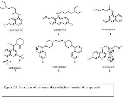
chloroquine. Quinacrine 2 (QN, Atabrine®), primaquine (PQ, Primaquine®) 3, and
mefloquine (MQ, Lariam®) 19 have been used as anti-malarial medicines. Working with
Dr. Noel McLaughlin, we derived three new classes of dimeric autophagy inhibitors from
these existing anti-malarial therapeutics. We wanted to test the hypothesis that dimeric
inhibitors synthesized from these heterocycles could be used as anti-cancer agents. As a
later independent effort, I designed and synthesized a series of inhibitors based on
piperaquine, the only clinically used dimeric chloroquine anti-malarial (Chapter 2,
Synthesis of Piperaquine Derivatives). We also synthesized dimeric forms of ferroquine,
a newly emerging anti-malarial compound which has demonstrated anti-proliferative
26
We designed a specific nomenclature to refer to dimeric inhibitors derived from
existing anti-malarial heterocycles. Dimeric inhibitors are described first by a two-letter
code, “D” for dimeric, followed by the first letter of the anti-malarial heterocycle. (DC
for dimeric chloroquine). Following the two-letter code is a three-digit number, where the
first digit corresponds to the length of the first alkyl chain of the triamine, followed by a
second digit corresponding to the length of the second alkyl chain of the triamine. The
final digit corresponds to whether the central nitrogen of the triamine is a secondary
amine (0) or a methylated tertiary amine (1). Utilizing this new nomenclature, Lys-05 9 is
27
defined as DC221 (Figure 2.5).
Spermidine was selected as a triamine scaffold to test the effect of dimerization on
the other heterocycles. The first three aryl halides were synthesized by Dr. Noel
McLaughlin according to literature precedent, each corresponding to either QN 2, PQ 3,
or MQ 19 (Figure 2.6). The acridine precursor of dimeric quinacrine inhibitors (DQ) was
synthesized as a 6,9-dichloro-2-methoxyacridine. Compound 22 was synthesized by an
Ullman coupling of p-anisidine 21 (Acros Organics) and 2,4-dichlorobenzoic acid 20
(Acros Organics). Ullman couplings are known to be directed and significantly
accelerated by an ortho-carboxylic acid moiety, which enabled the product 22 to be
isolated as a single isomer. The benzoic acid 22 derivative was then cyclized by
Friedel-Crafts acylation and chlorinated in situ with neat phosphorus(V) oxychloride, heated to
reflux, to afford 23. The resulting phenol 23 was chlorinated in situ to afford the
28
synthesized according to the method of Childers and coworkers.56 The heterocyclic
precursor of dimeric mefloquine (DM) was synthesized first from the
condensation/cyclization reaction of 2-trifluoromethyl aniline (Sigma Aldrich) and ethyl
4,4,4-trifluoro-3-oxobutanoate (Oakwood Chemical) in polyphosphoric acid, resulting in
29.57 4-hydroxyquinoline, 29 was chlorinated by phosphorus(V) oxychloride to give
compound 30.58
Palladium-catalyzed C-N arylation was then used to synthesize dimers from
commercially available spermidine as a linker. Spermidine 16 (Acros Organics) was
selected as a linker for this second-generation library because of its success in the case of
DC340 17. The Pd(OAc)2/BINAP catalyst system is compatible with spermidine, which
contains both primary amine and secondary amine functionality, due to the significantly
29
The Pd(OAc)2/BINAP catalyst in combination with potassium phosphate tribasic as a
base yields DC340 17 with excellent chemoselectivity. Dimeric forms of quinacrine 31
(DQ340) and mefloquine (DM340) 33 were synthesized in reasonable yields,
demonstrating the toleration of aryl chlorides by this catalyst system. Dimeric primaquine
(DP340) 32 was synthesized in only trace quantities using these reaction conditions.
Analysis of the crude reaction mixture by proton nuclear magnetic resonance (1H NMR)
showed the starting materials did not decompose. The base utilized in the reaction was
switched from potassium phosphate tribasic (K3PO4) anhydrous to anhydrous sodium
tert-butoxide (NaOtBu), a stronger base. The source of palladium was also changed to
Pd2(dba)3. These new reaction conditions afforded DP340 32 in 83% yield.
The compounds were first subjected to MTT anti-proliferative assays in A375P
BRAF mutant melanoma cells. In these assays, the new dimeric inhibitors were compared Figure 2.7: Synthesis of dimeric inhibitors with other anti-malarial
30
to their respective parent monomeric anti-malarial and to Lys-05, the state of the art in
dimeric autophagy inhibitors. As previously observed in MTT assays, cells treated with
DC340 17 exhibited reduced cell proliferation compared to cells treated with either
Lys-05 9 or CQ 1. DM340 33 treated cells exhibited reduced cell proliferation compared to
cells treated with the parent mefloquine 19 (MQ), with activity comparable to Lys-05.
DP340 32 treated cells exhibited reduced cell proliferation compared to cells treated with
the parent primaquine 3 (PQ), with activity comparable to Lys-05 9. Cells treated with
DQ340 31 exhibited reduced cell proliferation when compared to QN 2, and also relative
to Lys-05 9. When compared as a ratio of dimeric/monomeric, cells treated with DQ
exhibited the greatest decrease in cell proliferation. From this result we inferred that
DQ340 31 was the most promising lead molecule tested (Figure 2.8.).
Informed by these MTT results, a library of dimeric quinacrine inhibitors was
synthesized in collaboration with Dr. Noel McLaughlin, a postdoctoral researcher in the
Winkler Laboratory. The library was designed to explore two structural features of the Figure 2.8: MTT assay results for dimeric inhibitors derived from other
31
linker region of each dimeric inhibitor: the length of the aliphatic chains linking the
nitrogen atoms of the triamine, and the substitution of the nitrogen in the center of the
linker. The library was synthesized by arylation of various triamines with
6,9-dichloro-2-methoxyacridine. For each triamine linker, K3PO4 was found to be an adequate base in
combination with the Pd(OAc)2/BINAP catalyst system, suggesting that the previously
observed difference in reactivity between 8-chloro-6-methoxyquinoline and
6,9-dichloro-2-methoxyacridine lies in the substitution of the aryl halide. Many of the triamines
utilized in this library were commercially available (2/2, 3/3, 3/4, and 6/6) or were
previously synthesized (4/4, 5/5). However, the remaining triamines (7/7, 8/8, and 11/11)
were not known and required the development of a synthetic strategy.
2.2 Synthesis of a DQ library
Triamines based on heptyl and octyl aliphatic chains were prepared from
commercially available dibromoalkanes (1,7-dibromoheptane 34 and 1,8-dibromooctane
35). Sodium azide was selected as a protected nitrogen source because of its greater
reactivity as a SN2 nucleophile when compared to potassium phthalimide. Both
1,7-dibromoheptane 34 and 1,8-dibromooctane 35 were subjected to SN2 reaction with
sodium azide in dimethylsulfoxide (DMSO) at room temperature (23 °C). Use of the
dibromoalkanes in excess (2.2 equiv.) allowed for minimization of bis-SN2 product and
afforded the mono-substitution product 37 in reasonable yield. Benzylamine was selected
as a divalent nitrogen source to link the protected triamine. SN2 alkylation of
benzylamine with 37 in n-butanol afforded product 39 in serviceable yields. The azides
32
tertiary amine via hydrogenolysis catalyzed by 10 mol% palladium (0) on carbon support
(Pd/C) in methanol/ethyl acetate. The triamine products were then utilized in the
palladium catalyzed arylation reactions. (Figure 2.9)
The 11/11 linker was synthesized from 11-aminoundecanoic acid 42, a natural
product isolated from castor oil (Arkema). First, 11-aminoundecanoic acid 42 was
converted into 11-aminoundecanol 43 by lithium aluminum hydride reduction in
tetrahydrofuran (THF), upon heating the reaction to reflux. The resulting aminoalcohol
43 was protected by acylation with benzyl chloroformate to yield the Cbz-protected
aminoalcohol 44. Previous results demonstrated alkylation of benzylamine with alkyl
bromides was poor yielding. Fortunately, the existing alcohol functionality makes
oxidation followed by reductive alkylation of benzylamine to be step economical.
Therefore, alcohol 44 was oxidized under Swern conditions to yield 45. Benzylamine was
then reductively alkylated twice with the aldehyde, using sodium triacetoxyborohydride
as a reducing agent, to synthesize compound 46. We attributed the improved efficiency of
this transformation to the elimination of over alkylation. In contrast, SN2 alkylation of
primary amines often results in the production of tetraalkylammonium species in addition
to or to the exclusion of tertiary amines. Unlike sodium borohydride, the three acetoxy
substituents of this borohydride derivative reduce the hydridic character of the
33
hydrogen bonds, making the reduction selective for iminium ion intermediates formed in
34
The resulting protected triamine precursor, 46, was then deprotected in one step
utilizing hydrogenolysis in the presence of catalytic Pd/C. The reaction required 30 mol%
of catalyst for any consumption of starting material to occur. No consumption of starting
material was observed when using 10 mol% of Pd/C, even with prolonged reaction times.
Additionally, 1.2 equivalents of hydrochloric acid (HCl) were added in an attempt to
activate the benzylic amine toward hydrogenolysis. However still no starting material
was consumed in 24 hours. The protected triamine precursor was successfully
deprotected upon hydrogenolysis in the presence of 30 mol% Pd/C, rapidly affording the
product in good yield 47 (Figure 2.10). With the library of triamine precursors in hand,
palladium catalyzed arylation was utilized to synthesize the library of dimeric quinacrine
compounds (Figure 2.11).
35
Inhibitor Yield Compound.
DQ221 33% 52
DQ330 56% 53
DQ340 56% 31
DQ440 39% 54
DQ550 83% 55
DQ660 68% 56
DQ770 38% 57
DQ880 33% 58
DQ11110 30% 59
In Lys-05 9 the amine comprising the center of the linker is a methylated tertiary
amine. DC340 17 established the dimeric inhibitors containing secondary amines can also
be potent anti-cancer agents. A side-by-side comparison of secondary versus tertiary
amine-containing inhibitors was not yet available to establish the role of the alkylation of
this nitrogen in the potency of the inhibitors. Both previous work in our laboratory and
other studies have established that the aminoquinoline nitrogen in CQ 1 and our dimeric
lysosomal inhibitors to be a much weaker nucleophile than the linker central secondary
amine. Despite the presence of more than ten-fold molar excess of formaldehyde and
sodium triacetoxyborohydride, only the central linker amine is methylated at room
temperature (23 °C). This sequence allowed for the synthesis of both secondary and
tertiary amine linked dimers to proceed through the same common intermediates.