Head Injury Depth as an Indicator of Causes and
Mechanisms
WHAT’S KNOWN ON THIS SUBJECT: Young victims of abusive head trauma frequently manifest acute cardiorespiratory compromise, diffuse brain swelling, and poor outcomes. Deep, focal, hemorrhagic and axonal injuries were observed near the craniocervical junction in infants who died as a result of abuse.
WHAT THIS STUDY ADDS: For infants and young children, head injury depth is a useful indicator of head injury causes and mechanisms. These results have diagnostic, prognostic, and forensic significance.
abstract
OBJECTIVE:The goal was to measure differences in the causes, mech-anisms, acute clinical presentations, injuries, and outcomes of chil-dren⬍36 months of age with varying “greatest depths” of acute cra-nial injury.
METHODS:Children⬍36 months of age who were hospitalized with acute head trauma were recruited at multiple sites. Clinical and imag-ing data were collected, and caregivers underwent scripted inter-views. Neurodevelopmental evaluations were completed 6 months af-ter injury. Head trauma causes were categorized independently, and subject groups with varying greatest depths of injury were compared.
RESULTS:Fifty-four subjects were enrolled at 9 sites. Twenty-seven subjects underwent follow-up neurodevelopmental assessments 6 months after injury. Greatest depth of visible injury was categorized as scalp, skull, or epidural for 20 subjects, subarachnoid or subdural for 13, cortical for 10, and subcortical for 11. Compared with subjects with more-superficial injuries, subjects with subcortical injuries more fre-quently had been abused (odds ratio [OR]: 35.6;P⬍.001), more fre-quently demonstrated inertial injuries (P⬍ .001), more frequently manifested acute respiratory (OR: 43.9;P⬍.001) and/or circulatory (OR: 60.0;P⬍.001) compromise, acute encephalopathy (OR: 28.5;P⫽
.003), prolonged impairments of consciousness (OR: 8.4;P⫽ .002),
interhemispheric subdural hemorrhage (OR: 10.1;P⫽.019), and bilat-eral brain hypoxia, ischemia, or swelling (OR: 241.6;P⬍.001), and had lower Mental Developmental Index (P⫽.006) and Gross Motor Quotient (P⬍.001) scores 6 months after injury.
CONCLUSION:For children⬍3 years of age, head injury depth is a useful indicator of injury causes and mechanisms.Pediatrics2010;125: 712–720
AUTHORS:Kent P. Hymel, MD,aMichael A. Stoiko, MD,b
Bruce E. Herman, MD,cAmy Combs, LMSW,dNancy S.
Harper, MD,eDeborah Lowen, MD,f,gKatherine P. Deye,
MD,h,iKaren Homa, PhD,jand James A. Blackman, MD,
MPHk,l
aDepartment of Pediatrics, Dartmouth Medical School, Children’s Hospital at Dartmouth-Hitchcock Medical Center, Lebanon, New Hampshire;bDepartment of Pediatrics, Michigan State University School of Medicine, Spectrum Health Hospitals and Helen DeVos Children’s Hospital, Grand Rapids, Michigan; cDepartment of Pediatrics, University of Utah School of Medicine, Primary Children’s Medical Center, Salt Lake City, Utah;dCenter for Child Protection, Spectrum Health Hospitals and Helen DeVos Children’s Hospital, Grand Rapids, Michigan;eDepartment of Pediatrics, Texas A&M University, Driscoll Children’s Hospital, Corpus Christi, Texas;fDepartment of Pediatrics, University of Oklahoma College of Medicine, Tulsa, Oklahoma;gChildren’s Justice Center, Tulsa, Oklahoma;hDepartment of Pediatrics, Inova Fairfax Hospital for Children, Falls Church, Virginia; iDepartment of Pediatrics, National Children’s Medical Center, Washington, DC;jLeadership Preventive Medicine Residency Program, Dartmouth-Hitchcock Medical Center, Lebanon, New Hampshire;kDepartment of Pediatrics, University of Virginia Children’s Hospital, Charlottesville, Virginia; andlDepartment of Pediatrics, Kluge Children’s Rehabilitation Center,
Charlottesville, Virginia
KEY WORDS
depth of injury, head trauma, infants, abuse, accidents
ABBREVIATIONS
CI— confidence interval CT— computed tomography GMQ—Gross Motor Quotient MDI—Mental Developmental Index MVC—motor vehicle crash OR— odds ratio
PediBIRN—Pediatric Brain Injury Research Network SDH—subdural hematoma
www.pediatrics.org/cgi/doi/10.1542/peds.2009-2133 doi:10.1542/peds.2009-2133
Accepted for publication Nov 25, 2009
Address correspondence to Kent P. Hymel, MD, Dartmouth-Hitchcock Medical Center, Department of Pediatrics, One Medical Center Dr, Lebanon, NH 03756-0001. E-mail: kphymel@gmail.com
PEDIATRICS (ISSN Numbers: Print, 0031-4005; Online, 1098-4275). Copyright © 2010 by the American Academy of Pediatrics
FINANCIAL DISCLOSURE:The authors have indicated they have no financial relationships relevant to this article to disclose.
In young children, the traumatic cra-nial injuries that result from acciden-tal, short-distance falls tend to be su-perficial. Acute encephalopathy, acute respiratory or circulatory compro-mise, diffuse brain hypoxia-ischemia or swelling, and poor outcomes are rare in such cases.1–17
In contrast, the traumatic cranial in-juries that result from child abuse tend to be deeper and/or more dif-fuse.5,7,18–21 Neuropathological studies of abused infants revealed hemor-rhagic and axonal injuries in the base of the brain and in the upper spinal cord, which seem to trigger acute re-spiratory and/or circulatory compro-mise.22–24If not promptly recognized or treated, such compromise may initiate and/or exacerbate the development of secondary, diffuse, hypoxic-ischemic brain injuries, which have been linked to poor outcomes.5,7,18–21
Our research objective was to conduct a prospective, multicenter, cohort study that would measure differences in head injury causes, mechanisms, acute clinical presentations, and neu-rodevelopmental outcomes among in-fants and young children with acute traumatic injuries of varying “greatest depth” visible on neuroimaging scans. We hypothesized that deep, traumatic, cranial injuries in infants and young children could be linked to abusive causes and inertial injury mechanisms that increase the risk or incidence of acute respiratory or circulatory compromise, bilateral brain hypoxia-ischemia or swelling, and abnormal neurodevelopmental outcomes.
METHODS
Pediatric Brain Injury Research Network
The Pediatric Brain Injury Research Network (PediBIRN) is a consortium of clinical investigators who made a vol-untary commitment to conduct collab-orative, multicenter, clinical research
regarding traumatic brain injuries in infants and young children. The institu-tional review boards at all 9 participat-ing PediBIRN institutions approved this research study before local subject re-cruitment was initiated.
Participants
At every participating PediBIRN site, in-vestigators compared hospital admis-sion and computed tomography (CT) logs and then screened specific medi-cal records to identify eligible sub-jects. Inclusion criteria included age of
⬍36 months and CT evidence of any
acute, nonpenetrating, head or brain injury leading to inpatient evaluation or treatment. Exclusion criteria in-cluded preexisting central nervous system or brain disease, infection, hypoxia-ischemia, or trauma, birth in-jury, developmental delays, sensory deficits, bleeding disorders, and se-vere malnutrition. Screening for study exclusion criteria was completed after study enrollment, through detailed, fully scripted, parent interviews, plot-ting and interpretation of growth pa-rameters, and subsequent blinded re-views of the subjects’ initial cranial CT scans.
Procedures
Subjects were examined serially dur-ing the course of their acute hospital-izations. Local researchers reviewed their emergency medical services, emergency department, and inpatient medical records to capture extensive demographic, historical, clinical, labo-ratory, and neurosurgical data. Pri-mary caregivers (ie, the persons re-sponsible for the children when they were injured or became clearly and persistently ill with clinical signs later linked to their acute, traumatic, cra-nial injuries) were interviewed in fully scripted interviews designed to cap-ture extensive historical and clinical data from the scene of injury, in a con-sistent manner. More specifically,
ev-ery primary caregiver who consented to be interviewed was questioned sys-tematically regarding the specific tim-ing, cause (abusive versus nonabu-sive), and circumstances of the injury; the child’s clinical and mental status before and after acute clinical deterio-ration; and medical interventions at the scene of the injury. Qualified neu-roradiologists who were blinded with respect to all historical and clinical in-formation reviewed the subjects’ com-plete cranial CT and MRI studies, to document injuries and to identify the greatest depth of visible primary or secondary injury.
Follow-up Neurodevelopmental Assessments
The surviving subjects were invited to return 6 months after injury for an ex-tensive, outpatient, neurodevelopmen-tal assessment that included the Bay-ley Scales of Infant Development II. The fully qualified physical or occupational therapists, rehabilitation specialists, or developmental pediatricians who conducted the assessments at each participating site were blinded with re-spect to all historical, clinical, and ra-diologic information. The Mental Devel-opmental Index (MDI) and Gross Motor Quotient (GMQ) scores calculated dur-ing these follow-up assessments were used as outcome measures of overall cognitive and motor development, re-spectively. Both scores are standard-ized scores like IQ scores, with SDs of 15 and mean values of 100.
Statistical Analyses
To sort cases according to cause of in-jury, we applied definitional criteria specifically designed to reflect an emerging consensus, to be free of any references to specific injuries or injury severity, and to minimize circular rea-soning and inherent biases (Table 1). To sort cases according to required mechanisms of injury, every subject’s specific cranial injuries were listed and then classified (Table 2). Injuries resulting solely from tissue distortions induced by cranial impact and/or skull deformation were classified as con-tact injuries. Injuries resulting solely from tissue distortions induced by ro-tational cranial acceleration or
decel-eration were classified as inertial inju-ries. Cases then were categorized as revealing (1) isolated contact injuries, (2) isolated inertial injuries, (3) com-bined (ie, contact and inertial) injuries, or (4) isolated undetermined (ie, con-tract or inertial) injuries.
Fisher’s exact test was used to com-pare the frequencies of categorical variables among the 4 comparison groups defined according to the great-est depth of visible injury, categorized as scalp, skull, or epidural; subarach-noid or subdural; cortical brain; or subcortical brain. For continuous mea-sures, the Kruskal-Wallis test was used
to assess equality of the comparison groups. The Wilcoxon rank-sum test was used to compare subjects with subcortical greatest depth of visible injury with the other comparison groups, with more-superficial injuries. Finally, univariate logistic regression was used to calculate odds ratios (ORs) for specific findings linked to subcortical greatest depth of injury visible on neuroimaging scans.
RESULTS
Twenty-seven eligible subjects were re-cruited at the University of Virginia (Charlottesville, VA) or Inova Fairfax Hos-pital for Children (Falls Church, VA)
dur-TABLE 1 Criteria for Categorizing Causes of Head Injuries
Category Criteria
Abusive Cases in which primary caregiver admitted abusive acts that resulted in acute clinical signs linked to child’s acute traumatic cranial injuries Cases in which primary caregiver’s abusive acts were witnessed independently and resulted in acute clinical signs linked to child’s acute
traumatic cranial injuries
Cases in which child not yet cruising or walking first manifested acute clinical signs linked to acute traumatic cranial injuries while in the care of primary caregiver who specifically denied that child had experienced any trauma
Cases in which primary caregiver provided explanation for child’s acute clinical signs and acute traumatic cranial injuries that was clearly developmentally inconsistent with parents’ description of child’s developmental capabilities
Cases in which primary caregiver provided explanation for child’s acute clinical signs and acute traumatic cranial injuries that was clearly historically inconsistent with repetition over time
Cases in which child with acute head injury demonstratedⱖ2 categories of noncranial injuries considered moderately or highly specific for abuse Nonabusive Cases in which primary caregiver described accidental head injury event that was developmentally consistent with parents’ description of
child’s developmental capabilities, was historically consistent with repetition over time, could be linked to child’s acute clinical presentation for acute traumatic cranial injuries, and occurred in the absence of noncranial injuries considered moderately or highly specific for abuse
Cases in which accidental head injury event (eg, MVC) was witnessed independently and could be linked to child’s acute clinical presentation with acute traumatic cranial injuries
Undetermined Cases meeting criteria for both abusive and nonabusive causes Cases not meeting any criteria for either abusive or nonabusive causes Cases in which primary caregiver could not be interviewed
The primary caregiver was defined as the person responsible for the child when the child first became clearly and persistently ill with clinical signs later linked to his or her acute traumatic cranial injuries. Noncranial injuries considered moderately or highly specific for abuse included classic metaphyseal lesions; fractures of the ribs, scapula, sternum, spinous processes, or digits; vertebral body fractures or dislocations; epiphyseal separations; noncranial bruising, abrasions, or lacerations in locations other than the knees, shins, or elbows; patterned bruises or dry-contact burns; scalding burns with uniform depth, clear lines of demarcation, and a paucity of splash marks; intraabdominal injuries; retinal hemorrhage described by an ophthalmologist as dense, extensive, covering a large surface area of the retina, or extending to the periphery of the retina; and retinoschesis diagnosed by an ophthalmologist.
TABLE 2 Injury Classification Reflecting Required Mechanisms of Injury
Category Injuries
Primary cranial injuries that require contact mechanisms of injury Craniofacial soft-tissue injuries, subgaleal hematomas, cephalohematomas, skull fractures, epidural hematomas
Primary cranial injuries that require inertial mechanisms of injury Acute encephalopathy, diffuse axonal injuries Primary cranial injuries resulting from undetermined (ie, either
contact or inertial) mechanisms of injury
Subarachnoid hemorrhage, brain contusions, brain lacerations, any abnormal subdural collections
ing a 2.5-year period between 2003 and 2006. The remaining PediBIRN sites en-rolled a convenience sample of 27 addi-tional subjects during the same time pe-riod. Overall, 54 study subjects who met eligibility criteria were enrolled.
The greatest depth of cranial injury vis-ible on neuroimaging scans was cate-gorized as scalp, skull, or epidural for 20 subjects, subarachnoid or subdural for 13, cortical brain for 10, and cortical brain for 11. Among the sub-jects who met the definitional criteria for nonabusive head trauma were 4 children who sustained head injuries in motor vehicle crashes (MVCs) and 26 whose injuries were attributed to falls. The falls included 4 involving stairs, 3 from heights of⬎10 feet, and 2 from heights of 6 to 10 feet. The re-maining subjects (n⫽17) reportedly fell from heights of⬍6 feet.
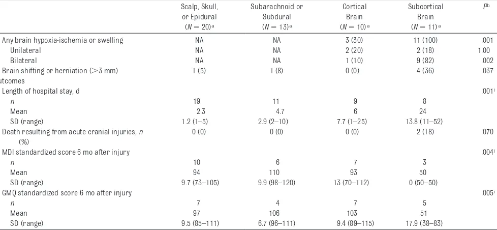
Demographic, etiologic, mechanistic, clinical, neuroimaging, and outcome data for the 4 comparison groups are summarized in Table 3. Fisher’s exact test revealed statistically significant differences in age and ethnicity, but not gender or race, between compari-son groups. Subjects with the most-superficial injuries tended to be older than subjects with deeper injuries.
Additional analyses revealed signifi-cant differences in head injury causes, mechanisms, acute clinical presenta-tions, and outcomes among the 4 com-parison groups. These differences were almost exclusively among sub-jects with subcortical injuries. Com-pared with all subjects with
more-superficial injuries, subjects with
subcortical greatest depth of visible injury (1) more frequently met defini-tional criteria for abuse (OR: 35.6 [95% confidence interval [CI]: 6.0 –209.0]; P⬍ .001; sensitivity: 0.73; specificity: 0.93); (2) more frequently manifested injuries requiring an inertial mecha-nism (sensitivity: 1.00; specificity: 0.72; Fisher’s exact test,P⬍.001); (3) had
lower initial Glasgow Coma Scale scores (OR: 0.59 [95% CI: 0.36 – 0.74];
P⬍.001); (4) more frequently
mani-fested acute respiratory compromise (OR: 43.9 [95% CI: 6.9 –277.8];P⬍.001; sensitivity: 0.82; specificity: 0.91), acute circulatory compromise (OR: 60.0 [95% CI: 8.7– 413.3]; P ⬍ .001; sensitivity: 0.82; specificity: 0.88), acute encepha-lopathy (OR: 28.5 [95% CI: 3.0 –224.2]; P⫽ .003; sensitivity: 1.00; specificity: 0.67), and prolonged impairments of
consciousness (OR: 8.4 [95% CI:
2.2–31.5]; P ⫽ .002; sensitivity: 0.73; specificity: 1.00); (5) more frequently demonstrated interhemispheric sub-dural bleeding (OR: 10.1 [95% CI: 1.5– 69.9];P⫽.019; sensitivity: 0.82; speci-ficity: 0.91) and bilateral brain hypoxia, ischemia, or swelling (OR: 241.6 [95% CI: 15.4 –2315.9];P⬍.001; sensitivity: 0.64; specificity: 0.91); and (6) had
lower MDI (Wilcoxon test, P ⫽ .006)
and GMQ (Wilcoxon test, P ⬍ .001)
standardized scores 6 months after in-jury. Both subjects who died had sub-cortical injuries.
Eight (73%) of 11 “abused” subjects
demonstrated subcortical greatest
depth of visible injury on neuroimag-ing scans. In contrast, only 2 (7%) of 30 “nonabused” subjects, both of whom were improperly restrained victims of a MVC, demonstrated subcortical greatest depth of injury. With exclusion of the 4 subjects who sustained head injuries in MVCs, all subjects with iso-lated scalp, skull, or epidural injuries
(n ⫽ 13) experienced a head injury
event categorized independently as nonabusive. The largest proportion (38%) of cases with undetermined causes of injury involved subjects with subarachnoid or subdural greatest depth of visible injury (Fig 1).
Contact injuries were confirmed for 9 (69%) of 13 subjects with subarach-noid or subdural greatest depth of vis-ible injury and for 8 (80%) of 10 sub-jects with cortical greatest depth of
visible injury. In contrast, 11 (100%) of 11 subjects with visible subcortical in-juries manifested inertial inin-juries, 7 (64%) of 11 manifested inertial injuries in isolation, and none manifested iso-lated contact injuries (Fig 2).
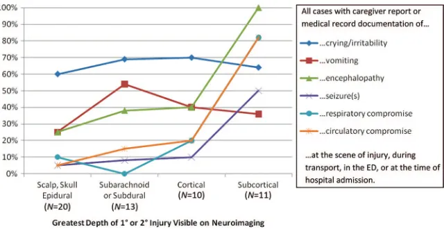
Crying and/or irritability before hospi-tal admission was documented for 7 (64%) of 11 subjects who later demon-strated subcortical injuries, including 6 (55%) of 11 who also experienced acute encephalopathy before hospital admission. All 11 subjects who eventu-ally demonstrated subcortical injuries on neuroimaging scans experienced at least brief impairment or loss of con-sciousness at the scene of the injury, and 8 (73%) experienced impairment or loss of consciousness that per-sisted for⬎24 hours. None of the sub-jects with more-superficial greatest depth of visible injury experienced such prolonged impairment or loss of consciousness. In contrast, 15 (75%) of 20 subjects with scalp, skull, or epi-dural greatest depth of visible injury experienced no impairment or loss of consciousness, as did 8 (62%) of 13 subjects with subarachnoid or sub-dural greatest depth of visible injury and 8 (80%) of 10 subjects with corti-cal greatest depth of visible injury (Figs 3 and 4).
Respiratory compromise before hospi-tal admission was observed or re-ported for 9 (82%) of 11 subjects with subcortical greatest depth of visible injury but only 4 (9%) of 43 subjects with more-superficial greatest depth of visible injury. Similarly, circulatory compromise was documented for 9 (82%) of 11 subjects with subcortical injuries but only 5 (12%) of 43 subjects with more-superficial greatest depth of injury (Fig 3).
TABLE 3 Demographic, Etiologic, Mechanistic, Clinical, Neuroimaging, and Outcome Data for Subject Comparison Groups Scalp, Skull,
or Epidural (N⫽20)a
Subarachnoid or Subdural (N⫽13)a
Cortical Brain (N⫽10)a
Subcortical Brain (N⫽11)a
Pb
Age at time of injury, mo .049
Median 15.5 3 3 5
Mean 14.5 5 8 8.5
SD (range) 10.5 (0.5–31) 7 (0.5–27) 10.5 (0.5–28) 8.5 (0.5–26)
Gender,n(%) .786
Female 10 (50) 6 (46) 6 (60) 4 (36)
Male 10 (50) 7 (54) 4 (40) 7 (64)
Race,n(%) .160
White 14 (70) 12 (92) 9 (90) 8 (73)
Black 3 (15) 1 (8) 0 (0) 0 (0)
Other 3 (15) 0 (0) 1 (10) 3 (27)
Ethnicity,n(%) .003
Hispanic 0 (0) 0 (0) 1 (10) 4 (36)
Non-Hispanic or unknown 20 (100) 13 (100) 9 (90) 7 (64)
Cause of injury,n(%)c ⬍.001
Abusive 0 (0) 1 (8) 2 (20) 8 (73)
Nonabusive 15 (75) 7 (54) 6 (60) 2 (18)
Undetermined 5 (25) 5 (38) 2 (20) 1 (9)
Inferred mechanisms of injury,n(%)d ⬍.001
Isolated contact 15 (75) 5 (38) 6 (60) 0 (0)
Isolated inertial 0 (0) 1 (8) 0 (0) 7 (64)
Combined 5 (25) 4 (31) 2 (20) 4 (36)
Undetermined 0 (0) 3 (23) 2 (20) 0 (0)
Acute clinical presentation,n(%)
Crying/irritability before admission 12 (60) 9 (69) 7 (70) 7 (64) .843 Vomiting before admission 5 (25) 7 (54) 4 (40) 4 (36) .378 Respiratory compromise before admissione 2 (10) 0 (0) 2 (20) 9 (82) ⬍.001
Circulatory compromise before admissionf 1 (5) 2 (15) 2 (20) 9 (82) ⬍.001
Severe pallor or cyanosis before admission 2 (10) 2 (15) 1 (10) 5 (45) .027
Seizures before admission 1 (5) 1 (8) 1 (10) 5 (50) .014
Acute encephalopathy before admissiong 5 (25) 5 (38) 4 (40) 11 (100) ⬍.001
Delayed impairment of consciousness 1 (5) 0 (0) 1 (10) 3 (27) .121 Decorticate or decerebrate posturing 0 (0) 0 (0) 0 (0) 4 (36) .002
Duration of impaired consciousness,n(%) ⬍.001
None reported or documented 15 (75) 8 (62) 8 (80) 0 (0) Only at scene of injury 4 (20) 5 (38) 1 (10) 3 (27) Upon admission; responsive by 24 h 1 (5) 0 (0) 1 (10) 0 (0)
⬎24 h, without deteriorationh 0 (0) 0 (0) 0 (0) 2 (18) ⬎24 h, with deteriorationh 0 (0) 0 (0) 0 (0) 6 (55)
Initial Glasgow Coma Scale score ⬍.001i
n 19 13 10 11
Mean 13.7 14.2 13.5 6.4
SD (range) 2.3 (6–15) 1.1 (12–15) 1.4 (11–15) 3.7 (3–15)
Pediatric Trauma Scale score .001i
n 17 11 9 10
Mean 8.8 8.5 8.4 5.0
SD (range) 1.6 (4–11) 1.1 (7–11) 0.9 (7–10) 2.5 (1–10) Injuries visible through neuroimaging,n(%)
Any craniofacial soft-tissue injuries 18 (90) 7 (54) 8 (80) 4 (36) .008
Any skull fractures 19 (95) 6 (46) 6 (60) 2 (18) ⬍.001
Isolated, nondiastatic, linear, parietal 10 (50) 4 (31) 2 (20) 0 (0) .021 Any “high-energy” skull fracturesj 2 (10) 1 (8) 2 (20) 1 (9) .111
Epidural hematoma 3 (15) 0 (0) 1 (10) 0 (0) .360
Any abnormal subdural collection NA 9 (69) 4 (40) 11 (100) .009
Interhemispheric NA 4 (31) 0 (0) 9 (82) ⬍.001
Bilateral NA 3 (23) 1 (10) 7 (64) .033
Unilateral NA 5 (38) 3 (30) 2 (18) .576
Small focal NA 2 (15) 3 (30) 0 (0) .191
Any subarachnoid hemorrhage NA 9 (69) 2 (20) 3 (27) .028
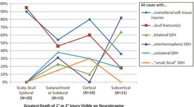
skull fractures were identified for 31 (72%). In contrast, only 4 (36%) of 11 subjects with subcortical greatest depth of visible injury demonstrated craniofa-cial soft-tissue injuries and only 2 (18%) demonstrated skull fractures.
Twenty-four (44%) of 54 study subjects demonstrated abnormal subdural col-lections of blood on CT and/or MRI scans. Among the 11 subjects with sub-cortical greatest depth of visible in-jury, 7 (64%) demonstrated subdural hematomas (SDHs) categorized as bi-lateral and 9 (82%) demonstrated SDHs categorized as interhemispheric. In contrast, only 2 (18%) of 11 subjects FIGURE 1
Causes of injury for children⬍3 years of age (N⫽54) with varying greatest depth of visible, acute, traumatic, cranial injury.
TABLE 3 Continued
Scalp, Skull, or Epidural (N⫽20)a
Subarachnoid or Subdural (N⫽13)a
Cortical Brain (N⫽10)a
Subcortical Brain (N⫽11)a
Pb
Any brain hypoxia-ischemia or swelling NA NA 3 (30) 11 (100) .001
Unilateral NA NA 2 (20) 2 (18) 1.00
Bilateral NA NA 1 (10) 9 (82) .002
Brain shifting or herniation (⬎3 mm) 1 (5) 1 (8) 0 (0) 4 (36) .037 Outcomes
Length of hospital stay, d .001i
n 19 11 9 8
Mean 2.3 4.7 6 24
SD (range) 1.2 (1–5) 2.9 (2–10) 7.7 (1–25) 13.8 (11–52) Death resulting from acute cranial injuries,n
(%)
0 (0) 0 (0) 0 (0) 2 (18) .070
MDI standardized score 6 mo after injury .004i
n 10 6 7 3
Mean 94 110 93 50
SD (range) 9.7 (73–105) 9.9 (98–120) 13 (70–112) 0 (50–50)
GMQ standardized score 6 mo after injury .005i
n 7 4 7 5
Mean 97 106 103 51
SD (range) 9.5 (85–111) 6.7 (96–111) 9.4 (89–115) 17.9 (38–83)
NA indicates not applicable.
aGreatest depth of primary or secondary cranial injury visible through neuroimaging.
bAs measured with Fisher’s exact test (for nominal variables) or Kruskal-Wallis test (for continuous variables). cSee Table 1 for a priori definitional criteria used to categorize each subject’s cause of injury.
dSee Table 2 for injury classification scheme used to categorize each subject’s inferred mechanisms of injury. Among subjects with visible contact injuries, cranial acceleration or
deceleration could not be excluded with certainty. However, visible and/or verifiable inertial injuries were not identified. Among subjects with visible or verifiable inertial injuries, cranial impact could not be excluded with certainty. However, visible contact injuries were not identified.
eRespiratory compromise was defined as labored or infrequent respirations, apnea, or any requirement for mouth-to-mouth breathing, bag-mask ventilation, or intubation occurring at the
scene of injury, during transport, in the emergency department, or at the time of hospital admission and specifically documented by medical personnel or reported by the child’s primary caregiver.
fCirculatory compromise was defined as bradycardia (ie,⬎35 beats per minute below normal mean heart rate for age), delayed capillary refill, absent or weak pulses, narrow pulse
pressure, hypotension, or any requirement for chest compressions, rapid volume expansion, or epinephrine therapy occurring at the scene of injury, during transport, in the emergency department, or at the time of hospital admission and specifically documented by medical personnel or reported by the child’s primary caregiver.
gAcute encephalopathy was defined as complete loss of consciousness or combined difficulty arousing from sleep and alteration of consciousness occurring at the scene of injury, during
transport, in the emergency department, or at the time of hospital admission and specifically documented by medical personnel or reported by the child’s primary caregiver.
hDeterioration included decorticate posturing, decerebrate posturing, and flaccidity.
iStatistical differences among the 4 subject comparison groups and posthoc comparisons of the subcortical group with the other 3 (combined) groups with more-superficial injuries
revealed significant differences in Glasgow Coma Scale scores, Pediatric Trauma Scale scores, length of stay, MDI scores, and GMQ scores.
with subcortical injuries demon-strated SDHs categorized as unilateral and none demonstrated SDHs catego-rized as small focal. Among the 13 sub-jects with subarachnoid or subdural greatest depth of visible injury and no visible underlying brain injury, 9 (69%) demonstrated SDHs, including 6 (67%) whose SDHs were categorized as bilat-eral and/or interhemispheric. Among the 10 subjects with cortical brain greatest depth of visible injury, 3 (30%) demonstrated SDHs categorized as unilateral and 3 (30%) demonstrated SDHs categorized as small focal. In contrast, only 1 (10%) of those 10
sub-jects demonstrated a SDH categorized as bilateral and none demonstrated SDHs categorized as interhemispheric (Fig 5).
Only 3 (27%) of 11 subjects with sub-cortical injuries had MDI scores calcu-lated 6 months after injury; each had a score of 50. In contrast, the 23 (53%) of 43 subjects with more-superficial inju-ries who returned for follow-up neuro-developmental assessments 6 months after injury had an average MDI score of 97.6 (SD: 12.7). Five (45%) of 11 sub-jects with subcortical injuries had GMQ scores calculated 6 months after
injury. Their average GMQ score was 54.0 (SD: 17.9). In contrast, the 18 (42%) of 43 subjects with more-superficial injuries who returned for follow-up neurodevelopmental assess-ments 6 months after injury had an av-erage GMQ score of 101.2 (SD: 9.2).
DISCUSSION
In 1974, Ommaya and Gennarelli25were the first to propose a specific causal re-lationship between inertial head injury mechanisms, injury depth, and clinical outcomes. Having observed that isolated inertial loading (ie, rotational cranial ac-celeration or deac-celeration) can induce traumatic cerebral concussion, they theorized that traumatic cerebral concussion represents “a graded set of clinical syndromes following head injury wherein increasing severity of disturbance in level and content of consciousness is caused by a mechanically-induced centripetal se-quence of disruptive effect on struc-ture and function.” In 1997, Levin et al26 verified that the centripetal theory for traumatic cerebral concussion
de-scribed by Ommaya and Gennarelli25
applies to children, linking the severity of impaired consciousness and out-comes to the depth of a child’s visible traumatic brain lesions.
In 2001, Geddes et al22,23described deep, focal, hemorrhagic and traumatic ax-onal injuries in the region of the cranio-cervical junction in infants who died as a result of suspected abusive head trauma. The authors opined that these deep, focal lesions represented primary traumatic injuries capable of inducing acute respiratory and/or circulatory compromise, which could initiate or ex-acerbate secondary brain injury.
Extrapolating from the work of
Om-maya and Gennarelli,25we theorized
that the deep traumatic cranial
inju-ries observed by Geddes et al22,23
resulted from inertial injury mech-anisms induced by perpetrators’ FIGURE 2
Injury mechanisms among children⬍3 years of age (N⫽54) with varying greatest depth of visible, acute, traumatic, cranial injury.
FIGURE 3
abusive actions. In this prospective, multicenter, cohort study, we sought to improve our understanding of these
broad pathophysiological
relation-ships by measuring differences in the causes, mechanisms, acute clinical presentations, injuries, and outcomes
of children ⬍36 months of age with
varying greatest depths of acute cra-nial injury.
Our results addressing this research objective support several conclusions that have diagnostic, prognostic, and forensic significance. Among infants and young children with closed-head trauma, (1) head injury causes and
mechanisms seem to be important de-terminants of head injury depth, (2) many subcortical brain injuries result from abusive events involving inertial in-jury mechanisms, (3) in the absence of underlying subcortical brain injury, many more-superficial cranial injuries result from nonabusive events in-volving contact injury mechanisms, (4) acute and/or prolonged encephalop-athy, acute respiratory and/or
circula-tory compromise, interhemispheric
and/or bilateral subdural hemorrhage, and bilateral brain hypoxia, ischemia, or swelling seem to be markers of subcor-tical brain injury, and (5) patients with
traumatic subcortical injuries unrelated to a MVC should undergo thorough eval-uation for abuse.
Our study’s primary strengths include its prospective, multicenter design; the breadth and depth of data capture; the novel use of fully scripted interviews to facilitate consistent data capture re-garding the scene of injury; and the a pri-ori application of criteria for abusive and nonabusive causes specifically designed to minimize circular reasoning and in-herent biases. Our study has numerous limitations. Variations in the frequency, timing, and/or modalities of cranial im-aging might have affected the validity of our conclusions regarding the greatest depth of visible injury. None of our sub-jects demonstrated macroscopic (CT or MRI) evidence of primary subcortical in-jury. Every study subject categorized as having subcortical greatest depth of vis-ible injury demonstrated only secondary brain hypoxia-ischemia or swelling in-volving the subcortical brain. Our inabil-ity to assess reliably the greatest depth of microscopic, primary, traumatic, cranial injuries represents a significant limitation of this study. To address this limitation, we attempted to link presumptive clinical signs of deep, mi-croscopic, primary, traumatic, brain in-jury (specifically, acute respiratory or circulatory compromise) with our sub-jects’ visible, subcortical, secondary, brain injury. The inherent assumptions in this method might be flawed.
Our schema for categorizing the re-quired mechanisms of injury was based solely on the subjects’ cranial injuries that were visible or verifiable. Some of our subjects categorized as having iso-lated contact injuries might have experi-enced inertial injury mechanisms that could not be verified. Conversely, some of our subjects categorized as having isolated inertial injuries might have ex-perienced cranial impacts and/or skull deformation that did not produce visible contact injuries.
FIGURE 4
Total duration of impaired consciousness among children⬍3 years of age (N⫽54) with varying greatest depth of visible, acute, traumatic, cranial injury.
FIGURE 5
Our definitional criteria for abusive and nonabusive head trauma causes very likely are imperfect. Only one-half of our subjects underwent follow-up neurodevelopmental assessments. Most importantly, our sample size was small, with significant risk of sampling bias. For all of these reasons, our re-sults should not be overinterpreted. Considered in isolation, this study does not verify a causal relationship between abuse, inertial mechanisms, and subcortical brain injury.
CONCLUSIONS
For infants and young children hospi-talized for evaluation and treatment of
acute, nonpenetrating, head trauma, assessment of head injury depth facil-itates the assessment of head injury causes and mechanisms. Acute en-cephalopathy, acute respiratory or circulatory compromise, and inter-hemispheric or bilateral subdural hemorrhage seem to be markers of subcortical brain injury. Infants and young children who demonstrate visi-ble subcortical injuries unrelated to a MVC require thorough evaluation for abuse. These results have diagnostic, prognostic, and forensic significance.
ACKNOWLEDGMENTS
This research was supported in part with resources provided by the
Na-tional Institutes of Health (grants R21HD043351-01 and R24HD39361-05), the American Academy for Cere-bral Palsy and Developmental
Medi-cine, the University of Virginia
Children’s Hospital Research Fund, and PediBIRN.
In addition to the authors, the follow-ing PediBIRN participants and sites made substantial contributions to re-search project design and data acqui-sition: Kathy L. Makoroff, MD, Univer-sity of Cincinnati; Antoinette L. Laskey, MD, MPH, University of Indiana; Carole Jenny, MD, MBA, Brown University; and Suzanne M. Edmunds, MD, Wake Forest University.
REFERENCES
1. Briss PA, Sacks JJ, Addiss DG, Kresnow M, O’Neil J. A nationwide study of the risk of injury associated with day care center at-tendance.Pediatrics.1994;93(3):364 –368 2. Chadwick DL, Bertocci G, Castillo E, et al.
Annual risk of death resulting from short falls among young children: less than 1 in 1 million.Pediatrics.2008;121(6):1213–1224 3. Chadwick DL, Salerno C. Likelihood of the
death of an infant or young child in a short fall of less than 6 vertical feet.J Trauma.
1993;35(6):968
4. Chang A, Lugg MM, Nebedum A. Injuries among preschool children enrolled in day-care centers. Pediatrics. 1989;83(2): 272–277
5. Duhaime AC, Alario AJ, Lewander WJ, et al. Head injury in very young children: mecha-nisms, injury types, and ophthalmologic findings in 100 hospitalized patients younger than 2 years of age.Pediatrics.
1992;90(2):179 –185
6. Helfer RE, Slovis TL, Black M. Injuries result-ing when small children fall out of bed. Pe-diatrics.1977;60(4):533–535
7. Hymel KP, Makoroff KL, Laskey AL, Conaway MR, Blackman JA. Mechanisms, clinical pre-sentations, injuries, and outcomes from in-flicted versus noninin-flicted head trauma during infancy: results of a prospective, multicentered, comparative study. Pediat-rics.2007;119(5):922–929
8. Landman PF, Landman GB. Accidental inju-ries in children in day-care centers.Am J Dis Child.1987;141(3):292–293
9. Leland NL, Garrard J, Smith DK. Injuries to preschool-age children in day-care centers:
a retrospective record review.Am J Dis Child.1993;147(8):826 – 831
10. Lyons TJ, Oates RK. Falling out of bed: a rel-atively benign occurrence.Pediatrics.1993; 92(1):125–127
11. Nimityongskul P, Anderson LD. The likeli-hood of injuries when children fall out of bed.J Pediatr Orthop.1987;7(2):184 –186 12. Plunkett J. Fatal pediatric head injuries
caused by short-distance falls.Am J Foren-sic Med Pathol.2001;22(1):1–12
13. Reiber GD. Fatal falls in childhood: how far must children fall to sustain fatal head in-jury? Report of cases and review of the lit-erature. Am J Forensic Med Pathol.1993; 14(3):201–207
14. Sacks JJ, Smith JD, Kaplan KM, Lambert DA, Sattin RW, Sikes RK. The epidemiology of in-juries in Atlanta day-care centers. JAMA.
1989;262(12):1641–1645
15. Tarantino CA, Dowd MD, Murdock TC. Short vertical falls in infants.Pediatr Emerg Care.
1999;15(1):5– 8
16. Williams RA. Injuries in infants and small children resulting from witnessed and cor-roborated free falls.J Trauma.1991;31(10): 1350 –1352
17. Willman KY, Bank DE, Senac M, Chadwick DL. Restricting the time of injury in fatal in-flicted head injuries.Child Abuse Negl.1997; 21(10):929 –940
18. Duhaime AC, Christian C, Moss E, Seidl T. Long-term outcome in infants with the shaking-impact syndrome. Pediatr Neuro-surg.1996;24(6):292–298
19. Duhaime AC, Durham S. Traumatic brain
in-jury in infants: the phenomenon of subdural hemorrhage with hemispheric hypodensity (“big black brain”).Prog Brain Res.2007; 161:293–302
20. Myhre MC, Grogaard JB, Dyb GA, Sandvik L, Nordhov M. Traumatic head injury in infants and toddlers. Acta Paediatr.2007;96(8): 1159 –1163
21. Zimmerman RA, Bilaniuk LT, Bruce D, Schut L, Uzzell B, Goldberg HI. Computed tomogra-phy of craniocerebral injury in the abused child.Radiology.1979;130(3):687– 690 22. Geddes JF, Hackshaw AK, Vowles GH,
Nick-ols CD, Whitwell HL. Neuropathology of in-flicted head injury in children, part I: pat-terns of brain damage.Brain.2001;124(7): 1290 –1298
23. Geddes JF, Vowles GH, Hackshaw AK, Nickols CD, Scott IS, Whitwell HL. Neuropathology of inflicted head injury in children, part II: mi-croscopic brain injury in infants.Brain.
2001;124(7):1299 –1306
24. Brennan LK, Rubin D, Christian CW, Duhaime AC, Mirchandani HG, Rorke-Adams LB. Neck injuries in young pediatric homicide vic-tims. J Neurosurg Pediatr. 2009;3(3): 232–239
25. Ommaya AK, Gennarelli TA. Cerebral concus-sion and traumatic unconsciousness: cor-relation of experimental and clinical obser-vations of blunt head injuries.Brain.1974; 97(4):633– 654
DOI: 10.1542/peds.2009-2133 originally published online March 29, 2010;
2010;125;712
Pediatrics
Deborah Lowen, Katherine P. Deye, Karen Homa and James A. Blackman
Services
Updated Information &
http://pediatrics.aappublications.org/content/125/4/712
including high resolution figures, can be found at:
References
http://pediatrics.aappublications.org/content/125/4/712#BIBL
This article cites 26 articles, 7 of which you can access for free at:
Subspecialty Collections
ub
http://www.aappublications.org/cgi/collection/child_abuse_neglect_s
Child Abuse and Neglect
son_prevention_sub
http://www.aappublications.org/cgi/collection/injury_violence_-_poi
Injury, Violence & Poison Prevention following collection(s):
This article, along with others on similar topics, appears in the
Permissions & Licensing
http://www.aappublications.org/site/misc/Permissions.xhtml
in its entirety can be found online at:
Information about reproducing this article in parts (figures, tables) or
Reprints
http://www.aappublications.org/site/misc/reprints.xhtml
DOI: 10.1542/peds.2009-2133 originally published online March 29, 2010;
2010;125;712
Pediatrics
Deborah Lowen, Katherine P. Deye, Karen Homa and James A. Blackman
Kent P. Hymel, Michael A. Stoiko, Bruce E. Herman, Amy Combs, Nancy S. Harper,
Head Injury Depth as an Indicator of Causes and Mechanisms
http://pediatrics.aappublications.org/content/125/4/712
located on the World Wide Web at:
The online version of this article, along with updated information and services, is
by the American Academy of Pediatrics. All rights reserved. Print ISSN: 1073-0397.