Open Access
Research article
Evaluation of procalcitonin for diagnosis of neonatal sepsis of
vertical transmission
José B López Sastre*
1, David Pérez Solís
1, Vicente Roqués Serradilla
2,
Belén Fernández Colomer
1, Gil D Coto Cotallo
1and Grupo de Hospitales
Castrillo
3Address: 1Service of Neonatology, Hospital Universitario Central de Asturias, Oviedo, Spain, 2Service of Neonatology, Hospital Universitario La
Fe, Valencia, Spain and 3Grupo de Hospitales Castrillo. Service of Neonatology, Hospital Universitario Central de Asturias, Oviedo, Spain
Email: José B López Sastre* - jose_lopez_sastre@hotmail.com; David Pérez Solís - david.perez@sespa.princast.es; Vicente Roqués Serradilla - roes5@ono.com; Belén Fernández Colomer - bcolomer@arrakis.es; Gil D
Coto Cotallo - gildaniel.coto@sespa.princast.es; Grupo de Hospitales Castrillo - gcastrillo@arrakis.es * Corresponding author
Abstract
Background: The results of recent studies suggest the usefulness of PCT for early diagnosis of neonatal sepsis, with varying results. The aim of this prospective multicenter study was to determine the behavior of serum PCT concentrations in both uninfected and infected neonates, and to assess the value of this marker for diagnosis of neonatal sepsis of vertical transmission.
Methods: PCT was measured in 827 blood samples collected prospectively from 317 neonates admitted to 13 acute-care teaching hospitals in Spain over one year. Serum PCT concentrations were determined by a specific immunoluminometric assay. The diagnostic efficacy of PCT at birth and within 12–24 h and 36–48 h of life was evaluated calculating the sensitivity, specificity, and likelihood ratio of positive and negative results.
Results: 169 asymptomatic newborns and 148 symptomatic newborns (confirmed vertical sepsis: 31, vertical clinical sepsis: 38, non-infectious diseases: 79) were studied. In asymptomatic neonates, PCT values at 12–24 h were significantly higher than at birth and at 36–48 h of life. Resuscitation at birth and chorioamnionitis were independently associated to PCT values. Neonates with confirmed vertical sepsis showed significantly higher PCT values than those with clinical sepsis. PCT thresholds for the diagnosis of sepsis were 0.55 ng/mL at birth (sensitivity 75.4%, specificity 72.3%); 4.7 ng/mL within 12–24 h of life (sensitivity 73.8%, specificity 80.8%); and 1.7 ng/mL within 36–48 h of life (sensitivity 77.6%, specificity 79.2%).
Conclusion: Serum PCT was moderately useful for the detection of sepsis of vertical transmission, and its reliability as a maker of bacterial infection requires specific cutoff values for each evaluation point over the first 48 h of life.
Published: 26 February 2007
BMC Pediatrics 2007, 7:9 doi:10.1186/1471-2431-7-9
Received: 4 July 2006 Accepted: 26 February 2007
This article is available from: http://www.biomedcentral.com/1471-2431/7/9
© 2007 López Sastre et al; licensee BioMed Central Ltd.
Background
Neonatal sepsis of vertical transmission remains a major problem associated with high morbidity and mortality in newborns, particularly amongst preterm infants [1-4]. Rapid diagnosis is problematic because the first signs of this disease may be minimal, and are similar to those of various non-infectious processes; furthermore, bacterial cultures are time-consuming, and other laboratory tests are either not available for routine use or lack sensitivity or specificity. In this situation, neonates with risk factors for infection or clinical suspicion of infection are empiri-cally treated with antibiotics. To avoid the unnecessary treatment of uninfected patients, an early, sensitive and specific laboratory test would be helpful to guide clini-cians in neonatal units in deciding whether or not to start administering antibiotics. Several leukocyte indices and acute-phase protein levels have been evaluated for the diagnosis of sepsis, and more recently, measurement of multiple plasma cytokines [5] and leukocyte activation markers [6] have showed promising results. However, to date, no single laboratory test has provided rapid and reli-able identification of early infected neonates. This inabil-ity has led to a search for new diagnostic markers [7,8].
It has been recently reported that procalcitonin (PCT), the prohormone of calcitonin, increases markedly in septic conditions [9] and it appears to be a good predictor of infection severity. Furthermore, the finding that PCT is released into the circulation within 3 h after endotoxin injection, plateaus at 6 h, and remains elevated for 24 h, makes PCT a promising new agent for early and sensitive identification of infected patients [10]. The results of recent studies suggest the usefulness of PCT for early diag-nosis of neonatal sepsis [11-23], although other investiga-tors have observed lack of accuracy for this marker [24-26]. Also, since markedly increased concentrations during the first 48 h of life in newborn infants without bacterial infection have been reported [12,23,27], there are con-flicting data regarding what constitutes an abnormal value.
The objective of this study was to determine the behavior of serum PCT concentrations in uninfected and infected neonates, and to assess the value of this marker for the diagnosis of neonatal sepsis of vertical transmission.
Methods
Since 1995 the neonatal services of 28 acute-care teaching hospitals distributed across 10 Autonomous Communi-ties in Spain ("Grupo de Hospitales Castrillo") have been involved in an ongoing prospective surveillance project to assess the incidence and characteristics of infection of ver-tical transmission during the newborn period. The neona-tal services of 13 hospineona-tals participated in the present study. Between January 2000 and January 2001, all
con-secutive neonates within 48 h of life were prospectively included in the study if blood samples were available for timed PCT measurement according to three postnatal periods: shortly after birth, within 12–24 h of life, and within 36–48 h of age, and complete neonatal and out-come data were collected to classify the newborn infant into two distinct populations. Exclusion criteria were infants born to mothers with gestational diabetes (because available data indicate that PCT values are higher for neonates born to diabetic mothers [12]) and refusal of parental consent for blood sampling. The study was approved by the Ethics Committees of the participating hospitals and the parents gave their written informed con-sent.
Two distinct populations were defined. The first popula-tion (group 1) included a group of asymptomatic new-born infants admitted during the first 24 h of life to the neonatal unit because of prematurity, low birth weight, or at least two risk factors for infection (table 1). They had no clinical signs of sepsis during their first week of life and had a negative blood culture. Patients in this group did not receive antibiotic treatment. Those who were dis-charged home before the seventh day of live were fol-lowed to assure they did not develop late-onset vertical sepsis.
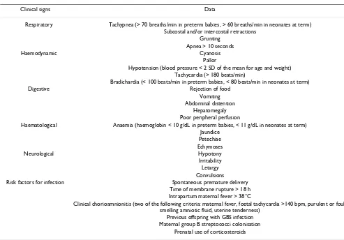
The second population included symptomatic neonates who were admitted to the neonatal unit and were evalu-ated for sepsis during the first 48 h of life. Patients in this population were later classified as group 2A (confirmed vertical sepsis), group 2B (vertical clinical sepsis), or group 2C (non-infectious disease). Criteria for confirmed vertical neonatal sepsis and vertical clinical sepsis were those previously established by "Grupo de Hospitales Castrillo" [1,2]. Clinical signs of neonatal sepsis and risk factors for vertical transmission are shown in Table 1.
- Group 2A: Confirmed vertical neonatal sepsis, defined as at least three clinical signs of infection in association with at least one bacteriological evidence of infection. Evidence of infection included positive blood culture, at least three positive surface swabs in the first 24 h of life for tradi-tional pathogens of vertical transmission – such as group B streptococci (GBS) and Escherichia coli-, or GBS culture-positive mother).
- Group 2C: Uninfected newborn infants with neonatal pathology other than an infectious process and with neg-ative blood culture.
PCT assay
Blood samples were centrifuged within 30 min of collec-tion. Serum was stored at -20°C before analysis. PCT was measured in duplicate by a specific immunoluminometric assay (LUMItest®, Brahms Diagnostica GmbH, Berlin, Germany), requiring 20 μL of serum and 2 h to complete. The detection limit of this immunoluminometric assay is 0.08 ng/mL. Luminescence was measured automatically on a Lumat LB 9507 tube luminometer (Berthold Tech-nologies GmbH & Co. KG, Bad Wildbad, Germany). Intraassay coefficients of variation were 5.9% at 31.0 ng/ mL and 13.8% at 0.81 ng/mL; interassay coefficients of variation were 10.8% and 25.9% for the same PCT con-centrations.
All PCT assays were performed by two centralised clinical chemistry laboratories of two participating hospitals.
Statistical analysis
Statistical analysis was done with SPSS version 11.0 (SPSS Inc., 2001, Chicago, Ill, USA) and EPIDAT version 3.0 (Servicio de Información sobre Saúde Pública de la Con-sellería de Sanidade e Servicios Sociais de la Xunta de Gali-cia, Spain, and the Health Situation Analysis Program of the Pan American Health Organization, Washington D.C., USA). PCT values are expressed as median and interquar-tile (25th–75th) ranges. Neither PCT values nor their log transformation were normally distributed according to kolmogorov-smirnov test, thus non-parametric tests were used for comparisons between groups (Kruskal-Wallis test and Mann-Whitney U test).
The association between different perinatal variables (cesarean section, rupture of membranes more than 18 h prior to delivery, clinical chorioamnionitis, resuscitation at birth, and prenatal steroid exposure) and PCT values in asymptomatic neonates was assessed with multiple linear regression analysis. As PCT variable was not normally dis-tributed, it was log-transformed and then extreme values
Table 1: Clinical signs of neonatal sepsis and risk factors for vertical transmission
Clinical signs Data
Respiratory Tachypnea (> 70 breaths/min in preterm babies, > 60 breaths/min in neonates at term) Subcostal and/or intercostal retractions
Grunting Apnea > 10 seconds
Haemodynamic Cyanosis
Pallor
Hypotension (blood pressure < 2 SD of the mean for age and weight) Tachycardia (> 180 beats/min)
Bradichardia (< 100 beats/min in preterm babies, < 80 beats/min in neonates at term)
Digestive Rejection of food
Vomiting Abdominal distention
Hepatomegaly Poor peripheral perfusion
Haematological Anaemia (haemoglobin < 10 g/dL in preterm babies, < 11 g/dL in neonates at term) Jaundice
Petechiae Echymoses
Neurological Hypotony
Irritability Letargy Convulsions
Risk factors for infection Spontaneous premature delivery Time of membrane rupture > 18 h Intrapartum maternal fever > 38°C
Clinical chorioamnionitis (two of the following criteria: maternal fever, foetal tachycardia >140 bpm, purulent or foul-smelling amniotic fluid, uterine tenderness)
Previous offspring with GBS infection Maternal group B streptococci colonisation
were removed, so the variable showed adequate homoce-dasticity for this analysis. In order to report the effect of the perinatal variables, the regression coefficients and 95% confidence limits were exponentiated. R-square val-ues were calculated to estimate how well the model explained PCT variability.
The reliability of serum PCT concentration for the diagno-sis of sepdiagno-sis of vertical transmission was calculated by receiver-operating characteristics (ROC) curves. Youden's index (sensitivity + specificity - 1) was used for determina-tion of optimal cutoff values of the diagnostic tests in the different postnatal periods. Sensitivity, specificity, and the likelihood ratio of positive and negative results with a 95% confidence interval (CI) were calculated. Statistical significance was set at P < 0.05.
Results
The study included 827 blood samples collected from 317 neonates who met the inclusion criteria. Group 1 included 169 asymptomatic newborns. There were 148 symptomatic neonates distributed in group 2 as follows: 31 newborns with confirmed vertical sepsis (group 2A), 38 with vertical clinical sepsis (group 2B) and 79 in the group of non-infectious diseases (group 2C). Non-infec-tious diseases were all of respiratory origin (hyaline mem-brane disease: 60, transient respiratory distress of the newborn: 12, meconium aspiration syndrome: 5, pneu-mothorax: 2). Causative pathogens of confirmed vertical sepsis included GBS in 19 cases, E. coli in 8, Streptococcus bovis in 1, Klebsiella oxytoca in 1, Haemophilus infuenzae in 1, and Candida albicans in 1. Clinical and anthropometric characteristics are shown in Table 2. There were 2 deaths in group 2A, no deaths in group 2B, and 3 deaths in group 2C. Patients in group 2C had significantly lower birth weights and gestational ages (P < 0.0001, analysis of vari-ance) than those in groups 1, 2A and 2B.
Pairwise comparisons of serum PCT concentrations in group 1 at each postnatal period confirmed higher serum PCT values (median [interquartile range]) at 12–24 h of life (1.54 [0.68–3.78] ng/mL) than those at birth (0.35 [0.23–0.64] ng/mL, P < 0.0001, Mann-Whitney U test) and at 36–48 h of life (0.73 [0.45–1.48] ng/mL, P < 0.0001, Mann-Whitney U test) as shown in Figure 1.
Multiple linear regression analyses identified some varia-bles independently associated to PCT values in group 1. PCT concentrations were associated with resuscitation at birth (defined as the necessity of tracheal intubation) at 36–48 h of life, and with chorioamnionitis at birth (Table 3). Nevertheless, variables specified in the model accounted for just a small part of the PCT variability (10.1% at birth, 2.0% at 12–24 h, and 9.4% at 36–48 h of life).
Serum PCT concentrations of patients in groups 2A, 2B, and 2C were higher than those of patients in group 1 at each postnatal period, as showed in Table 4 (P < 0.0001, Mann-Whitney U test). Patients of group 2A (confirmed vertical sepsis) had consistently higher serum PCT values than those of group 2B (vertical clinical sepsis) at birth (P
= 0.006), at 12–24 h (P = 0.002), and 36–48 h of life (P = 0.032). There were no significative differences between the PCT concentrations of neonates with respiratory dis-eases (group 2C) and those with clinical sepsis.
We elaborated ROC curves considering groups 2A and 2B as infected and group 1 as non-infected. ROC of PCT at birth and 12–24 h and 36–48 h of life for the diagnosis of sepsis of vertical transmission are shown in Figure 2. The areas under the ROC curve for the three periods were 0.754 (95% CI, 0.671 to 0.838), 0.802 (95% CI, 0.735 to 0.869), and 0.820 (95% CI, 0.750 to 0.891), respectively. Cutoff levels with the optimum diagnostic efficiency derived from the ROC curves were ≥ 0.55 ng/mL at birth
Table 2: Obstetric history and somatometric characteristics of patients included in the study
Group 1 Group 2A Group 2B Group 2C
No. patients 169 31 38 79
Gestational age, weeks, mean (SD) 36.2 (3.7) 34.5 (5.4) 35.2 (4.8) 30.3 (6.2) Weight at birth, g., mean (SD) 2622 (860) 2496 (1107) 2573 (997) 1542 (880) GBS colonisation, no. (%) 44 (26.0) 13 (41.9) 0 (0) 11 (13.9) Prenatal steroid exposure, no. (%) 40 (23.7) 9 (29.0) 9 (23.7) 38 (48.1) Membrane rupture > 18 h, no. (%) 70 (41.4) 18 (22.8) 5 (13.2) 11 (35.5) Intrapartum fever, no. (%) 15 (8.9) 7 (22.6) 8 (21.1) 3 (3.8) Chorioamnionitis, no. (%) 10 (5.9) 4 (12.9) 3 (7.9) 8 (10.1) Intrapartum antibiotics, no. (%) 105 (62.1) 15 (48.4) 12 (31.6) 22 (27.8) Resuscitation at birth, no. (%) 10 (5.9) 8 (25.8) 8 (21.1) 33 (41.8)
Males, no. (%) 93 (55.0) 15 (48.4) 16 (42.1) 43 (54.4)
(sensitivity 75.4%, specificity 72.3%), ≥ 4.7 ng/mL at 12– 24 h of life (sensitivity 73.8%, specificity 80.8%), and ≥ 1.7 ng/mL at 36–48 h of life (sensitivity 77.6%, specificity 79.2%) (Table 5). We also selected other cutoff points in order to get better sensitivity (>90%) as seen in Table 6.
Discussion
This study shows that increased serum PCT concentra-tions in newborn infants are related to different factors other than neonatal sepsis. Elevated PCT levels were apparent 12–24 h after birth in asymptomatic uninfected infants. The physiological peak of serum PCT concentra-tions in healthy neonates has been previously reported
[12,23], and increased PCT concentrations have also been found in neonates with very low probability of infection [27,28]. Although PCT may cross the placental barrier, the findings of higher PCT concentrations in cord sera com-pared with maternal samples, with even larger differences at 24 and 48 h of age, cannot be explained on the grounds of maternal transfer alone; therefore, the postnatal surge of PCT most likely represents endogenous synthesis [29]. This phenomenon might be attributed to direct stress on the baby during the perinatal period or to the adaptation to the extrauterine environment. The normal birth process and the extrauterine adaptation stimulates an acute phase reaction in the newborn infant with a release of C-reactive protein (CRP), interleukin-6 (IL-6), and serum amyloid A [30,31].
The higher concentrations of PCT observed in uninfected infants with respiratory disorders (mostly hyaline mem-brane disease) compared with asymptomatic infants is also consistent with previous reports. Other authors have shown elevated PCT values during the first 10 days of life in uninfected newborns who presented respiratory dis-tress syndrome (RDS) [26,32]. Because no significant effect related to the severity of RDS could be detected, Monneret et al. [32] suggested that hypoxemia (a com-mon event to each etiology of RDS and which is transient during delivery) could be responsible for these increased PCT values, providing further support for the hypothesis of pulmonary PCT synthesis. Yet it is not possible to assure these patients were really uninfected, since evi-dence of no infection was based on negative bacterial cul-tures, absence of fever, and a normal CRP value, and these criteria may fail in an appreciable number of cases [33]. Our study has a similar problem, as intrapartum antibiot-ics had been used in 22/79 neonates with respiratory dis-orders, and 66/79 were treated with antibiotics after birth. This is also the reason why we excluded group 2C from ROC curves.
Elevated PCT levels in newborn infants who required resuscitation at birth may be related to the stressful situa-tion or tissular hypoxia. Similar findings have been
Table 3: Independent effects of some perinatal variables on serum procalcitonin values at birth, at 12–24 h and 36–48 h of life.
Risk factor Serum PCT values, ng/mL, median (interquartile 25th–75th range)
At birth 12–24 h of life 36–48 h of life
Cesarean section 1.08 (0.58–2.02) 0.73 (0.26–2.02) 0.91 (0.54–1.51) Membrane rupture > 18 h 0.67 (0.37–1.20) 1.32 (0.51–3.45) 0.96 (0.59–1.56) Chorioamnionitis 18.61 (2.43–142.5)* 1.08 (0.14–8.15) 1.78 (0.54–5.84) Resuscitation at birth 1.30 (0.39–4.40) 5.58 (0.49–63.17) 8.71 (2.40–31.57)* Prenatal steroid exposure 1.84 (0.91–3.73) 0.58 (0.17–1.95) 0.93 (0.49–1.76)
Includes data of 169 asymptomatic newborns (group 1). Data are expressed as regression coefficients (95% confidence interval). * P < 0.01.
Changes in PCT from birth to 48 h of life in asymptomatic newborns
Figure 1
Changes in PCT from birth to 48 h of life in asympto-matic newborns. Box plot comparing serum PCT concen-trations in asymptomatic newborns (group 1) at birth, at 12– 24 h and 36–48 h of life. The boxes are limited by the 75th
and 25th percentiles of the data. The middle lines represent
the median. Values plotted with cross markers are outside values (more than 1.5 times the interquartile range over 75th
or under 25th percentiles). Values plotter with plus markers
are far out values (more than 3 times the interquartile range over 75th or under 25th percentiles).
Birth 12-24 h 36-48 h 1000
100
10
1
0.1
0.01
Procalcitonin
Table 6: Performance of procalcitonin at birth, at 12–24 h, and 36–48 h of life for the diagnosis of neonatal sepsis of vertical transmission (cutoff points selected to achieve 90% sensitivity).
At birth n = 205 12–24 h of life n = 212 36–48 h of life n = 193
Cutoff, ng/ml ≥0.15 ≥1.2 ≥0.75
Sensitivity, % (95% CI) 91.2 (81.1 – 96.2) 90.2 (80.2 – 95.4) 91.8 (80.8 – 96.8%) Specificity (95% CI) 10.8 (6.8 – 16.8) 43.0 (35.4 – 51.0) 51.4 (43.3 – 59.4)
Youden's index 0.02 0.33 0.43
PPV (95% CI) 28.3 (22.3 – 35.2) 39.0 (31.3 – 47.2) 55.9 (44.1 – 67.1) NPV (95% CI) 76.2 (54.9 – 89.4) 91.5 (82.8 – 96.1) 91.2 (84.9 – 95.0) LR+ (95% CI) 1.02 (0.93 – 1.13) 1.58 (1.35 – 1.86) 1.89 (1.57 – 2.28) LR- (95% CI) 0.81 (0.32 – 2.07) 0.23 (0.10–0.50) 0.16 (0.06 – 0.41)
PPV: positive predictive value, NPV: negative predictive value, LR+: likelihood ratio of a positive result, LR-: likelihood ratio of a negative result.
Table 4: Serum procalcitonin values at birth, at 12–24 h and 36–48 h of life in the groups included in the study.
Study group Serum PCT values, ng/mL median (interquartile 25th–75th range)
At birth 12–24 h of life 36–48 h of life
Group 1, n = 169 0.35 (0.23–0.64) 1.54 (0.68–3.78) 0.73 (0.45–1.48) Group 2A, n = 31 13.59 (0.50–101.3) 30.65 (6.38–95.9) 6.74 (2.85–29.15) Group 2B, n = 38 1.09 (0.54–3.04) 5.88 (1.53–11.92) 2.52 (1.44–8.71) Group 2C, n = 79 1.12 (0.38–7.06) 4.79 (1.22–16.89) 2.96 (0.97–7.11)
Group 1: asymptomatic newborn; group 2A: confirmed vertical neonatal sepsis; group 2B: vertical clinical sepsis; group 2C: neonatal pathology other than an infectious process (respiratory diseases).
Table 5: Performance of procalcitonin at birth, at 12–24 h, and 36–48 h of life for the diagnosis of neonatal sepsis of vertical transmission (cutoff points selected according to Youden's index).
At birth n = 205 12–24 h of life n = 212 36–48 h of life n = 193
Cutoff, ng/ml ≥0.55 ≥4.7 ≥1.7
Sensitivity, % (95% CI) 75.4 (62.9 – 84.8) 73.8 (61.6 – 83.2) 77.6 (64.1 – 87.0%) Specificity (95% CI) 72.3 (64.6 – 78.9) 80.8 (73.8 – 86.3) 79.2 (71.8 – 85.0)
Youden's index 0.48 0.55 0.57
PPV (95% CI) 51.2 (40.7 – 61.6) 60.8 (49.4 – 71.1) 55.9 (44.1 – 67.1) NPV (95% CI) 88.4 (81.5 – 93.0) 88.4 (82.0 – 92.7) 91.2 (84.9 – 95.0) LR+ (95% CI) 2.72 (2.02 – 3.67) 3.84 (2.68 – 5.50) 3.72 (2.62 – 5.29) LR- (95% CI) 0.34 (0.21 – 0.55) 0.32 (0.21–0.50) 0.28 (0.17 – 0.48)
reported in perinatal asphyxia [32] and need for delivery room intubation [13]. Chorioamnionitis has been also independently associated with increased PCT values [13]. These findings in our study must be considered cau-tiously, since variables selected for the multiple linear regression analysis explained just a small piece of total PCT variability. It is noteworthy that we found no effect of perinatal variables on PCT at 12–24 h of life, when PCT values were higher.
In this study, serum PCT concentrations showed a moder-ate diagnostic efficiency in the detection of sepsis of verti-cal transmission. When considering a potentially serious illness as neonatal sepsis, the selection of cutoff values for a diagnostic marker must emphasize sensitivity over spe-cificity [34]. But with our ROC curves, improvements in sensitivity implied big reductions in specificity, and the overall efficacy was quite better using Youden's index to select the cutoff points (Tables 5 and 6). Specific cutoff values for each evaluation point over the first 48 h of life were needed to improve the diagnostic accuracy of this marker of bacterial infection. In 1998, Chiesa et al. [12] determined reference ranges for PCT across the range of postnatal hours from 0 to 48 h and, in a further study, these authors showed that PCT sensitivity and specificity were greater than those of CRP or interleukin 6 if different
cutoff points at birth and at 24 h and 48 h of life were used [13]. The sensitivity and specificity values found in our study according to specific cutoff points for each evalua-tion point over the first 48 h of life are within the range of 57–100% sensitivity and 50–100% specificity previously reported for thresholds ranging between 0.5 and 100 ng/ mL [9,11-17,24-26]. The heterogeneity in the study design, particularly the definition of sepsis, and the ent nature of the control groups might explain the differ-ences in sensitivity and specificity. Neonatal sepsis of vertical transmission was diagnosed according to criteria defined by our group for infections diagnosed within the first 3 days after delivery [1,2]. However, the present results refer to early-onset neonatal sepsis because the study population was limited to neonates within 48 h of life.
A limitation of this study is that the reliability of PCT was not compared with CRP or other infection markers, such the leukocyte count, because these analyses were not standardised and performed at every biochemical labora-tory of the participating hospitals. Some studies have shown that PCT is more reliable than CRP as a test for the diagnosis of early-onset neonatal sepsis [13-15], but other studies have not found any advantage of PCT over CRP [11,23-25]. However, it is important to consider that large sample sizes are needed to assess differences in the accu-racy of diagnostic tests, e.g., according to an estimated 25% prevalence of sepsis and an absolute difference of 15% between sensitivities of PCT and CRP, approximately 300 patients would be needed for 80% statistical power. Another limitation of our study is that we used a control group formed by asymptomatic infants without evidence of infection, which may overestimate the reliability of PCT as diagnostic test when used in a population of neonates with clinical suspicion of sepsis [35].
To be able to establish precise conclusions regarding the usefulness of PCT and other inflammatory response mark-ers, some methodological issues should be solved [36], specially regarding a sufficiently reliable "gold standard" of neonatal sepsis. Although a recent international con-sensus conference has adapted diagnostic criteria for sep-sis to several pediatric age groups, including newborn infants, premature infants were not included [37]; so these criteria for sepsis must be evaluated before being used in the neonatal intensive care unit. On the other side, although, in the field of neonatology, the concept of "clin-ical sepsis" is widely used, a uniform definition of this common diagnosis is lacking, as is a standard criterion for the inclusion or exclusion of probable/suspected sepsis from the analysis [38]. Moreover, the risk of neonatal sep-sis is likely to be different with varying scenarios related to the neonatal population [7]. An optimal design of these studies should allow assessing the pre-test probability of ROC curves
Figure 2
ROC curves. ROC curves of PCT at birth, and at 12–24 h and 36–48 h of life for the diagnosis of neonatal sepsis of ver-tical transmission.
1,0
0.8
0.6
0.4
0.2
0.0
0.0 0.2 0.4 0.6 0.8 1.0
Sensitivity
1 - Specificity
sepsis according to different clinical features and risk fac-tors for infection.
Conclusion
We conclude that the serum PCT concentration showed a moderate diagnostic value for the detection of sepsis of vertical transmission, with better results after 12 h of birth. The reliability of PCT as a maker of bacterial infec-tion requires specific cutoff values for each evaluainfec-tion time point beyond the first 48 h of life.
Abbreviations
CI: confidence interval.
CRP: C-reactive protein.
EPIDAT: epidemiological analysis of tabulated data.
GBS: group B streptococci
GHC: Grupo de Hospitales Castrillo
PCT: procalcitonin.
RDS: respiratory distress syndrome
ROC: receiver-operating characteristics.
Competing interests
The author(s) declare that they have no competing inter-ests.
Authors' contributions
JBLS had primary responsibility for protocol develop-ment, outcome assessdevelop-ment, preliminary data analysis and writing of the manuscript.
DPS participated in the development of the protocol, lit-erature search, performed the final data analyses, and had primary responsibility for writing of the manuscript.
VRS had primary responsibility for protocol development, patient enrolment and preliminary data analysis, super-vised the design and execution of the study, and resuper-vised the manuscript for scientific content.
BFC and GDCC participated in the development of the protocol, patient enrollment, and contributed to the writ-ing of the manuscript.
Members of the GHC who participated in the develop-ment of the protocol and patient enrolldevelop-ment are listed in the Acknowledgements section.
Acknowledgements
Members of the "Grupo de Hospitales Castrillo" who participated in the study as coordinators of the participating centres: Xavier Krauel Vidal (Hospital Sant Joan de Déu, Barcelona, Spain), Eduardo Narbona López (Hospital Universitario San Cecilio, Granada, Spain), Manuel García del Río (Hospital Regional Universitario Carlos Haya, Málaga, Spain), Manuel Sánchez Luna (Hospital Universitario Gregorio Marañón, Madrid, Spain), Concepción de Alba Romero (Hospital Universitario Doce de Octubre, Madrid, Spain), Manuel Moro Serrano (Hospital Clínico San Carlos, Madrid, Spain), Alfonso Urbón Artero (Complejo Hospitalario de la Seguridad Social, Segovia, Spain), Emilio Álvaro Iglesias (Hospital de León, León, Spain), Ángel Cotero Lavín (Hospital de Cruces, Barakaldo, Spain), Eduardo Martínez Vilalta (Hospital Universitario Virgen de la Arrixaca, Murcia, Spain), and Bartolomé Jiménez Cobos (Hospital General Universitario de Alicante, Alicante, Spain)
ATOM S.A./BRAHMS Diagnostica GmbH supported the study by providing PCT diagnostic kits and logistic support. Funding for the study was also pro-vided by participating centers and GHC. Manuscript preparation was sup-ported by GHC and Consejería de Salud y Servicios Sanitarios (Principado de Asturias, Spain).
We thank Marta Pulido, MD, for editing the manuscript and for editorial assistance. We also thank Patricio Suárez Gil and Tania Iglesias Cabo from Consejería de Salud y Servicios Sanitarios (Principado de Asturias, Spain) for statistical counselling. We are also grateful to Blanca Méndez Suárez and the staff of the Biosanitary Research Office of Asturias (Principado de Astu-rias, Spain) for their help in the final draft of the manuscript.
References
1. Lopez Sastre JB, Fernandez Colomer B, Coto Cotallo GD, Ramos Aparicio A: Trends in the epidemiology of neonatal sepsis of vertical transmission in the era of group B streptococcal pre-vention. Acta Paediatr 2005, 94:451-457 [http://PM:16092460]. 2. López Sastre JB, Coto Cotallo GD, Fernández Colomer B, Grupo de
Hospitales Castrillo: Neonatal sepsis of vertical transmission: an epidemiological study from the "Grupo de Hospitales Castrillo". J Perinat Med 2000, 28:309-315.
3. Oddie S, Embleton ND: Risk factors for early onset neonatal group B streptococcal sepsis: case-control study. BMJ 2002, 325:308.
4. Stoll BJ, Gordon T, Korones SB, Shankaran S, Tyson JE, Bauer CR, Fanaroff AA, Lemons JA, Donovan EF, Oh W, Stevenson DK, Ehrenk-ranz RA, Papile LA, Verter J, Wright LL: Early-onset sepsis in very low birth weight neonates: a report from the National Insti-tute of Child Health and Human Development Neonatal Research Network. J Pediatr 1996, 129:72-80.
5. Hodge G, Hodge S, Haslam R, McPhee A, Sepulveda H, Morgan E, Nicholson I, Zola H: Rapid simultaneous measurement of mul-tiple cytokines using 100 microl sample volumes--association with neonatal sepsis. Clin Exp Immunol 2004, 137:402-407. 6. Hodge G, Hodge S, Han P, Haslam R: Multiple leucocyte
activa-tion markers to detect neonatal infecactiva-tion. Clin Exp Immunol
2004, 135:125-129.
7. Polin RA: The "ins and outs" of neonatal sepsis. J Pediatr 2003, 143:3-4.
8. Weinberg GA, Powell KR: Laboratory aids for diagnosis of neo-natal sepsis. In Infectious diseases of the fetus and newborn infant fifth edition. Edited by: Remington JS and Klein JO. Philadelphia, Saunders; 2001:1327-1344.
9. Gendrel D, Bohuon C: Procalcitonin as a marker of bacterial infection. Pediatr Infect Dis J 2000, 19:679-687.
10. Müller B, White JC, Nylén ES, Snider RH, Becker KL, Habener JF: Ubiquitous expression of the calcitonin-i gene in multiple tis-sues in response to sepsis. J Clin Endocrinol Metab 2001, 86:396-404.
Publish with BioMed Central and every scientist can read your work free of charge "BioMed Central will be the most significant development for disseminating the results of biomedical researc h in our lifetime."
Sir Paul Nurse, Cancer Research UK
Your research papers will be:
available free of charge to the entire biomedical community
peer reviewed and published immediately upon acceptance
cited in PubMed and archived on PubMed Central
yours — you keep the copyright
Submit your manuscript here:
http://www.biomedcentral.com/info/publishing_adv.asp
BioMedcentral cell count for diagnosis of culture-proven neonatal sepsis.
Scand J Infect Dis 2002, 34:620-622.
12. Chiesa C, Panero A, Rossi N, Stegagno M, De Giusti M, Osborn JF, Pacifico L: Reliability of procalcitonin concentrations for the diagnosis of sepsis in critically ill neonates. Clin Infect Dis 1998, 26:664-672.
13. Chiesa C, Pellegrini G, Panero A, Osborn JF, Signore F, Assumma M, Pacifico L: C-reactive protein, interleukin-6, and procalcitonin in the immediate postnatal period: influence of illness sever-ity, risk status, antenatal and perinatal complications, and infection. Clin Chem 2003, 49:60-68.
14. Guibourdenche J, Bedu A, Petzold L, Marchand M, Mariani-Kurdjian P, Marie F, Hurtaud-Roux O, Aujard Y, Porquet D: Biochemical markers of neonatal sepsis: value of procalcitonin in the emergency setting. Ann Clin Biochem 2002, 39:130-135.
15. Joram N, Boscher C, Denizot S, Loubersac V, Winer N, Roze JC, Gras-Le GC: Umbilical cord blood procalcitonin and C reac-tive protein concentrations as markers for early diagnosis of very early onset neonatal infection. Arch Dis Child Fetal Neonatal Ed 2006, 91:F65-F66.
16. Maire F, Héraud MC, Loriette Y, Normand B, Bègue RJ, Labbé A: Intérêt de la procalcitonine dans les infections néonatales.
Arch Pediatr 1999, 6:503-509.
17. Resch B, Gusenleitner W, Muller WD: Procalcitonin and inter-leukin-6 in the diagnosis of early-onset sepsis of the neonate.
Acta Paediatr 2003, 92:243-245.
18. Enguix A, Rey C, Concha A, Medina A, Coto D, Dieguez MA: Com-parison of procalcitonin with C-reactive protein and serum amyloid for the early diagnosis of bacterial sepsis in critically ill neonates and children. Intensive Care Med 2001, 27:211-215. 19. Gendrel D, Assicot M, Raymond J, Moulin F, Francoual C, Badoual J,
Bohuon C: Procalcitonin as a marker for the early diagnosis of neonatal infection. J Pediatr 1996, 128:570-573.
20. Vazzalwar R, Pina-Rodrigues E, Puppala BL, Angst DB, Schweig L: Procalcitonin as a screening test for late-onset sepsis in pre-term very low birth weight infants. J Perinatol 2005, 25:397-402. 21. Lopez Sastre JB, Perez Solis D, Roques Serradilla V, Fernandez Colomer B, Coto Cotallo GD, Krauel Vidal X, Narbona Lopez E, Gar-cia del Rio M, Sanchez Luna M, Belaustegui Cueto A, Moro Serrano M, Urbon Artero A, Alvaro Iglesias E, Cotero Lavin A, Martinez Villa-lta E, Jimenez Cobos B: Procalcitonin is not sufficiently reliable to be the sole marker of neonatal sepsis of nosocomial ori-gin. BMC Pediatr 2006, 6:16.
22. Petzold L, Guibourdenche J, Boissinot C, Benoist JF, Luton D, Deme-lier JF, Porquet D: Apport de la procalcitonine dans le diagnos-tic des infections materno-faetales. Ann Biol Clin (Paris) 1998, 56:599-602.
23. Pérez Solís D, López Sastre JB, Coto Cotallo GD, Diéguez Junquera MA, Deschamps Mosquera EM, Crespo Hernández M: Procalci-tonina para el diagnóstico de sepsis neonatal de transmisión vertical. An Pediatr (Barc ) 2006, 64:341-348.
24. Franz AR, Kron M, Pohlandt F, Steinbach G: Comparison of pro-calcitonin with interleukin 8, C-reactive protein and differen-tial white blood cell count for the early diagnosis of bacterial infections in newborn infants. Pediatr Infect Dis J 1999, 18:666-671.
25. Koskenvuo MM, Irjala K, Kinnala A, Ruuskanen O, Kero P: Value of monitoring serum procalcitonin in neonates at risk of infec-tion. Eur J Clin Microbiol Infect Dis 2003, 22:377-378.
26. Lapillonne A, Basson E, Monneret G, Bienvenu J, Salle BL: Lack of specificity of procalcitonin for sepsis diagnosis in premature infants. Lancet 1998, 351:1211-1212.
27. Sachse C, Dressler F, Henkel E: Increased serum procalcitonin in newborn infants without infection. Clin Chem 1998, 44:1343-1344.
28. Monneret G, Labaune JM, Isaac C, Bienvenu F, Putet G, Bienvenu J: Procalcitonin and C-reactive protein levels in neonatal infec-tions. Acta Paediatr 1997, 86:209-212.
29. Assumma M, Signore F, Pacifico L, Rossi N, Osborn JF, Chiesa C: Serum procalcitonin concentrations in term delivering mothers and their healthy offspring: a longitudinal study. Clin Chem 2000, 46:1583-1587.
30. Chiesa C, Signore F, Assumma M, Buffone E, Tramontozzi P, Osborn JF, Pacifico L: Serial measurements of C-reactive protein and interleukin-6 in the immediate postnatal period: reference
intervals and analysis of maternal and perinatal confounders.
Clin Chem 2001, 47:1016-1022.
31. Marchini G, Berggren V, Djilali-Merzoug R, Hansson LO: The birth process initiates an acute phase reaction in the fetus-new-born infant. Acta Paediatr 2000, 89:1082-1086.
32. Monneret G, Labaune JM, Isaac C, Bienvenu F, Putet G, Bienvenu J: Increased serum procalcitonin levels are not specific to sep-sis in neonates [letter]. Clin Infect Dis 1998, 27:1559-1560. 33. Chiesa C, Panero A, Pacifico L: Increased serum procalcitonin
levels are not specific to sepsis in neonates [letter:reply]. Clin Infect Dis 1998, 27:1560-1561.
34. Mehr S, Doyle LW: Cytokines as markers of bacterial sepsis in newborn infants: a review. Pediatr Infect Dis J 2000, 19:879-887. 35. Jaeschke R, Guyatt G, Sackett DL: Users' guides to the medical
literature. III. How to use an article about a diagnostic test. A. Are the results of the study valid? Evidence-Based Medi-cine Working Group. JAMA 1994, 271:389-391.
36. Chiesa C, Panero A, Osborn JF, Simonetti AF, Pacifico L: Diagnosis of neonatal sepsis: a clinical and laboratory challenge. Clin Chem 2004, 50:279-287.
37. Goldstein B, Giroir B, Randolph A: International pediatric sepsis consensus conference: definitions for sepsis and organ dys-function in pediatrics. Pediatr Crit Care Med 2005, 6:2-8. 38. Malik A, Hui CP, Pennie RA, Kirpalani H: Beyond the complete
blood cell count and C-reactive protein: a systematic review of modern diagnostic tests for neonatal sepsis. Arch Pediatr Adolesc Med 2003, 157:511-516.
Pre-publication history
The pre-publication history for this paper can be accessed here: