INTRODUCTION
Dimethyl sulfoxide (DMSO) is a colorless liquid which is an important polar aprotic solvent that dissolves both polar and nonpolar compounds and is miscible in a wide range of organic solvents as well as water. It has the distinctive property of penetrating the skin very readily, so that one can taste it soon after it comes in contact with the skin. DMSO is used in the polymerase chain reaction to inhibit the formation of secondary structures in the DNA template or DNA primers. It is added to the polymerase chain reaction mix before reacting, where it intereferes with the self complementarity of the DNA, preventing the occurrence of interfering reactions.1 However, use of DMSO in polymerase chain reaction increases the mutation rate. DMSO is also used as a cryoprotectant, added to cell media in order to prevent the cells from dying as they are being frozen2 dimethyl sulfide is an important sulfur containing trace gas produced by some classes of
Spectroscopic investigations of dimethyl sulfoxide
C.YOHANNAN PANICKER
1*, HEMA TRESA VARGHESE
2, SHEENA MARY Y.
3,
K. RAJU
3, P.S. AMALA DEVI
4, P. GEETHA
5and MANIKANTAN NAIR
31Department of Physics, TKM College of Arts and Science, Kollam - 691 005 (India). 2Department of Physics, Fatima Mata National College, Kollam - 691 001 (India).
3Department of Physics, Univeristy College, Thiruvananthapuram (India). 4Department of Physics, S.N.College, Chempazhanthy, Thiruvananthapuram (India).
5Department of Physics, Government Arts and Science College, Calicut (India).
(Received: April 12, 2010; Accepted: June 04, 2010)
ABSTRACT
The vibrational wavenumbers of dimethyl sulfoxide molecule were calculated using Gaussian03 software package at different levels and the fundamental modes are assigned. The predicted infrared and Raman activities are reported. The first hyperpolarizability is calculated and the dimethyl sulfoxide molecule is an attractive object for future studies of non linear optics. The calculated wavenumbers and geometrical parameters are in agreement with the reported experimental values.
Key words: HF, DFT, sulfoxide, hyperpolarizability.
Computational details
The vibrational wavenumbers were calculated using the Gaussian03 software package on a personal computer.11 The computations were performed at HF/6-31G*, HF/6-311G* and B3LYP/6-31G* levels of theory to get the optimized geometry (Fig. 1) and vibrational wavenumbers of the normal modes of the title compound. DFT calculations were carried out with Becke’s three-parameter hybrid model using the Lee-Yang-Parr correlation functional (B3LYP) method. Molecular geometries were fully optimized by Berny’s optimization algorithm using redundant internal coordinates. Harmonic vibrational wavenumbers were calculated using analytic second derivatives to confirm the convergence to minima in the potential surface. The DFT hybrid B3LYP functional tends also to overestimate the fundamental modes; therefore scaling factors have to be used for obtaining a considerably better agreement with experimental data. Thus, a scaling factor of 0.9613 has been uniformly applied to the B3LYP and 0.8929 for HF methods calculated wavenumbers.12 The observed disagreement between theory and experiment could be a consequence of the anharmonicity and of the general tendency of the quantum chemical methods to overestimate the force constants at the exact equilibrium geometry. The obtained geometrical parameters are given in Tables 1-3.
RESULTS AND DISCUSSION
The calculated (scaled) wavenumbers and assignments are given in Table 4. In the spectra of methyl esters, the CH vibrations13 are expected in the region 3050-2840 cm-1. The asymmetric and symmetric deformations of the methyl group13,14 are
expected in the region1485-1435 cm-1 and 1450-1420 cm-1, respectively. The methyl rock νCH
3 has been observed13 at 1185 ± 35 cm-1 and at 1155 ± 35 cm-1. In the present work, all the methyl modes are found in this regions and assigned (table 4).
The υS-O mode is reported at 1026 cm-1 for sulfamic acid,14 at around 1020 cm-1 for amino benzenesulfonic acids15 and at 1040 cm-1 for p-toluene sulfonic acid.16 For the title compound, the
νS-O is assigned at 1059 cm-1 theoretically.17 The C-S stretching vibration appears moderately to strongly in the region 500-700 cm-1 for majority of investigated molecules.13 The bands calculated (DFT) at 639 and 610cm-1 are assigned to C-S stretching vibration.
According to literature, the S-O bond lengths are 1.386 Å for pyridine sulfuric acid complex,18 1.446-1.448 Å for sulfuric acid,19 1.448-1.472 Å for some monohydrated sulfuric acid.20 In
Table 1: Bond lengths (Å)
HF/6-31G* HF/6-311G* B3LYP/6-31G*
S1-O2 1.7259 1.4828 1.5117
S1- C3 1.8522 1.7965 1.8376
S1-C4 1.8522 1.7965 1.8376
C3-H5 1.0773 1.0807 1.0927
C3-H6 1.0781 1.0817 1.0941
C3-H7 1.0782 1.0825 1.0945
C4-H8 1.0773 1.0807 1.0927
C4-H9 1.0782 1.0825 1.0945
C4-H10 1.0781 1.0817 1.0941
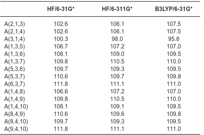
Table 2. Bond angles (°)
HF/6-31G* HF/6-311G* B3LYP/6-31G*
A(2,1,3) 102.6 106.1 107.5
A(2,1,4) 102.6 106.1 107.5
A(3,1,4) 100.3 98.0 95.8
A(1,3,5) 106.7 107.2 107.0
A(1,3,6) 108.1 109.0 109.5
A(1,3,7) 109.8 110.5 110.0
A(5,3,6) 109.7 109.3 109.5
A(5,3,7) 110.6 109.7 109.8
A(6,3,7) 111.8 111.1 111.0
A(1,4,8) 106.6 107.2 107.0
A(1,4,9) 109.8 110.5 110.0
A(1,4,10) 108.1 109.1 109.5
A(8,4,9) 110.6 109.6 109.8
A(8,4,10) 109.7 109.3 109.5
A(9,4,10) 111.8 111.1 111.0
Table 3. Dihedral Angles (º)
HF/6-31G* HF/6-311G* B3LYP/6-31G*
D(2,1,3,5) 70.7 68.3 66.9
D(2,1,3,6) -47.3 -49.9 -51.7
D(2,1,3,7) -169.4 -172.2 -173.9
D(4,1,3,5) 176.2 177.7 177.4
D(4,1,3,6) 58.3 59.5 58.7
D(4,1,3,7) -63.9 -62.9 -63.4
D(2,1,4,8) -70.7 -68.3 -66.9
D(2,1,4,9) 169.4 172.3 173.9
D(2,1,4,10) 47.3 49.9 51.7
D(3,1,4,8) -176.2 -177.7 -177.4
D(3,1,4,9) 63.9 62.9 63.4
D(3,1,4,10) -58.2 -59.5 -58.7
present case, from DFT calculations, the S-O bond length as 1.5117 Å. The reported values of C-S bond lengths are 1.8807, 1.8316Å (DFT calculations)21 and in the present case, the DFT calculations give the CS bond lengths as 1.8376 Å.
The first hyperpolarizability (â0) of this novel molecular system is calculated using theoretically, based on the finite field approach. In the presence of an applied electric field, the energy
The theoretically calculated thermal energy (KCal/Mol), specific heat capacity at constant volume (Cal/Mol-Kelvin), entropy (Cal/Mol-Kelvin)
Table 4: Calculated (scaled) wavenumbers and assignments
HF/6-31G* HF/6-311G* B3LYP/6-31G*
Assig-υυυυυ(cm-1) IR Raman υυυυυ(cm-1) IR Raman υυυυυ(cm-1) IR Raman nments
intensity activity intensity activity intensity activity
3003 0.77 101.48 2949 9.58 102.73 3046 4.75 107.52 νasMe
3003 0.19 46.47 2948 5.94 43.70 3046 3.32 39.86 νasMe
2997 5.90 110.89 2945 25.39 110.27 3038 14.37 104.01 νasMe
2996 0.06 12.03 2940 0.33 10.91 3034 0.04 8.32 νasMe
2895 14.90 212.39 2860 14.54 216.62 2944 7.26 206.03 νsMe
2893 11.69 1.39 2856 12.49 0.37 2942 4.63 0.13 δsMe
1455 35.58 3.55 1442 23.14 1.35 1449 19.97 2.73 δasMe
1444 2.89 33.16 1425 0.28 22.50 1431 0.47 32.50 δasMe
1442 7.21 27.46 1421 3.69 18.45 1427 3.07 26.60 δasMe
1432 13.98 4.76 1409 9.69 2.34 1414 9.37 3.95 δasMe
1365 1.43 0.95 1345 7.84 3.28 1318 6.49 0.62 δsMe
1342 1.01 0.08 1323 1.18 0.16 1295 0.77 1.33 δaMe
1048 20.55 9.34 1023 100.88 3.29 1059 111.2 6.69 νSO
1005 5.97 6.99 1020 38.68 3.07 999 22.87 7.00 ρMe
970 0.49 9.85 948 50.48 6.05 934 13.01 9.03 ρMe
936 0.73 0.76 933 9.08 3.72 908 5.86 6.47 ρMe
671 0.01 38.70 888 1.02 0.35 870 2.15 1.02 ρMe
637 3.40 38.70 694 11.80 18.09 639 20.40 15.81 νCS
568 8.02 29.43 659 2.77 31.83 610 10.04 26.69 νCS
267 3.39 2.39 358 13.92 1.41 346 7.52 1.51 tMe
219 6.32 3.41 310 17.08 2.99 284 7.11 3.47 δCSO
211 11.80 6.28 277 0.56 1.83 273 0.62 1.67 δCSC
165 0.90 0.01 228 0.816 0.05 222 0.49 0.07 tMe
141 0.25 0.00 188 0.00 0.13 177 0.00 0.06 tSCMe
ν-stretching; δ-bending; ρ-rocking; t-torsion; subscripts: as-asymmetric; s-symmetric; Me-methyl.
are respectively, 56.770, 17.578, 71.962 for HF/6-31G*, 56.770, 17.578, 71.962 for HF/6-311G* and 53.670, 18.930, 73.521 for B3LYP/6-31G* methods.
REFERNCES
1. R.Chakrabarti, R., and Schutt, C.E., Gene doi:10.1016/S0378-1119(01)00621-7. 2. Pegg, D.E., Methods Mol. Biol. 368: 39 (2007). 3. Francesca, C., and Albert, G., J. Statistical
Comp. Stim. 57: 337 (1997).
4. Finlayson-Pitts, B.J, and Pitts, J.N., Chemistr y of the Upper and Lower Atmosphere, Academic Press, San Diego (1999).
5. Charlson, R.J., Lovelock, J.E., Andreae, M.O., and Warren, S.G., Nature 326: 655 (1987).
Panel on Climate Change, Cambridge University Press, New York (2001).
7. Bates, T.S., Kiene, R.P., Wolfe, G.V., Matrai, P.A., Chavez, R.P., Buck, K.R., Blomquist, B.W., and Cuhel, R.L., J. Geophs. Res. 99: 7835 (1994).
8. Kiene, R.P., and Bates, T.S., Nature 345: 702 (1990).
9. Kiene, R.P., Oremland, R.S., Catena, A., Miller, L.G., and Capone, D.G., Appl. Environ. Microbiol. 52: 2037 (2986).
10. Tanimoto, Y., and Bak, F., Appl. Environ. Microbiol. 60: 2450 (1994).
11. Frisch, M.J., et al., Gaussian03, Revision C.02., Gaussian Inc., Wallingford, CT (2004). 12. Foresman, J.B., in: Frisch, E., (Ed.), Exploring Chemistry with Electronic Structure Methods: A Guide to Using Gaussian, Pittsburg, PA, (1996).
13. Roeges, N.P.G., A Guide to the Complete Interpretation of Infrared Spectra of Organic Structures, Wiley, New York (1994).
14. Silverstein, R.M., Bassler, G.C., and Morrill, T.C., Spectrometric Identification of Organic Compounds, ed.5, Singapore (1991). 15. Ganguly, L., Jose, C.I., and Biswas, A.B.,
Spectrochim. Acta, 24A: 215 (1968). 16. Edwards, H.G.M., Brown, D.R, Dale, J.R.,
and Plant, S., J. Mol. Struct. 595, 111 (2001). 17. Panicker, C.Y., Varghese, H.T., Philip, D., and Nogueria, H.I.S., Spectrochim. Acta 64A: 744 (2006).
18. Baran, J., Dega-Szafran, Z., Jaskolski, M., Marchewka, M.K., Ratajczak, H., and Szafran. M., J. Mol. Struct. 406: 127 (1997). 19. Miller, Y., Chaban, G.M., and Gerber. R.B.,
J. Phys. Chem. A 109: 6565 (2005). 20. Re, S., Osamura, Y., and Morokuma, K., J.
Phys. Chem. A 103, 3535 (1999).
21. Chowdhury, J., Sarkar, J., De, R., Ghosh, M., and Talapatra, G.B,. Chem. Phys., 330: 172 (2006).