Original Research Article
Bacterial etiology and antibiotic resistance pattern of neonatal sepsis: a
study in a tertiary care hospital, in Bangladesh
Mahfuza Shirin
1*, M. Monir Hossain
1, Manifa Afrin
2, Mohammad Abdullah Al Mamun
3INTRODUCTION
Sepsis is a leading cause of mortality and is responsible for nearly 25% of deaths among the neonates
worldwide.1,2 Most of these deaths occur in the
developing world.3 It also remains a significant cause of
morbidity.3,4 The clinical diagnosis of neonatal sepsis is
difficult due to its non-specific presentations. So, early
1Department of Neonatal Medicine and NICU, Bangladesh Institute of Child Health, Dhaka Shishu (Children)
Hospital, Dhaka, Bangladesh
2Department of Paediatrics, BIHS General Hospital Dhaka, Bangladesh
3Department of Paediatric Cardiology, Bangladesh Institute of Child Health, Dhaka Shishu (Children) Hospital Dhaka,
Bangladesh
Received: 14 June 2019 Revised: 24 June 2019
Accepted: 28 June 2019
*Correspondence:
Dr. Mahfuza Shirin,
E-mail: mahfuzashirin@gmail.com
Copyright: © the author(s), publisher and licensee Medip Academy. This is an open-access article distributed under the terms of the Creative Commons Attribution Non-Commercial License, which permits unrestricted non-commercial use, distribution, and reproduction in any medium, provided the original work is properly cited.
ABSTRACT
Background: Neonatal sepsis is a leading cause of neonatal mortality and morbidity. The objective of the study was to detect causative microorganisms of neonatal sepsis and their antimicrobial resistance patterns.
Methods: This prospective cross-sectional study was conducted from July 2017 to June 2018 in the Department of Neonatal Medicine and NICU of Dhaka Shishu (Children) Hospital (DSH). Neonates diagnosed with probable sepsis were studied. After enrollment, 1 mL blood was taken and sent to Microbiology department of DSH for culture and sensitivity. With baseline characteristics, clinical examination findings and outcome, were also recorded.
Results: Rate of isolation of single organism was 9.2% (84/913). Out of 84 isolates, gram negative bacteria were 77.4% with Klebsiella pneumonae being the commonest (35, 41.7%), gram positive bacteria were 11.9% with Staphylococcus aureus and Streptococcus were equal (5, 5.95% each) and the remaining (9, 10.7%) isolated organism was Candida. Most of the isolated gram-negative bacteria were resistant to ampicillin, gentamicin, and ceftazidime; but gram-positive bacteria preserved 20-80% sensitivity. Klebsiella was more resistant than Acinetobacter to amikacin, netilmicin, ciprofloxacin and levofloxacin. Around 45-65% of gram-negative bacteria were resistant to imipenem and meropenem but gram-positive bacteria showed lesser resistance. Among the gram-negative bacteria, Klebsiella and Acinetobacter were resistant to piperacillin as same as carbapenem group, but gram-positive bacteria were 100% sensitive to piperacillin. All the gram-negative bacteria showed more resistance to 4th generation cephalosporin, cefepime than carbapenem. Out of culture positive 84 neonates, 63 (75.0%) were cured but 21 (25.0%) died. Among the 21 expired neonates, 47.6% (10/21) were infected with Klebsiella.
Conclusion: This study observed that gram-negative bacteria causing neonatal sepsis predominantly, with emergence of Candida. All the isolated gram-positive and gram-negative organisms were mostly resistant to available antibiotics.
Key words: Antibiotic resistance pattern, Bacterial etiology, Neonatal sepsis, Sensitivity
diagnosis is crucial to start early and appropriate treatment for improvement of survival of neonates with sepsis. For that correct identification of causative organisms and their antibiotic sensitivity patterns are necessary. Neonatal sepsis is classified as early onset if it occurs within first 72 hours of life and as late onset if occurs after 72 hours of age until the end of the neonatal period. Early onset sepsis is conventionally regarded as maternally acquired, whereas late onset sepsis is considered environmental in origin, either hospital or community acquired.5
The bacteriological profile of septicemia keeps changing with the passage of time from region to region and hospital to hospital, in the same city or country. In developing countries, gram-negative organisms are the predominant causative agents of neonatal sepsis. But it is changing worldwide from predominant gram-negative to a predominant gram-positive bacteria isolation. Studies from Bangladesh over the time of two decades reported that though majority of identified organisms were gram negative bacteria but it has shifted from E. coli, 6 to
Klebsiella and Acinetobacter.7 Several recent studies
have reported that emergence of organisms such as CONS, NFGO, and Candida spp. occurred as a cause of neonatal septicemia.8-10 Continuous evolution of drug
resistant pathogens causing neonatal sepsis is becoming a potentially devastating problem. The situation is getting worse in developing countries and reports of multi-resistant bacteria causing neonatal sepsis in developing countries are increasing.4,11 Klebsiella and Enterobacter
species are often found to be resistant.
In developing countries, wide availability of over the counter antibiotics, irrational use of antibiotics and poor hygiene are important factors of development of this condition. Also, improvement of neonatal care service especially establishment of NICU care, has led to neonates born premature or needing intensive care for more than 48 hours to be more risk prone. Spread of resistant organisms in hospitals is a recognized problem.
Most Gram-negative organism are resistant to ampicillin and cloxacillin, and many are becoming resistant to gentamicin. In many units the antibiotic policy has been changed to include third generation cephalosporin. But the organisms have reduced susceptibility to them and even quinolones. In many developing countries S. aureus is a common cause and methicillin resistant strains (MRSA) are widespread.11
In this respect, it has become necessary to engage in appropriate epidemiological surveillance to identify the etiological agents and their antibiotics susceptibilities so that the emergence of new pathogens and their resistance patterns can be monitored. So, this study was conducted to find out the most important organisms causing sepsis in neonates, their changing pattern and the antibiotic resistance.
METHODS
This was a prospective cross-sectional study conducted over the period of one year from July 2017 to June 2018 in the Department of Neonatal Medicine and NICU of Dhaka Shishu (Children) hospital (DSH) to identify the current pattern of organisms causing sepsis in neonates and their resistance pattern. Neonates diagnosed with probable sepsis at the time of admission were enrolled in this study after taking informed consent from the parents. Neonates who were critically ill, expired within 10 hours of admission and did not give consent were excluded from the study.
After enrollment, blood samples for culture were collected and sent to the Microbiology department of DSH. For each neonate to collect blood for culture, the skin site for venipuncture was cleaned thoroughly with diluted iodine solution followed by 2.5% chlorhexidine solution and 1 mL blood was drawn by using a butterfly needle attached to a 2 ml disposable syringe. Culture and identification of the isolates was done in Bact /Alert FAN media. Bacterial isolates were tested for susceptibility to various antimicrobial agents and categorized as sensitive or resistant. Printed reports were sent to the designated wards.
For each neonate, information including gender, gestation, age, weight, birth history, maternal perinatal history, were recorded in a questionnaire from mother or attendant. Clinical examination findings at the time of enrollment, report of blood culture with sensitivity pattern and outcome were also recorded.
Data analysis, including descriptive statistics such as frequency tabulation, mean, standard deviation, were done by using SPSS version 20.
RESULTS
A total of 1359 neonates with sepsis were admitted in DSH during this period, of them from 891 neonate were included in this study. Amongst these 891 neonates, 68.2% were male and 31.8% were female, gestational age was 36.5±2.8 weeks, of them 51.5% were preterm. At the time of admission, mean age of them was 8.9±6.6 days and out of them, 34.9% neonates were admitted within first 72 hours of age. Admission weight was 2357.7±719.9 gm and 16.2% of them were very low birth weight (<1500gm) baby (Table 1).
Table 1: Baseline characteristics of neonates with sepsis (n=891).
Neonatal variables Gender
Male: female 2.1:1 Male 608 (68.2%) Female 283 (31.8%)
Gestational age 36.52.8 weeks (23-42 weeks)
Preterm 459 (51.5%) Term 432 (48.5%)
Admission weight 2357.7719.9gm (800-4600) <1500gm 144(16.2%)
1500-<2500gm 396(44.4%) 2500-4000gm 344(38.6%) >4000gm 7 (0.8%)
Age on admission 8.9±6.6 days (14 hours - 28 days)
Age at enrollment <3days 157 (17.6%) 3-7 days 303 (34.0%) >7 days 431 (48.4%)
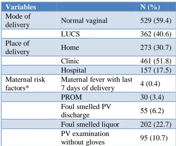
Table 2: Birth history and maternal risk factors (n=891).
Variables N (%)
Mode of
delivery Normal vaginal 529 (59.4)
LUCS 362 (40.6)
Place of
delivery Home 273 (30.7)
Clinic 461 (51.8)
Hospital 157 (17.5)
Maternal risk factors*
Maternal fever with last
7 days of delivery 4 (0.4)
PROM 30 (3.4)
Foul smelled PV
discharge 55 (6.2)
Foul smelled liquor 202 (22.7) PV examination
without gloves 95 (10.7) *Multiple responses
From all (891, 100%) neonates blood culture was sent for 2nd time. So, total 913 samples were sent for blood
culture, out of them only 9.2% (84/913) culture specimens had growth of single organism. Out of 84 positive cultures, 76 were yielded from first time blood culture, 8 were isolated from 2nd time blood culture and 3 were isolated from both the time. Out of 84 isolates, gram negative bacteria were found in 77.4% with Klebsiella being the commonest (35, 41.7%), gram positive bacteria in 11.9% with Staphylococcus aureus and Streptococcus were equal (5, 5.95% each) and the
remaining (9, 10.7%) isolated organism was Candida (Figure 2).
Figure 1: Type of sepsis of the study participants (n=891).
Table 3: Clinical findings at the time of enrollment (n=891).
Clinical findings* N (%)
Lethargy 103 (11.6)
Abdominal distension 35 (3.9)
Vomiting 160 (18.0)
Jaundice 203 (22.8)
Tachypnoea 40 (4.5)
Apnoea 109 (12.2)
Convulsion 108 (12.1)
Prolonged CRT 527 (59.2)
Hypothermia 397 (44.6)
Fever 129(14.5)
Hypoglycemia 12 (1.3)
Figure 2: Distribution of isolated organisms in blood culture (n=84).
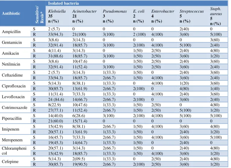
Susceptibility pattern of isolated bacteria is shown in Table IV. Most of the gram-negative bacteria were 90%-100% resistant to ampicillin, gentamicin, and ceftazidime; but gram-positive bacteria Streptococcus
21 35
3 2 4 5 5
9
and Staphylococcus preserved 20-80% sensitivity, except staphylococcus was 100% resistant to ampicillin and Streptococcus was 100% resistant to gentamicin. Klebsiella was more resistant than Acinetobacter to amikacin (89% vs 86%), netilmicin (91 vs 52%), ciprofloxacin (86% vs 62%), levofloxacin (85% vs 67%) and cotrimoxazole (77% vs 52%), but Acinetobacter was more resistant to chloramphenicol (86% vs 43%). Around 45-65% of gram-negative organisms were resistant to imipenem and meropenem but gram-positive bacteria showed lesser resistance (0-20%). Among the gram-negative bacteria, Klebsiella and Acinetobacter were resistant to broad spectrum β-lactam antibiotic, piperacillin as same as carbapenem group, but gram-positive bacteria were 100% sensitive to piperacillin. All the gram-negative bacteria showed more resistance (86%-100%) to 4th generation cephalosporin, cefepime than
carbapenem.
Figure 3: Outcome of neonates with positive blood culture (n=84).
Table 4: Sensitivity pattern of isolated organisms (n=75).
Antibiotic
S
en
si
ti
ve
/
Re
si
s*
Isolated bacteria
Klebsiella
35 n (%)
Acinetobacter
21 n (%)
Pseudomonas
3 n (%)
E. coli
2 n (%)
Enterobacter
4 n (%)
Streptococcus
5 n (%)
Staph. aureus
5 n (%)
Ampicillin S 2 (5.7) 0 0 0 0 2(40) 0
R 33(94.3) 21(100) 3(100) 2 (100) 4(100) 3(60) 5(100)
Gentamicin S 3(8.6) 3(14.3) 0 0 0 0 3(60)
R 32(91.4) 18(85.7) 3(100) 2(100) 4(100) 5(100) 2(40)
Amikacin S 4(11.4) 3(14.3) 0 1(50) 2(50) 2(40) 4(80)
R 31(88.6) 18(85.7) 3(100) 1(50) 2(50) 3(60) 1(20)
Netilmicin S 3(8.6) 10(47.6) 0 1(50) 2(50) 2(40) 3(60)
R 32(91.4) 11(52.4) 3(100) 1(50) 2(50) 3(60) 2(40)
Ceftazidime S 2 (5.7) 3(14.3) 1(33.3) 1(50) 0 2(40) 3(60)
R 33(94.3) 18(85.7) 2(66.7) 1(50) 4(100) 3(60) 2(40)
Ciprofloxacin S 5(14.3) 8(38.1) 1(33.3) 0 4(100) 1(20) 3(60)
R 30(85.7) 13(61.9) 2(66.7) 2(100) 0 4(80) 1(40)
Levofloxacin S 11(31.4) 7(33.3) 1(33.3) 0 4(100) 2(40) 3(60)
R 24 (84.6) 14(66.7) 2(66.7) 2(100) 0 3(60) 2(40)
Cotrimoxazole S 8(22.9) 10(47.6) 1(33.3) 1(50) 2(50) 0 4(80)
R 27(77.1) 11(52.4) 2(66.7) 1(50) 2(50) 5(100) 1(20)
Piperacillin S 14(40.0) 6(28.6) 3(100) 2(100) 4(100) 5(100) 5(100)
R 21(60.0) 15(71.4) 0 0 0 0 0
Imipenem S 15(42.9) 8(38.1) 2(66.7) 1(50) 4(100) 3(60) 4(80)
R 20(57.1) 13(61.9) 1(33.3) 1(50) 0 2(40) 1(20)
Meropenem S 16(45.7) 7(33.3) 2(66.7) 1(50) 4(100) 3(60) 5(100)
R 19(45.3) 14(64.7) 1(33.3) 1(50) 0 2(40) 0
Chlorampheni col
S 20(57.1) 3(14.3) 2(66.7) 1(50) 0 2(40) 4(80)
R 15(42.9) 18(85.7) 1(33.3) 1(50) 4(100) 3(60) 1(20)
Cefepime S 5(14.3) 2(09.5) 1(33.3) 0 2(50) 2(40) 4(80)
R 30(85.7) 19(90.5) 2(66.7) 2(100) 2(50) 3(60) 1(20)
*S=Sensitive; R=Resistant
Out of 84 neonates with positive blood culture, 63 (75.0%) were cured but 21 (25.0%) died. Out of the 21
expired neonates, 47.6% (10/21) were infected with Klebsiella (Figure 3).
Cured 63 (75.0%)
Expired, 25.0%
DISCUSSION
This study was conducted to find out the causative organisms, their changing pattern and the antibiotic resistance in neonatal sepsis. Authors found that the rate of isolation of single organism was about 9.2%. In a previous study reported from Bangladesh, the culture positivity rate was 36%.7 In studies done in India, it has
ranged from 16%-54%.4,12,13 One study from Africa also
reported similar rate of culture positivity, approximately 44-47%.14,15 Rate of culture positivity in this study might
be lower than that of actual, because DSH is solely a children hospital, all of the enrolled neonates were out born, no options to get inborn babies and many of them got antibiotics beforehand. Most of the neonatal septicemia cases now are either LBW or preterm.16 In this
study, 60% were LBW and 51.5% were preterm neonates. Authors found foul smelled liquor (22.7%) and PV examination without gloves (10.7%) among the maternal risk factors for occurrence of EOS. In this study LOS (51%) was more than EOS (34.9%) but percentage of EOS was more than that of other studies.17
The bacteriological profile has changed worldwide from predominant negative to a predominant gram-positive bacteria isolation.18-21 But among the positive
cultures Authors found more gram-negative bacteria (77.4%) contrasted with gram-positive bacteria (11.9%) and a good number of Candida spp. (10.7%). Our finding was similar to that of previous studies reported from Bangladesh.6,7 Several recent studies also have reported
that emergence of organisms such as CONS, NFGO, and Candida spp. occurred as a cause of neonatal septicemia.8-10
Regarding changes in distribution of organism it was clearly evident from this study that about 11% Candida was isolated, which should be taken into account.
The pattern of bacterial organisms is constantly changing with time and place. Previously sensitive organisms are rapidly becoming resistant to commonly used antibiotic due to indiscriminate use thus making the treatment difficult and costly.22 Reports showed that almost all
isolated Enterobacteriaceae were either completely resistant to early beta-lactam antibiotics including ampicillin, amoxicillin, carbenicillin alone or in combination with beta-lactam inhibitor clavulanic acid along with resistant to second generation cephalosporin and to some extent to third generation drugs such as ceftriaxone.8,23 Authors found most of the isolated
gram-negative bacteria were resistant to ampicillin, gentamicin, and ceftazidime; but gram-positive bacteria streptococcus and staphylococcus preserved 20-80% sensitivity to those antibiotics. But Authors found that staphylococcus was 100% resistant to ampicillin and Streptococcus was 100% resistant to gentamicin. Similar findings of high resistance to ampicillin (71%) was reported by Bhat et al.9 It was also reported by Bhat et al 9 that Klebsiella
pneumoniae showed resistance to all antibiotics tested
except imipenem. Authors found that Klebsiella was more resistant than Acinetobacter to amikacin, netilmicin, ciprofloxacin, levofloxacin, cotrimoxazole but sensitivity was still present. This high resistance pattern could be attributed to easy availability and widespread use of broad-spectrum antibiotics in the presumptive treatment of infections prevailing in our country. Blood culture facilities are often not available in most of the settings in Bangladesh. In such scenarios, clinicians have to depend on empirical antibiotic regimens. The high prevalence of resistance to ampicillin makes it out of use in neonatal sepsis in our hospital. The increasing resistance of Gram-negative organisms to extended spectrum cephalosporins and carbapenems makes the choice of antibiotics difficult.
This was a prospective cross-sectional study in a single center. So, the study results might not be reflected throughout the whole country.
CONCLUSION
This study concluded that gram-negative bacteria causing neonatal sepsis predominantly with emergence of Candida. It was also evident that all isolated gram-negative and gram-positive organisms were mostly resistant to available antibiotics. So, Authors recommend to take strict policy to prevent sepsis, and use available antibiotics rationally, to reduce the risk of death from neonatal sepsis.
Funding: No funding sources Conflict of interest: None declared Ethical approval: Not required
REFERENCES
1. WHO. Newborns: Reducing Mortality. WHO.
Available at:
http://www.who.int/mediacentre/factsheets/fs333/en /.
2. Lawn JE, Cousens S, Zupan J. Lancet Neonatal Survival Steering Team. 4 million neonatal deaths: When? Where? Why? Lancet. 2005;365:891-900. 3. Jain A, Awasthi AK, Kumar M. Etiological and
antimicrobial susceptibility profile of nosocomial blood stream infections in neonatal Intensive Care Unit. Indian J Med Microbiol. 2007;25:299-300. 4. Thaver D, Zaidi AK. Burden of neonatal infections
in developing countries: A review of evidence from community-based studies. Pediatr Infect Dis J. 2009;28:S3-S9.
5. Kumhar GD, Ramachandran VG, Gupta P. Bacteriological analysis of blood culture isolates from neonates in a tertiary care hospital in India. J Health Popul Nutr. 2002;20:343-7.
hospital in Bangladesh. Ind Pediatr. 2002;39:1034-9.
7. Hossain MM, Afroza S, Shirin M, Chowdhury NA, Saha SK. Bacterial aetiology of neonatal sepsis in a tertiary care hospital in Bangladesh. Bang J Child Health. 2004;28:81-5.
8. Sundaram V, Kumar P, Dutta S, Mukhopadhyay K, Ray P, Gautam V, et al. Blood culture confirmed bacterial sepsis in neonates in a North Indian tertiary care center: Changes over the last decade. Jpn J Infect Dis. 2009;62:46-50.
9. Bhat Y R, Lewis LE, Ke V. Bacterial isolates of early-onset neonatal sepsis and their antibiotic susceptibility pattern between 1998 and 2004: An audit from a center in India. Ital J Pediatr. 2011;37:32.
10. Sharma P, Kaur P, Aggarwal A. Staphylococcus aureus – The predominant pathogen in the neonatal ICU of a tertiary care hospital in Amritsar, India. J Clin Diagn Res. 2013;7:66-9.
11. Deorari AK. Neonatal sepsis: Manageable daunting issue for India. J Neonatol. 2009;23:7-11.
12. Zakariya BP, Bhat V, Harish BN, Arun Babu T, Joseph NM. Neonatal sepsis in a tertiary care hospital in South India: Bacteriological profile and antibiotic sensitivity pattern. Indian J Pediatr. 2011;78:413-17.
13. Kaistha N, Mehta M, Singla N, Garg R, Chander J. Neonatal septicemia isolates and resistance patterns in a tertiary care hospital of North India. J Infect Dev Ctries. 2009;4:55-7.
14. Shitaye D, Asrat D, Woldeamanuel Y, Worku B. Risk factors and etiology of neonatal sepsis in Tikur Anbessa University Hospital, Ethiopia. Ethiop Med J. 2010;48(1):11-21.
15. G/eyesus T, Moges F, Eshetie S, Yeshitela B, Abate E. Bacterial etiologic agents causing neonatal sepsis and associated risk factors in Gondar, Northwest Ethiopia. BMC Pediatrics. 2017 June;17(1):137. 16. Hornik CP, Fort P, Clark RH, Watt K, Benjamin
DK Jr, Smith PB, et al. Early and late onset sepsis in very-low-birth-weight infants from a large group of
neonatal Intensive Care Units. Early Hum Dev 2012; 88(2):S69-S74.
17. Mhada TV, Fredrick F, Matee MI, MassaAuthorsA. Neonatal sepsis at Muhimbili National Hospital, Dar es Salaam, Tanzania: Aetiology, antimicrobial sensitivity pattern and clinical outcome. BMC Public Health. 2012;12:904.
18. Darmstadt GL, Saha SK, Choi Y, El Arifeen S, Ahmed NU, Bari S, et al. Population-based incidence and etiology of community-acquired neonatal bacteremia in Mirzapur, Bangladesh: An observational study. J Infect Dis. 2009;200:906-15. 19. Monjur F, Rizwan F, Asaduzzaman M, Nasrin N,
Ghosh NK, Apu AS, et al. Antibiotic sensitivity pattern of causative organisms of neonatal septicemia in an urban hospital of Bangladesh. Indian J Med Sci. 2010;64:265-71.
20. Kohli-Kochhar R, Omuse G, Revathi G. A ten-year review of neonatal bloodstream infections in a tertiary private hospital in Kenya. J Infect Dev Ctries. 2011;5:799-803.
21. Kumar DVP, Mohan J, Rakesh PS, Prasad J, Joseph LK. Bacteriological profile of neonatal sepsis in a secondary care hospital in rural Tamil Nadu, Southern India. J Family Med Prim Care. 2017;6(4):735-8.
22. Thakur S, Thakur K, Sood A, Chaudhary A. Bacteriological profile and antibiotic sensitivity pattern of neonatal septicaemia in a rural tertiary care hospital in North India. Indian J Medical Microbiol. 2016;34(1):67-71.
23. Deman P, Verhoeven BA, Verburgh HA. An antibiotic policy to prevent emergence of resistance bacilli. Lancet. 2000;355:973-8.