Original Research Article
Study of clinical profile of acute lower respiratory tract infection in
children aged 2 months to 5 years
Dhivyanarayani M.*, Raju V., Jeyachandran P.
INTRODUCTION
Acute Lower Respiratory Tract Infection (ALRI) is the leading cause of under-5 childhood morbidity in the world, with nearly 156 million new episodes each year, of which India ac-counts for a bulk of 43 million.1 The
mortality burden is 1.9 million per year, out of which India accounts for around four hundred thousand deaths per year.2 The WHO Programme for ARI Control
guidelines define Pneumonia as cough in the presence of tachypnoea (respirato-ry rate >50/ min in children aged 2 months to 12 months and >40/min in children aged 13 months to 60 months) and Severe and Very Severe Pneumonia as the presence of chest in-drawing and
central cyanosis, lethargy, convulsions and refusal of feeds of respectively.3 Tachypnoea and lower chest
indrawing, when applied by health workers and paediatricians as a diagnostic tool, had the sensitivity of 70% and 81% respectively.4
On an average, children below 5 years of age about suffer 5 episodes of ARI per child per year, thus accounting for about 238 million attacks. Consequently, although most of the attacks are mild and self-limiting episodes, ARI is responsible for about 30-50 percent of visits to health facilities and for about 20-40 percent of admissions to hospitals.5 Streptococcus pneumoniae is a major cause of
illness and death in children, as well as in adults.
ABSTRACT
Background: Acute respiratory infections are a leading cause of morbidity and mortality in under-five chil-dren in developing countries. Hence, the present study was undertaken to study the various risk factors, clinical profile and outcome of acute lower respiratory tract infections (ALRI) in children aged 2 months to 5 years. To study the risk factors associated with ALRI in these children. To ascertain the association of the various cultural practices prevalent in this area with ALRI.
Methods: 100 ALRI cases fulfilling WHO criteria for pneumonia, in the age group of 2 months to 5 years were evaluated for potential risk factors, clinical profile and outcome as per a predesigned proforma in a rural medical college.
Results: Socio-demographic risk factors like parental illiteracy, overcrowding, partial immunization and low socioeconomic status were potential risk factors; similarly, nutritional risk factors like early and late weaning, anemia, and malnutrition were associated with ALRI. Significant environmental risk factors were the use of biomass fuels, inadequate ventilation at home, and lack of separate kitchen.
Conclusions: The present study has identified various socio-demographic, nutritional and environmental risk factors for ALRI which can be tackled by effective health education of the community and effective training of peripheral health personnel.
Keywords: Environmental risk factors, Malnutrition risk factors, Respiratory infections Department of Paediatrics, Sri Muthu Kumaran Medical College, Mangadu, Tamil Nadu, India
Received: 22 May 2018
Accepted: 26 May 2018
*Correspondence:
Dr. Dhivyanarayani M, E-mail: cmafedz@gmail.com
Copyright: © the author(s), publisher and licensee Medip Academy. This is an open-access article distributed under the terms of the Creative Commons Attribution Non-Commercial License, which permits unrestricted non-commercial use, distribution, and reproduction in any medium, provided the original work is properly cited.
According to a 2002 WHO estimate, about 1.6 million cases of the fatal pneumococcal disease occur worldwide, mostly in infants and elderly. In addition, immunocompromised individuals of all ages are at increased risk.6 Likewise, Haemophilus influenza type b
(Hib) bacteria is estimated to cause 3 million cases of severe pneumonia and meningitis, and approximately 386,000 deaths per year in children under 5 years of age.7
METHODS
A prospective study of ALRI in children aged 2 months to 5 years conducted at Department of Paediatrics, Sri Muthu Kumaran Medical College. Totally 100 Children admitted to our hospital with a clinical diagnosis of ALRI as per WHO criteria from January 2016 TO December 2016.Inclusion Criteria-Children with ALRI from 2 months to 60 months. Exclusion Criteria-Children less than 2 months and more than 60 months. Children with any underlying chronic respiratory or cardiac illness. Children in the age group of 2 months to 5 years admit-ted with ALRI during the study period were enrolled in the study as cases. A detailed history and physical
examination were done according to a predesigned proforma to elicit various potential risk factors and another relevant history. Age of the child was recorded in completed months and age of parents in a completed year.
Distress was assessed in each child. Pallor and other signs of vitamin deficiencies were recorded. A detailed systemic examination was done in both cases and controls. Routine hematological investigations were done in all cases to know the degree of anemia and blood counts; chest x-ray was done in all cases to categorize the ALRI into clinical entities and to detect complications if any. Appropriate tables and graphical representations were used to display the data. Chi-square test was used. A “p” value <0.05 was taken as significant.
RESULTS
Among the 100 cases of ALRI studied, majority were infant age group with male predominance. However, no significant association was found between age and ALRI severity. Chi2 =1.46 DF=2 p=0.481.
Table 1: The age of pediatrics cases
Age Pneumonia Severe pneumonia V. severe pneumonia Total
2-12 months 10 34 17 61
13-60 months 6 26 7 39
Total 16 60 24 100
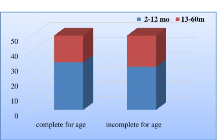
Figure 1: The immunization statutes of paediatrics cases.
Of the 100ALRI cases studied, 50% were incompletely immunized for age, the main reason was parent’s low socioeconomic status.
The highly significant association was found between ALRI severity and immunization status, thus immuunization plays a bigger role in ALRI. Overcrowding was 70% among the 100 ALRI cases. No significant association of over-crowding with ALRI severity was found. Chi2 0.591 DF=2 p=0.744. Of the 100 ALRI cases, 47% were anemic, due to late weaning No significant association was found between anemia and ALRI severity.
Table 2: Overcrowding of paediatrics cases.
Overcrowding Pneumonia Severe pneumonia V. severe pneumonia Total
Present 12 38 20 70
Absent 4 22 4 30
Total 16 60 24 100
0 10 20 30 40 50
complete for age incomplete for age
Among the 100 ALRI cases, 46% were found to be malnourished main reason was low socioeconomic status, no significant association was found between ALRI severity and nutrition status.
Table 3: The prevalence of anemia among ALRI pediatrics cases.
Anaemia 2-12 months 13-60 months Total
Present 30 17 47
Absent 31 22 53
Total 61 39 100
Among the 100 ALRI cases, 40% were found to be living in houses with inadequate ventilation due to overcrowding.
No significant association was found between ALRI severity and ventilation. Chi2 =3.06 DF=2 p=0.217. Of the 100 ALRI cases, 30% had a history of at least one family member having or having had a URI in the preceding 2 weeks.
Table 4: The prevalence of malnutrition among ALRI pediatrics cases.
2-12 months 13-60 months Total
Absent 30 24 54
Grade 1 9 6 15
Grade 2 9 4 13
Grade 3 12 4 16
Grade 4 1 1 2
Total 61 39 100
No significant association was found between family history of URI and ALRI severity. Chi2 =0.823 DF=2 p=0.646. The occurrence of leukocytosis was 22% among the ALRI cases, an association with severity of ALRI was found to be significant.
Chi2 =7.22 DF=2 p=0.027. Among the 100 ALRI cases,
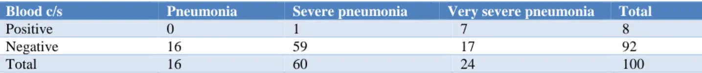
only 8% had a positive blood culture. However, the highly significant association was found between ALRI severity and blood culture Chi2 =19.3 DF=2 p=0.0001.
Table 5: Ventilation among ALRI pediatrics cases
Ventilation Pneumonia Severe pneumonia V. severe pneumonia Number
Adequate 4 28 8 60
Inadequate 12 32 16 40
Total 16 60 24 100
Table 6: History of upper respiratory tract infection in the family (<2 weeks).
H/o URI Pneumonia Severe pneumonia V. severe pneumonia Number
Present 6 16 8 30
Absent 10 44 16 70
Total 16 60 24 100
Table 7: Shows the total leukocyte count among ALRI paediatrics cases.
TLC Pneumonia Severe pneumonia V. severe pneumonia Total
Normal 13 51 14 78
Raised 3 9 10 22
Total 16 60 24 100
Table 8: The blood culture among ALRI paediatrics cases
Blood c/s Pneumonia Severe pneumonia Very severe pneumonia Total
Positive 0 1 7 8
Negative 16 59 17 92
Total 16 60 24 100
The most common diagnosis was bronchiolitis (41%), and the other major diagnoses were lo-bar pneumonia and bronchopneumonia (26% and 17% respectively).
Table 9: Shows the final diagnosis ALRI pediatrics cases.
Clinical diagnosis Number Bronchiolitis 41 Lobar pneumonia 26 Bronchopneumonia 17
WLRI 10
Empyema thoracic 1
Croup 5
Total 100
DISCUSSION
Acute lower respiratory infections (ALRI) are among the commonest causes of morbidity and mortality among children under 5 years of age, especially in developing countries. The preventive role of immunization in ALRI prevention has been stressed upon extensively. The present study shows 50% were partially immunized children, and this is higher than the Janssen R et al.8
Letterio JJ et al studies which showed 21.15% and 38.2% partially immunized children respectively. However, the Broor the spread of infection via respiratory droplets may be aggravated by overcrowding. In our study, 70% cases were associated with overcrowding, which is similar to the results (71.6%).9 Martineau AR et studies showed that
slightly more cases are associated with overcrowding, that is, 91.35% and 80.87% respectively.10
Muhe Let al in Malaysia showed a significant association between ALRI and overcrowding. However, no significant association was found between overcrowding and ALRI severity. The spread of infection via respiratory droplets may be aggravated by overcrowding. No significant association was found between anemia and ALRI severity. The role anemia plays in infection is still not exactly established. A proposed pathophysiologic mechanism is that neutrophils have reduced the ability to kill Staphylococcus aureus due to reduced activity of myeloperoxidase, and the T-cells in circulation are reduced in number and they have defective DNA synthesis due to the attenuated activity of ribonucleotide reductase.11
Malnutrition causes children to have defective cell-mediated immunity secondary to thymolymphatic depletion leading to Gram-negative bacterial infections and sepsis. There may also be qualitatively deficient immunoglobulins and impairment of leukocytic enzymes involved in the bactericidal activity. As the secretory IgA is generally reduced, the recovery from infections is delayed and infections tend to be severe in malnourished subjects. The period of infection is prolonged. Because of increased duration of replication and shedding of pathogens, the systemic spread is also more likely.12
Also, the skin and mucous membranes do not offer effective physical barriers against infection. Malnutrition is invariably associated with deficiency of vitamins like A, B and D. Vitamin A is mainly necessary for
maintaining the integrity of the epithelial cells, deficiency of vitamin D may lead to deformity in the thoracic cavity which predisposes for ALRI.13 Malnutrition was found in
46% of ALRI cases in our study.
In their study, Smith et al showed a higher incidence of inadequate ventilation. No significant association was found between ventilation and ALRI severity. Inadequate ventilation tends to accentuate the effect of the indoor air pollutants as proper egress from the house is not possible. Positive blood culture was obtained in only 8% of cases; however, a significant association was found between blood culture and ALRI severity.
The most common organism was Staphylococcus aureus. The reason why this was the most common isolate in this study might be because the majority of children with bacteremia were severely malnourished and
Staphylococcus aureus bacteremia is commonly
associated with malnutrition Mechanical ventilation was required by 8% cases, all classified as very severe pneumonia.14 This constituted 33% of the very severe
pneumonia cases and 9.5% of the cases graded severe pneumonia and higher. The study by reported higher rates of ventilation among children with severe pneumonia (20.5%). Udani PM There was one death among the 100 cases, and the other 99 recovered and were dis-charged uneventfully. The complication rate was 1% in our study.15
CONCLUSION
Regarding the laboratory profile, leukocytosis and blood culture positivity were observed in a small percentage, but significant association with ALRI severity was observed for both. Thus, clinical signs, and not invasive blood tests are better diagnostic tools, though the latter may provide additional therapeutic and prognostic information in severe disease. Early diagnosis and treatment initiation helps improve the morbidity and mortality profile, as evidenced by the relatively low rates of mechanical ventilation and mortality in the present study.
ACKNOWLEDGEMENTS
Authors Would Like to Thank the Pediatric department Faculty of Sri Muthu Kumaran Medical College, Mangadu.
Funding: No funding sources Conflict of interest: None declared
Ethical approval: The study was approved by the Institutional Ethics Committee
REFERENCES
Respiratory Tract Infection in US children. Pediatrics. 2006;117:425-32.
2. Cowgill KD, Ndiritu M, Nyiro J, Slack MP, Chiphatsi S, Ismail A et al. Effectiveness of Haemophilus influenzae type b conjugate vaccine introduction into routine childhood immunization in Kenya. JAMA. 2006;296:671-8.
3. Ganong WF. Gas transport between the lungs and the tissue. In: Ganong WF. Review of medical physiology. 22nd ed. New York. McGraw Hill.
2005:666-9.
4. Ganz T. Defensins: antimicrobial peptides of innate immunity. Nat Rev Immunol.2003;3(9):710-20. 5. Guyton and Hall Textbook of medical physiology.
11th ed. Philadelphia. Saunders Elsevier. 2006:507-
8.
6. Hawkes JS, Neumann MA, Gibson RA. The effect of breastfeeding on lymphocyte subpopulations in healthy term infants at 6 months of age. Pediatr Res. 1999;45:648-51.
7. Jain L, Vidyasagar D, Xanthou M, Ghai V, Shimada S, Blend M. In vivo distribution of human milk leukocytes after ingestion by newborn baboons. Arch Dis Child. 1989;64:930-3.
8. Janssen R, Bont L, Siezen CL, et al. Genetic susceptibility to respiratory syncytial virus bronchiolitis is predominantly associated with innate immune genes. J Infect Dis. 2007;196 (6):826-34.
9. Letterio JJ, Geiser AG, Kulkarni AB, Roche NS, Sporn MB, Roberts AB. Maternal rescue of transforming growth-beta 1 null mice. Science. 1994;264:1936-8.
10. Martineau AR, Wilkinson RJ, Wilkinson KA, Newton SM, Kampmann B, Hall BM et al. A single dose of vitamin D enhances immunity to mycobacteria. Am J Respir Crit Care Med. 2007; 176(2):208-13.
11. Muhe L, Lulseged S, Mason KE, Simoes EA. Case-control study of the role of nutritional rickets in the risk of developing pneumonia in Ethiopian children. Lancet. 1997;349(9068):1801-4.
12. Ogawa J, Sasahara A, Yoshida T, Sira MM, Futatani T, Kanegane H, Miyawaki T. Role of transforming growth factor-beta in breast milk for initiation of IgA production in newborn infants. Early Hum Dev. 2004;77:67-75.
13. Pabst HF, Spady DW, Pilarski LM, Carson MM, Beeler JA, Krezolek M. Differential modulation of the immune response by breast or formula feeding of infants. Acta Paediatr. 1997;86:1291-7.
14. Rousset F, Garcia E, Defrance T, Péronne C, Vezzio N, Hsu DH, Kastelein R, Moore KW, Banchereau J. human B lymphocytes. Proc Natl Acad Sci USA. 1992;89:1890-3.
15. Udani PM. Vitamin D and deficiency Rickets. In: Udani PM. Textbook of Pediatrics. 1st ed. New
Delhi. Jaypee brothers.1998:631-666.
Cite this article as: Dhivyanarayani M, Raju V, Jeyachandran P. Study of clinical profile of acute lower respiratory tract infection in children aged 2 months to 5 years. Int J Contemp Pediatr