R E S E A R C H
Open Access
VEGF isoforms have differential effects
on permeability of human pulmonary
microvascular endothelial cells
Khadija Ourradi
1, Thomas Blythe
1, Caroline Jarrett
1, Shaney L. Barratt
1, Gavin I. Welsh
2and Ann B. Millar
1*Abstract
Background:Alternative splicing of Vascular endothelial growth factor-A mRNA transcripts (commonly referred as VEGF) leads to the generation of functionally differing isoforms, the relative amounts of which have potentially significant physiological outcomes in conditions such as acute respiratory distress syndrome (ARDS). The effect of such isoforms on pulmonary vascular permeability is unknown. We hypothesised that VEGF165a and VEGF165b isoforms would have differing effects on pulmonary vascular permeability caused by differential activation of intercellular signal transduction pathways.
Method: To test this hypothesis we investigated the physiological effect of VEGF165a and VEGF165b on Human Pulmonary Microvascular Endothelial Cell (HPMEC) permeability using three different methods: trans-endothelial electrical resistance (TEER), Electric cell-substrate impedance sensing (ECIS) and FITC-BSA passage. In addition, potential downstream signalling pathways of the VEGF isoforms were investigated by Western blotting and the use of specific signalling inhibitors.
Results: VEGF165a increased HPMEC permeability using all three methods (paracellular and transcellular) and led to associated VE-cadherin and actin stress fibre changes. In contrast, VEGF165b decreased paracellular permeability and did not induce changes in VE-cadherin cell distribution. Furthermore, VEGF165a and VEGF165b had differing effects on both the phosphorylation of VEGF receptors and downstream signalling proteins pMEK, p42/44MAPK, p38 MAPK, pAKT and peNOS. Interestingly specific inhibition of the pMEK, p38 MAPK, PI3 kinase and eNOS pathways blocked the effects of both VEGF165a and VEGF165b on paracellular permeability and the effect of VEGF165a on proliferation/migration, suggesting that this difference in cellular response is mediated by an as yet unidentified signalling pathway(s).
Conclusion:This study demonstrates that the novel isoform VEGF165a and VEGF165b induce differing effects on permeability in pulmonary microvascular endothelial cells.
Keywords:Vascular permeability, Vascular endothelial growth factor (VEGF), Cell signalling
Background
VEGF was originally identified by its properties as both a permogen and a mitogen, key elements in the function of the alveolar-capillary membrane, leading to interest in its role in many forms of lung disease particularly ARDS [1–3]. We and others found that VEGF levels were com-partmentalised between the alveolar space and the vas-cular bed [4, 5]. Low levels of intrapulmonary VEGF
were found in patients with ARDS with increasing intra-pulmonary VEGF levels associated with recovery [5]. In contrast, plasma levels in patients with ARDS were ele-vated compared with normal, at-risk, or ventilated con-trol subjects, with falling levels associated with recovery [6]. These data suggest that VEGF is beneficial in the al-veolar space but detrimental in the vascular space. To explore the significance of these observations, it is neces-sary to understand the mechanisms that regulate VEGF bioactivity. VEGF exerts its biological effect through spe-cific receptors, VEGF-R1 and VEGF-R2 and co-receptors, neuropilin-1 and neuropilin-2 [7]. In addition, alternative * Correspondence:ann.millar@bristol.ac.uk
1Academic Respiratory Unit, School of Clinical Sciences, University of Bristol,
Bristol, UK
Full list of author information is available at the end of the article
splicing of VEGF transcripts leads to the generation of sev-eral functionally different isoforms [8, 9]. We have previ-ously explored changes in VEGFxxx-isoforms and receptor expression as mechanisms for regulating VEGF bioactivity and suggested that both these factors may contribute [10] but do not fully explain the reported contradictory find-ings. The VEGFxxxb isoform family consists of peptides of the same length as other forms but with a different C-terminal six amino acids-SLTRKD rather than CDKPRR [11]. The receptor binding and dimerisation domains are intact, but VEGFxxxb stimulates a unique pattern of VEGF-R2 tyrosine residue phosphorylation, contrasting with those activated by conventional isoforms [9]. Two specific isoforms, VEGF165a and VEGF165b isoforms were shown to have contrasting effects on the epithelial and endothelial sides of the alveolar-capillary membrane [12]. These data suggest a pneumotropic effect which could be beneficial within the alveolar space following ARDS. However, the effect of these isoforms on vascular permeability another key element of ARDS is unknown.
We hypothesised that VEGF165a and VEGF165b activate different signalling pathways mediating cell permeability, a potential explanation for the conflicting observations on effects in the vascular space. To explore this theory, we used three methods of assessing vascular barrier function and found contrasting effects with VEGF165a increasing permeability and VEGF165b decreasing permeability. We then explored the relationship of downstream pathways to these functional differences. We compared the effects of specific signalling pathway inhibitors of MEK/p38MAPK/ PI3K and eNOS on permeability, cell migration and prolif-eration to identify a mechanism by which increased per-meability could be resolved whilst maintaining beneficial cell proliferation and migration.
Methods
A detailed description of materials and methods is given in the online data supplement.
Primary cell culture
Human Pulmonary microvascular endothelial cell (HPMEC) cryopreserved from passage 2 (PromoCell, Heidelberg, Germany) were cultured in endothelial cell basal medium MV2 (C-22221, PromoCell, Germany) complemented with supplement pack (C-39221, PromoCell, Germany) according to manufacturer’s instructions.
For all experiments cells were grown to 80% conflu-ence, quiesced (MV2 media only) and stimulated with combinations of VEGF165a and VEGF165b (20 ng/ml as considered physiologically relevant in circulating plasma) [4, 6] in the presence or absence of specific signalling pathway inhibitors (U0126, SB203580, LY294002 (Cell Signalling, UK) or L-NAME (Calbiochem, UK).
Measurement of TEER by Endohm
Measurement of trans-endothelial electrical resistance (TEER) of the cell monolayer was performed using an Endohm 12 electrode chamber and an endothelial volt/ ohm meter EVOM2 (World precision Instruments, USA) as previously described by Bevan and al [13].
ECIS
Cells were plated at 20000 cells/cm2 into 8-well arrays (8W10E+; Wolf laboratories Ltd). Data was automatically and continuously collected every 2 min and recorded by computer. Experiments were performed after cells reached confluence with basal TEER values > 1500 Hz.
FITC-BSA passage
Transendothelial permeability to macromolecules was assessed by the passage of FITC-conjugated BSA (relative molecular mass 66,000) across cell monolayers in tissue culture inserts as previously described [14].
Scratch assay (Migration and proliferation)
Cells were seeded with 100μl of cell suspension (5 × 10⁵ cells/ml) in an Ibidi culture-chamber (Ibidi GmbH Mun-ich, Germany). Cells were pre-incubated with or without inhibitor for 1 hour before removal of the chamber. Cells were then incubated in MV2 medium alone or MV2 medium with 20 ng/ml of recombinant protein VEGF165a or VEGF165b. Images were captured and ana-lysed at 0 and 24 h.
Western blotting analysis
Cell lysates were separated on sodium dodecyl sulphate– polyacrylamide gel electrophoresis (SDS-PAGE) and immunoblotted. Blots were blocked with 5% bovine serum albumin (BSA) (Fischer Scientific UK,) and incubated overnight at 4 °C with primary antibodies
Immunocytochemistry
HPMEC were stimulated with 100 ng/ml of VEGF165a, VEGF165b, VEGF165a + b or without any stimulation (con-trol) for 10 min. They were then fixed, permeabilised and immunostained for VE-cadherin (Sigma, UK) and Alexa Fluor® 568 Phalloidin (Invitrogen, UK) for staining actin structures.
Results
VEGF165a increases and VEGF165b decreases permeability
in HPMEC
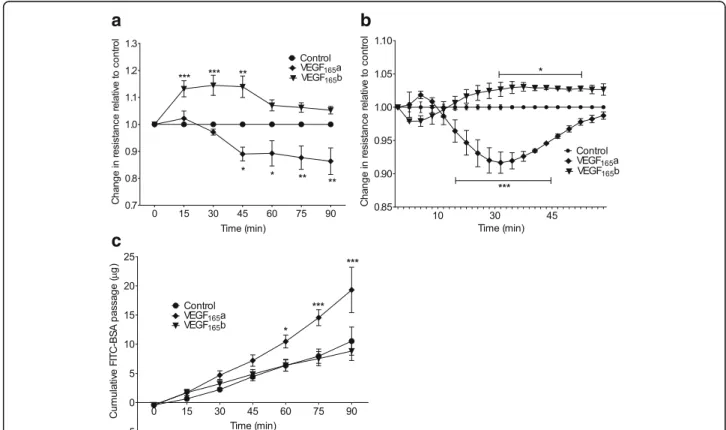
monolayers. VEGF165a significantly reduced the cell resistance (increased permeability) **p< 0.01, from 45 min onwards, in contrast, VEGF165b significantly increased re-sistance (decreased permeability) **p< 0.01 between 15 to 45 min compared with unstimulated control cells (Fig. 1a).
ECIS was utilised as another method to evaluate the ef-fect of VEGF isoforms on cell permeability. A similar re-sponse to that seen using the Endohm system was observed for HPMEC. VEGF165a induced an increase in cell monolayer permeability (***p< 0.001) in contrast to VEGF165b which induced a significant decrease (*p< 0.05) in the cell monolayer permeability (Fig. 1b).
The last experimental technique used to assess perme-ability was FITC-coupled BSA passage. The permeperme-ability of the HPMEC monolayer for FITC-BSA was monitored every 15 min up to 90 min by measuring the fluores-cence intensity of the medium in the lower compart-ment. For unstimulated control cells, the level of BSA in
the lower compartment increased slowly over time in a comparable manner to those treated with VEGF165b; no significant difference was observed between them in HPMEC (Fig. 1c). When the cells were cultured in the presence of VEGF165a, a significant (*p< 0.05) time-dependant increase in BSA permeability was detected after 45 min and persisted for at least 90 min, suggesting an increase in the cell monolayer permeability.
VEGF165a induces changes in VE-cadherin distribution
pattern and actin stress fibres in HPMEC compared to VEGF165b
Changes in cell permeability are closely related to changes in the cell-cell junction structures [18]. We went on to analyse potential cytoskeleton remodelling induced by VEGF isoforms. VE-cadherin has been particularly associ-ated with endothelial cell adherens junctions but may also contribute to change in tight junction activity [19, 20].
a
c
b
Fig. 1HPMEC stimulated with 20 ng/ml of VEGF165a, VEGF165b or without any stimulation (control).aParacellular permeability was quantified by
TEER using endohm-12 system in HPMEC cultured in inserts. VEGF165a reduced resistance (increased permeability) *p< 0.05 (45 min onwards) and
VEGF165b increased resistance (decreased permeability) ***p< 0.001 (15 to 45 min) compared to control.bECIS measurements on HPMEC show
that VEGF165a reduced the resistance (increased permeability) significantly***p< 0.001 in comparison to control; whereas VEGF165b had an opposite
effect with a significant increase (*p< 0.05) in the resistance (reduced permeability). Data expressed as a mean fold-change compared to control over time.cThe passage of FITC-coupled BSA across the monolayer of HPMEC was monitored over a period of 90 min. The fluorescence intensity of the aliquots was quantified and data was expressed as cumulative FITC-BSA over time. Concentration of FITC-BSA in the lower chamber slowly increased in both control cells and those stimulated with VEGF165b; in contrast cells stimulated with VEGF165a showed a significant increase in the passage of
Previously, a changed distribution to a zig-zag pattern in VEGF165a treated human umbilical vascular endothelial cells (HUVEC) was demonstrated using 100 ng/ml stimu-lation [20]. We repeated this experiment using HPMEC and showed similar results suggesting a significant change in VE-cadherin distribution between the cells stimulated with VEGF165a (Fig. 2a). Visible gaps ap-peared between the cells with an apparent zig-zag pat-tern of VE-cadherin distribution between adjacent cells. In contrast, stimulation with the VEGF165b isoform in-duced minimal change compared to untreated control cells suggesting it did not induce VE-cadherin reorgani-sation (Fig. 2a). In addition, changes in the actin struc-ture were observed between the stimulated and the unstimulated cells demonstrating an increase of stress fibres across the cells and more actin filopodia with VEGF165a. Again, in contrast, there was only a partial induction of actin filaments in the cell periphery and stress fibres by VEGF165b (Fig. 2b). These data led to the hypothesis that VEGF165a and VEGF165b activate different signalling pathways involved in permeability in HPMEC.
VEGF-R2 has been reported to induce most down-stream signalling effects through the tyrosine sites tyr1175 and tyr1214 [21, 22]. These two tyrosine sites play a crucial and direct role in the recruitment of adaptor proteins that activate multiple signalling path-ways such as proliferation, survival, migration and
permeability [23, 24]. Therefore, we studied VEGF iso-form induced phosphorylation of those tyrosine sites in HPMECs.
VEGF165a and VEGF165b induce differential
phosphorylation of the VEGF receptors at tyrosine 1175 and 1214 in HPMEC
VEGF-R2 phosphorylation at the tyrosine (tyr) 1175 site, was significantly induced by VEGF165a at 5 min and 10 min (p< 0.05) in HPMEC (Fig. 3a) and returned to control levels at 60 min post stimulation. In contrast, VEGF165b stimulation did not induce significant phos-phorylation at this site. Phosphos-phorylation of VEGF-R2 at the tyr1214 also reached a maximum at 5 min (p< 0.01) with VEGF165a and subsequently decreased as repre-sented in Fig. 3b. VEGF165b also induced significant phosphorylation of tyr1214 site at 5 min (p< 0.05) in HPMEC, in contrast to tyr1175. These differences in re-ceptor phosphorylation support the potential for differ-ential binding of other adaptor proteins in addition to the previously reported changes in co-receptor binding that has been shown to contribute to VEGF-induced vascular permeability.
Having identified these phosphorylation differences we wanted to identify specific pathways leading to cell per-meability which is less well characterised than those of other VEGF functional effects [7]. Further Western blot-ting of HPMEC was undertaken, to investigate the effect
a
b
Fig. 2aHPMEC were stimulated with 100 ng/ml of VEGF165a, VEGF165b or without stimulation (control) for 10 min. Then they were fixed, permeabilised
and immunostained for VE-cadherin. Control and VEGF165b: straight and linear distribution of VE-cadherin at the cell junctions were observed (seearrows).
VEGF165a: distribution of VE-cadherin in a zig-zag pattern with the appearance of gaps between the cells (seearrows) (magnification x40).bActin and
nucleus staining in HPMEC in different conditions. Control: actin filament distributed mainly at the cell periphery between the cell-cell junctions. VEGF165a:
disruption of the cortical actin frame with filopodia and stress fibre formation across the cells (arrows). VEGF165b: mixed field with the appearance of actin
of VEGF isoforms on the phosphorylation of several pro-teins previously suggested to be involved in specific functional downstream cellular effects. In parallel, we continued to use the Endohm assay to assess paracellu-lar permeability and utilised the scratch assay combining both migration and proliferation processes [25, 26] to assess the specificity of the functional effect of individual signalling pathway inhibitors.
Specific inhibition of pMEK1/2, p42/44MAPK and p38MAPK induction by VEGF165a and VEGF165b in
HPMEC does not have a differential effect on permeability and migration/proliferation pathways
The MEK and mitogen activated protein kinase (MAPK) pathways are among the most widely studied in VEGF biology and considered to have critical roles in cell pro-liferation and cell growth and differentiation [27, 28].
a
b
Fig. 3Activation of VEGF-R2 was determined with a phospho-specific antibody recognising tyrosine (Y) site 1175 and site 1214 in graph (a) and (b) respectively for cells stimulated with 20 ng/ml of VEGF165a (a), VEGF165b (b) or without stimulation (Ctl). Representative Western blot showing
We initially sought to determine whether inhibition of these proteins would have a specific functional effect i.e. inhibit proliferation/migration but not permeability as previously suggested [29].
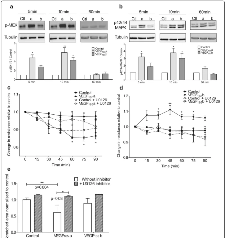
HPMEC showed a similar activation pattern of pMEK1/2 and p42/44MAPK proteins with an increase in the phosphorylation when treated with VEGF165a for 5 and 10 min that subsequently returned towards base-line at 60 min (Fig. 4-a&b respectively). VEGF165b had a delayed effect with significant phosphorylation of both proteins occurring at 10 min. The pMEK1/2 phosphoryl-ation in response to VEGF isoforms showed a significant increase at 5 and 10 min (p< 0.01) for the cells stimu-lated with VEGF165a compared to control. The max-imum response was observed at 10 min for both proteins. However, VEGF165b induced a significant in-crease of pMEK1/2 and p42/44MAPK phosphorylation at 10 min only. At 60 min the level of both phospho-proteins in the stimulated cells returns toward the con-trol levels.
U0126 is a compound reported to be a highly selective inhibitor of MEK1 and MEK2 and utilised to block the classical MAPK cascade in cells that leads to cell prolifera-tion [30]. U0126 inhibited the permeability effects of both VEGF165a and VEGF165b on HPMEC (Fig. 4c and d respectively). VEGF165a, but not VEGF165b, induced a significant increase in the cell migration into the“scratch” (p= 0.004) in comparison to control (Fig. 4e) that was inhibited byU0126. These data suggest that the MEK pathway is activated by both VEGF165a and VEGF165b but any functional divergence must occur downstream.
In addition, phosphorylation of p38MAPK was assessed as studies reported that VEGF stimulation on endothelial cells, also lead to activation of the Cdc42/p38MAPK pathway which triggers cytoskeletal modifications [31]. Similarly, to the other proteins studied, p38 MAPK phosphorylation was significantly increased by VEGF165a in HPMEC at 5 and 10 min (p< 0.05). Cells stimulated with VEGF165b showed a significant increase in p38MAPK phosphorylation (p= 0.04) compared to the untreated cells (Fig. 5a). Nevertheless, this phosphorylation was transient and returned to baseline levels by ten minutes. SB203580 is reported to specifically inhibit the activation of SAPK2/ p38 MAPK in cell-based assays but no other related kin-ase protein including MAPK family members [30]. When HPMEC were incubated with SB203580 (p38MAPK in-hibitor) again there was inhibition of VEGF165a and VEGF165b permeability effects (Fig. 5b and c). Cell prolif-eration/migration into the “scratch” was significantly in-creased (p= 0.002) only with VEGF165a stimulation and this effect was inhibited by SB203580 (Fig. 5d).
Having identified significant differences in the activa-tion kinetics of MEK, p42/44MAPK and p38MAPK by VEGF165a compared to VEGF165b (that reflected changes
in permeability but not specific to it alone), we then looked at the effect of VEGF isoforms on AKT also known as protein kinase B (PKB) protein. AKT has been shown to promote the proposed cell survival pathway mediated by the activation of PI3-kinase protein kinase [32] but it has been also associated with eNOS produc-tion which is closely associated with permeability [33].
Specific inhibition of AKT induction by VEGF165a and
VEGF165b in HPMEC does not have a differential effect on
permeability and survival pathways
VEGF165b induces a rapid and robust phosphorylation of pAKT at 5 min and 10 min (p< 0.01) in HPMEC with maximal AKT phosphorylation detected at 5 min (Fig. 5e). Stimulation with VEGF165a also induced a sig-nificant phosphorylation of AKT at 5 (p< 0.05) and 10 min (p< 0.01) and returned to control levels at 60 min. LY294002 compounds is an inhibitor of phos-phatidylinositol 3 kinase (PI3K) and inactivation of PI3K have been reported to lead to dephosphorylation of Akt that subsequently stop G1 cycle in cell growth and ul-timately lead to cell apoptosis [34, 35]. Co-culture with LY294002 to block AKT protein inhibited both the per-meability effects of VEGF165a and VEGF165b on HPMEC (Fig. 5 f and g). In contrast with the permeability assay where the cell monolayer was stable over the experiment (2 h), inhibition of AKT for long periods (24 h) induced cell death so we were unable to undertake the scratch assay as a migration/proliferation model.
Finally, we investigated the potential of the eNOS path-way for differential effects. There is a significant body of evidence to suggest that endothelial nitric oxide (synthe-sised by eNOS) may have a crucial role in causing hyper-permeability in response to pro-inflammatory agents such as VEGF [36, 37]. Therefore, the effects of the different VEGF isoforms on the eNOS phosphorylation in HPMEC were studied.
Specific inhibition of eNOS induction by VEGF165a and
VEGF165b in HPMEC does not have a differential effect on
permeability and nitric oxide signalling pathways
a
b
c
d
e
Fig. 4HPMEC were stimulated with 20 ng/ml of VEGF165a, VEGF165b or without any stimulation (Ctl/control). Representative western blots
showing VEGF stimulation at 5, 10 and 60 min plus loading control (α-tubulin) with densitometry (n= 3).a&bWestern blot of phosphorylated MEK1/2 (a) and phosphorylated p42/p44MAPK (b) in HPMEC. Stimulation with VEGF165a increased phosphorylation of MEK1/2 and p42/p44 at 5
and 10 min in contrast with VEGF165b which induced increased phosphorylation only at 10 min for both proteins.c&dMeasurement of cells
resistance by Endohm system. Dotted lines correspond to cell pre-incubated with pMEK inhibitor UO126. UO126 inhibited the effect of VEGF165a
(c) and the effect of VEGF165b (d).eGraphs represent the mean of the scratched area in HPMEC treated with UO126 inhibitor at 24 h relative to
control. VEGF165a but not VEGF165b stimulation significantly diminished scratched area (p= 0.004) and was inhibited by UO126 (p= 0.03). Densitometry
a
b
c
d
e
f
g
cells stimulated by VEGF165b (Fig. 6d). The inhibition of those effects induced by both VEGF165a and VEGF165b by L-NAME suggests that eNOS is not involved in the dif-ferential effect on permeability induced by these VEGF isoforms.
Discussion
The pulmonary endothelium is crucial to the regulation of the passage of solutes and molecules between the blood and the interstitial space of the lung, enabling close proximity of the vascular bed to the alveolar space for gaseous exchange to occur. Despite this, there is a
very limited understanding of the mechanisms involved in the regulation of pulmonary endothelial cell (EC) bar-rier function integrity, which is so essential for maintain-ing this critical function of the lung.
Vascular endothelial growth factor (VEGF) was originally described as both an angiogenic and a permeability factor [39] and its effects have previously been studied using the human umbilical vein endothelial cell (HUVEC) as the archetypal EC. Large organ functional differences are reflected in the variability of endothelial cell junction struc-ture and composition particularly relevant in the functional differences between the pulmonary and systemic circulation (See figure on previous page.)
Fig. 5HPMEC were stimulated with 20 ng/ml of VEGF165a, VEGF165b or without stimulation (Ctl/control). Representative western blots showing
VEGF stimulation at 5, 10 and 60 min plus loading control (α-tubulin) with densitometry (n= 3).aWestern blot of phosphorylated p-38MAPK in HPMEC. VEGF165a significantly increased phosphorylation of p-38MAPK at 5 and 10 min where VEGF165b only induced increased phosphorylation at
5 min.b&c: Measurement of HPMEC resistance by Endohm system. Dotted lines correspond to cell pre-incubated with p38 inhibitor SB203580. SB203580 inhibited the effect of VEGF165a (b) and the effect of VEGF165b (c).dGraphs represent the mean of the scratched area for HPMEC treated with
SB203580 inhibitor at 24 h relative to control. VEGF165a but not VEGF165b stimulation significantly diminished scratched area (p= 0.002) and was
inhib-ited by SB203580 (p= 0.02).eWestern blot of phosphorylated AKT in HPMEC. VEGF165a and VEGF165b significantly increased phosphorylation of p-Akt at
5 and 10 min.f&gMeasurement of HPMEC resistance by Endohm system. Dotted lines correspond to cell pre-incubated with AKT inhibitor LY294002. LY294002 inhibited the effect of VEGF165a (f) and the effect of VEGF165b (g). Densitometry and scratch assay analysed by Kruskal-Wallis test with post
hoc Dunn’s analysis. TEER measurement analysed using two-way ANOVA and Bonferroni post-test. All data were plotted as mean ± SEM (n= 3-6)
a b
c d
Fig. 6aRepresentative Western blot of phosphorylated p-eNOS in HPMEC (n= 3). VEGF165a significantly increased phosphorylation of eNOS at 5
and 10 min in contrast with VEGF165b stimulation which did not induce any changes.b&cMeasurement of HPMEC resistance by Endohm system. Dotted
lines correspond to cell pre-incubated with inhibitor. L-NAME inhibited the effect of VEGF165a (b) and the effect of VEGF165b (c).dGraphs represent the
mean of the scratched area for HPMEC treated with L-NAME inhibitor at 24 h relative to control. VEGF165a but not VEGF165b stimulation significantly
[40, 41]. To explore our hypothesis and its relationship to previous clinical studies (5, 6) it was important to study the response of human pulmonary microvascular endothelial cell (HPMEC) to VEGF isoforms.
Among all the pathological processes involved in. ARDS increases in lung vessel permeability are critical and non-redundant [42]. The measurement of perme-ability in this study has been undertaken only in-vitro models with self-evident limitations [43]. Transport of plasma proteins, cell and solutes across monolayers oc-curs paracellularly via specialised endothelial cell-cell junctions, or transcellularly by special transport mechan-ism including transcytosis, via transcellular channels or cell membrane transporter proteins.
Two types of inter-endothelial junction are present in the endothelium, adherens and tight junctions, the former being dominant in most vascular beds. The in-tegrity of the adherens junction is particularly critical for regulating paracellular permeability via homophilic ad-hesions between VE-cadherin molecules [19, 20]. Dis-ruptions of these domains lead to downstream events that result in organisational changes in the actin cyto-skeleton [44]. The transcellular pathway is responsible for the transport of larger molecules such as albumin across endothelial cell monolayers, classically via trans-cellular pores associated with caveolae and lipid rafts [45]. Traditionally these pathways have been considered independent but there is now a body of evidence show-ing interdependence [46]. Followshow-ing on from this, the in-vitro methods of measuring permeability that we have used, TEER (thought to reflect only paracellular perme-ability) and FITC-BSA (thought to only reflect transcel-lular mechanism) are recognised to have influences from crosstalk between both pathways [15].
We have demonstrated for the first time that VEGF165a and VEGF165b induce differing effects on the permeability of pulmonary microvascular endothelial cells. Specifically, VEGF165a induced an increase and VEGF165b a decrease in permeability. The receptor bind-ing and dimerisation domains are intact in the VEGFxxxb family of VEGF isoforms. However, in porcine aortic endothelial cells, VEGF165b has been shown to stimulate a unique pattern of VEGF-R2 tyrosine residue phosphor-ylation, contrasting with those activated by conventional isoforms suggesting activation of differing downstream signalling pathways in addition to partial agonist activity and changes in neuroplin-1 binding [22, 47, 48]. In this study, differing phosphorylation kinetics were clearly ob-served following stimulation by VEGF165a and VEGF165b using what we considered to be physiologically relevant concentrations of VEGF.
The differential effects of VEGF165a and VEGF165b on the vascular permeability in addition to those we have pre-viously shown and published on proliferation in HPMEC
(also repeated in the scratch experiments) and human al-veolar epithelial cells offer a potential paradigm to explain the apparent compartmentalisation of VEGF between the alveolar and vascular space and the apparent disparity of data relating to the role of VEGF in ARDS [5, 6, 12].
We identified that VEGF165a and VEGF165b lead to differential functional outcomes with VEGF165a increas-ing cell permeability in methods suggested to reflect both para and transcellular permeability and VEGF165b reducing paracellular permeability only. This suggested that the differences were due to divergence of signalling pathways and therefore potential targets for amelioration of outcome e.g. reducing permeability whilst preserving a pneumotropic effect. To verify this hypothesis, dif-ferent protein inhibitors have been used to look at their effect on the change in resistance reflecting the para-cellular permeability pathway of pulmonary microvascu-lar ECs.
The VEGF165a signalling pathways have been studied extensively in HUVEC although studies in HPMEC are limited [41]. We chose to use inhibitors of pMEK, P38 MAPK, PI3 kinase and eNOS proteins as these proteins have been suggested to be involved in VEGF signalling pathways and look at both permeability and proliferation/ migration in an attempt to identify a divergence in func-tionality and thus an opportunity for selective inhibition.
The use of L-NAME (eNOS inhibitor) and LY294002 (PI3K inhibitor) on HPMEC inhibited the effect of both VEGF165a and VEGF165b. Being part of the same sig-nalling pathway these results suggest that VEGF cell paracellular permeability involves the phosphoinositide 3-kinase–AKT pathway, which then further phosphory-lates and activates endothelial nitric oxide synthase (eNOS) [49]. Also, the inhibition of pMEK and p38 MAPK did not affect VEGF isoforms activity on the pro-liferation and paracellular permeability pathway in HPMEC. In summary, the inhibitors chosen did not allow for the identification of specific differential path-ways between VEGF165a and VEGF165b. Further studies of other pathways are required in order to unravel the molecules responsible for the differential permeability effects of VEGF isoforms such as Src kinase pathway and its role in the regulation of endothelial-barrier integ-rity as demonstrated recently by Gao et al. [50].
Conclusion
of the ACM barrier function, of particular significance in the lung environment. On the other hand, depending on the isoforms present, VEGF may also be a protective agent, the data presented showing a decrease in perme-ability with VEGF165b exposure. Further work will need to be undertaken to clearly identify the divergence in the per-meability signalling pathway induced by both isoforms and the potential protective proprieties of VEGF165b.
Abbreviations
°C:Degrees celsius; ACM: Alveolar capillary membrane; AKT: AKT8 virus oncogene cellular homolog; ANOVA: Analysis of variance; ARDS: Acute respiratory distress syndrome; BSA: Bovine serum albumin; Ctl: Control; DAPI: 4′,6-diamidino-2-phenylindole; EC: Endothelial cell; ECIS: Electrical cell impedance system; eNOS: Endothelial nitric oxide synthase; EVOM: Epithelial voltohmmeter; FITC: Fluorescein isothiocyanate; HPMEC: Human pulmonary microvascular endothelial cell; HUVEC: Human umbilical vein endothelial cell; L-NAME: NG-Nitro-L-arginine Methyl Ester; MAPK: Mitogen-activated protein kinase; MEK: Mitogen/extracellular signal-regulated kinase; Min: Minutes; p38MAPK: p38 mitogen-activated protein kinase; PI3K: Phosphoinositide 3-kinase; SDS-PAGE: Sodium dodecyl sulphate polyacrylamide gel electrophoresis; SEM: Standard error of the mean; SRC: V-scr sarcoma (Schimidt-Ruppin A-2) viral oncogene homolog; TEER: Transendothelial electrical resistance; Tyr: Tyrosin; UK: United Kingdom; USA: United States of America; VE-Cadherin: Vascular endothelial cadherin; VEGF: Vascular endothelial growth factor; VEGF-R1: Vascular endothelial growth factor– receptor 1; VEGF-R2: Vascular endothelial growth factor–receptor 2; WB: Western blotting
Acknowledgements
Not applicable.
Funding
This study was funded by an unrestricted Novartis PhD Studentship to KO and a Welcome Clinical training fellowship to SL. The funding organisations had no role in the design of the study, collection, analysis, and interpretation of the data, or writing of the manuscript.
Availability of data and materials
All data generated during this study are included in this published article [and its supplementary information files].
Authors’contributions
KO, GIW and ABM designed the experiments; KO and TB carried out experiments and collected data. CJ, SBO helped with analysis and interpretation of the data. KO, GIW and ABM contributed to data analysis and prepared the manuscript.
Competing interests
The authors declare that they have no competing interests.
Consent for publication
Not applicable.
Ethics approval and consent to participate
Not applicable.
Publisher’s Note
Springer Nature remains neutral with regard to jurisdictional claims in published maps and institutional affiliations.
Author details
1Academic Respiratory Unit, School of Clinical Sciences, University of Bristol,
Bristol, UK.2Bristol Renal, School of Clinical Sciences, University of Bristol,
Bistol, UK.
Received: 20 February 2017 Accepted: 30 May 2017
References
1. Abadie Y, Bregeon F, Papazian L, Lange F, Chailley-Heu B, Thomas P, Duvaldestin P, Adnot S, Maitre B, Delclaux C. Decreased VEGF concentration in lung tissue and vascular injury during ARDS. Eur Respir J. 2005;25:139–46. 2. Maitre B, Boussat S, Jean D, Gouge M, Brochard L, Housset B, Adnot S,
Delclaux C. Vascular endothelial growth factor synthesis in the acute phase of experimental and clinical lung injury. Eur Respir J. 2001;18:100–6. 3. Kaner RJ, Ladetto JV, Singh R, Fukuda N, Matthay MA, Crystal RG. Lung
overexpression of the vascular endothelial growth factor gene induces pulmonary edema. Am J Respir Cell Mol Biol. 2000;22:657–64.
4. Kaner RJ, Crystal RG. Compartmentalization of vascular endothelial growth factor to the epithelial surface of the human lung. Mol Med. 2001;7:240–6. 5. Thickett DR, Armstrong L, Millar AB. A role for vascular endothelial growth factor in acute and resolving lung injury. Am J Respir Crit Care Med. 2002; 166:1332–7.
6. Thickett DR, Armstrong L, Christie SJ, Millar AB. Vascular endothelial growth factor may contribute to increased vascular permeability in acute respiratory distress syndrome. Am J Respir Crit Care Med. 2001;164:1601–5.
7. Koch S, Tugues S, Li X, Gualandi L, Claesson-Welsh L. Signal transduction by vascular endothelial growth factor receptors. Biochem J. 2011;437:169–83. 8. Houck KA, Ferrara N, Winer J, Cachianes G, Li B, Leung DW. The vascular
endothelial growth factor family: identification of a fourth molecular species and characterization of alternative splicing of RNA. Mol Endocrinol. 1991;5: 1806–14.
9. Harper SJ, Bates DO. VEGF-a splicing: the key to anti-angiogenic therapeutics? Nat Rev Cancer. 2008;8:880–7.
10. Medford AR, Ibrahim NB, Millar AB. Vascular endothelial growth factor receptor and coreceptor expression in human acute respiratory distress syndrome. J Crit Care. 2009;24:236–42.
11. Bates DO, Cui TG, Doughty JM, Winkler M, Sugiono M, Shields JD, Peat D, Gillatt D, Harper SJ. VEGF165b, an inhibitory splice variant of vascular endothelial growth factor, is down-regulated in renal cell carcinoma. Cancer Res. 2002;62:4123–31.
12. Varet J, Douglas SK, Gilmartin L, Medford AR, Bates DO, Harper SJ, Millar AB. VEGF in the lung: a role for novel isoforms. Am J Physiol Lung Cell Mol Physiol. 2010;298:L768–774.
13. Bevan HS, van den Akker NM, Qiu Y, Polman JA, Foster RR, Yem J, Nishikawa A, Satchell SC, Harper SJ, Gittenberger-de Groot AC, Bates DO. The alternatively spliced anti-angiogenic family of VEGF isoforms VEGFxxxb in human kidney development. Nephron Physiol. 2008;110:p57–67. 14. Van Nieuw Amerongen GP, Van Hinsbergh VW. Determination of the
endothelial barrier function in vitro. Methods Mol Biol. 1999;96:183–9. 15. Wegener J, Seebach J. Experimental tools to monitor the dynamics of
endothelial barrier function: a survey of in vitro approaches. Cell Tissue Res. 2014;355:485–514.
16. Cereijido M, Gonzalez-Mariscal L, Contreras RG, Gallardo JM, Garcia-Villegas R, Valdes J. The making of a tight junction. J Cell Sci Suppl. 1993;17:127–32. 17. Wegener J, Sieber M, Galla HJ. Impedance analysis of epithelial and
endothelial cell monolayers cultured on gold surfaces. J Biochem Biophys Methods. 1996;32:151–70.
18. Forster C. Tight junctions and the modulation of barrier function in disease. Histochem Cell Biol. 2008;130:55–70.
19. Wallez Y, Huber P. Endothelial adherens and tight junctions in vascular homeostasis, inflammation and angiogenesis. Biochim Biophys Acta. 1778; 2008:794–809.
20. Esser S, Lampugnani MG, Corada M, Dejana E, Risau W. Vascular endothelial growth factor induces VE-cadherin tyrosine phosphorylation in endothelial cells. J Cell Sci. 1998;111(Pt 13):1853–65.
21. Smith GA, Fearnley GW, Tomlinson DC, Harrison MA, Ponnambalam S. The cellular response to vascular endothelial growth factors requires co-ordinated signal transduction, trafficking and proteolysis. Biosci Rep. 2015;35:e00253. 22. Kawamura H, Li X, Harper SJ, Bates DO, Claesson-Welsh L. Vascular
endothelial growth factor (VEGF)-A165b is a weak in vitro agonist for VEGF receptor-2 due to lack of coreceptor binding and deficient regulation of kinase activity. Cancer Res. 2008;68:4683–92.
24. Dayanir V, Meyer RD, Lashkari K, Rahimi N. Identification of tyrosine residues in vascular endothelial growth factor receptor-2/FLK-1 involved in activation of phosphatidylinositol 3-kinase and cell proliferation. J Biol Chem. 2001;276: 17686–92.
25. Zahm JM, Kaplan H, Herard AL, Doriot F, Pierrot D, Somelette P, Puchelle E. Cell migration and proliferation during the in vitro wound repair of the respiratory epithelium. Cell Motil Cytoskeleton. 1997;37:33–43.
26. Liang CC, Park AY, Guan JL. In vitro scratch assay: a convenient and inexpensive method for analysis of cell migration in vitro. Nat Protoc. 2007;2:329–33. 27. Zachary I. VEGF signalling: integration and multi-tasking in endothelial cell
biology. Biochem Soc Trans. 2003;31:1171–7.
28. Crews CM, Alessandrini A, Erikson RL. The primary structure of MEK, a protein kinase that phosphorylates the ERK gene product. Science. 1992; 258:478–80.
29. Issbrucker K, Marti HH, Hippenstiel S, Springmann G, Voswinckel R, Gaumann A, Breier G, Drexler HC, Suttorp N, Clauss M. p38 MAP kinase–a molecular switch between VEGF-induced angiogenesis and vascular hyperpermeability. FASEB J. 2003;17:262–4.
30. Davies SP, Reddy H, Caivano M, Cohen P. Specificity and mechanism of action of some commonly used protein kinase inhibitors. Biochem J. 2000; 351:95–105.
31. Lamalice L, Houle F, Huot J. Phosphorylation of Tyr1214 within VEGFR-2 triggers the recruitment of Nck and activation of Fyn leading to SAPK2/p38 activation and endothelial cell migration in response to VEGF. J Biol Chem. 2006;281:34009–20.
32. Song G, Ouyang G, Bao S. The activation of Akt/PKB signaling pathway and cell survival. J Cell Mol Med. 2005;9:59–71.
33. Fulton D, Gratton JP, McCabe TJ, Fontana J, Fujio Y, Walsh K, Franke TF, Papapetropoulos A, Sessa WC. Regulation of endothelium-derived nitric oxide production by the protein kinase Akt. Nature. 1999;399:597–601. 34. Bondar VM, Sweeney-Gotsch B, Andreeff M, Mills GB, McConkey DJ.
Inhibition of the phosphatidylinositol 3′-kinase-AKT pathway induces apoptosis in pancreatic carcinoma cells in vitro and in vivo. Mol Cancer Ther. 2002;1:989–97.
35. Vlahos CJ, Matter WF, Hui KY, Brown RF. A specific inhibitor of phosphatidylinositol 3-kinase, 2-(4-morpholinyl)-8-phenyl-4H-1-benzopyran-4-one (LY294002). J Biol Chem. 1994;269:5241–8.
36. Fukumura D, Gohongi T, Kadambi A, Izumi Y, Ang J, Yun CO, Buerk DG, Huang PL, Jain RK. Predominant role of endothelial nitric oxide synthase in vascular endothelial growth factor-induced angiogenesis and vascular permeability. Proc Natl Acad Sci U S A. 2001;98:2604–9.
37. Hatakeyama T, Pappas PJ, Hobson 2nd RW, Boric MP, Sessa WC, Duran WN. Endothelial nitric oxide synthase regulates microvascular hyperpermeability in vivo. J Physiol. 2006;574:275–81.
38. Kubes P, Suzuki M, Granger DN. Nitric oxide: an endogenous modulator of leukocyte adhesion. Proc Natl Acad Sci U S A. 1991;88:4651–5.
39. Ferrara N, Gerber HP, LeCouter J. The biology of VEGF and its receptors. Nat Med. 2003;9:669–76.
40. Aird WC. Endothelial cell heterogeneity. Cold Spring Harb Perspect Med. 2012;2:a006429.
41. Becker PM, Verin AD, Booth MA, Liu F, Birukova A, Garcia JG. Differential regulation of diverse physiological responses to VEGF in pulmonary endothelial cells. Am J Physiol Lung Cell Mol Physiol. 2001;281:L1500–1511. 42. Herold S, Gabrielli NM, Vadasz I. Novel concepts of acute lung injury and
alveolar-capillary barrier dysfunction. Am J Physiol Lung Cell Mol Physiol. 2013;305:L665–681.
43. Bates DO. Vascular endothelial growth factors and vascular permeability. Cardiovasc Res. 2010;87:262–71.
44. Giannotta M, Trani M, Dejana E. VE-cadherin and endothelial adherens junctions: active guardians of vascular integrity. Dev Cell. 2013;26:441–54. 45. Garrido-Urbani S, Bradfield PF, Lee BP, Imhof BA. Vascular and epithelial
junctions: a barrier for leucocyte migration. Biochem Soc Trans. 2008;36:203–11. 46. Komarova Y, Malik AB. Regulation of endothelial permeability via paracellular
and transcellular transport pathways. Annu Rev Physiol. 2010;72:463–93. 47. Cebe Suarez S, Pieren M, Cariolato L, Arn S, Hoffmann U, Bogucki A, Manlius
C, Wood J, Ballmer-Hofer K. A VEGF-a splice variant defective for heparan sulfate and neuropilin-1 binding shows attenuated signaling through VEGFR-2. Cell Mol Life Sci. 2006;63:2067–77.
48. Becker PM, Waltenberger J, Yachechko R, Mirzapoiazova T, Sham JS, Lee CG, Elias JA, Verin AD. Neuropilin-1 regulates vascular endothelial growth factor-mediated endothelial permeability. Circ Res. 2005;96:1257–65.
49. Zachary I, Gliki G. Signaling transduction mechanisms mediating biological actions of the vascular endothelial growth factor family. Cardiovasc Res. 2001;49:568–81.
50. Gao F, Sabbineni H, Artham S, Somanath PR. Modulation of long-term endothelial-barrier integrity is conditional to the cross-talk between Akt and Src signaling. J Cell Physiol. 2017. doi:10.1002/jcp.25791. [Epub ahead of print]
• We accept pre-submission inquiries
• Our selector tool helps you to find the most relevant journal • We provide round the clock customer support
• Convenient online submission • Thorough peer review
• Inclusion in PubMed and all major indexing services • Maximum visibility for your research
Submit your manuscript at www.biomedcentral.com/submit