The anatomy of the body wall and appendages in Arenicola marina L., Arenicola claparedii Levinson and Arenicola ecaudata Johnston
45
0
0
Full text
(2) G. P. WELLS. 2. genera. only,. Arenicolaand Branchiomalda1lP. The first is made up of eight. species, to which Berkeley & Berkeley (1939) have subsequently added a ninth; they fall quite clearly, from an anatomical point of view, into three groups. The second consists only of a single species of small and aberrant animals. Now to describe only Arenicola marina would result in a work of very limited usefulness, more particularly as the species is restricted to the cooler shores of the northern hemisphere; on the other hand, by including the three species marina L., claparedii Levinsen1 and ecaudata Johnston, each representing one of the sections into which the genus naturally falls, an idea can be given of the general organization and also the range of variation found in this world-wide group. I hope later to publish notes on the other species; briefly, grubii most nearly resembles ecaudata, assimilis is most closely related to claparedii, though differing from it in several important points, and the rest group themselves with marina. The descriptions are based mainly on material collected by myself-marina at Plymouth, at Bangor, North Wales, and at Thorpe Bay on the Thames Estuary; claparedii at Naples, where my expenses -were partly defrayed by a grant from the Challenger Society; and ecaudata at Plymouth. I have also a small number of claparedii from the Pacific Coast of U.S.A., sent, very illpreserved, by an American dealer. By the courtesy of Mr H. W. Parker I have been allowed to examine the collection of Arenicolidae in the British Museum (Natural History); this remains, with a few additions, substantially as catalogued by Ashworth (1912). The following study is concerned with gross morphology, not histological detail. As far as possible, some indication will be given of the functional significance of the structures described. With marina, the habits of the worm are well known and such interpretations can often be made. This species lives in great numbers in muddy sand flats, where the piles of sandy cylinders, its faeces, and the saucer-shaped or funnel-shaped depressions, caused by its eating away the sand below, are familiar to everybody. It sometimes occurs in less typical habitats, for example, in gravel or among stones, provided there is a certain amount of muddy material to eat. Under favourable conditions, it inhabits the same burrow for weeks or even months at a stretch, swallowing the sand at one end of the burrow and depositing it as faeces at the other. The. form of the burrow, and the behaviour of the worm, have been described in detail elsewhere (Wells, 1945, 1949b, 1950; Newell, 1948). 1 A. claparedii I.evinsen is probably identical with A. pusilla Quatrefages. The original definition of the latter species is inadequate and conflicts with the characters of the. type specimen. The type was examined by Ashworth, who identified it as an incomplete specimen of claparedii, atypical as to the number of nephridia. He therefore merged both species under the older name of pusilla (Ashworth, 1912). The great majority oflater writers term it claparedii, ~d Fauvel (1927) explicity rejects Ashworth's proposal, not denying the identity of the two species, but because of the inadequacy of the definition of pusilla and the aberration of the type specimen..
(3) ANATOMY. OF 4RENICOLA. 3. Of claparedii, Ashworth (1912) wrote: 'The general habits of this species, which the writer had an opportunity-of observing in Naples for some weeks, are similar to those of A. marina. Examples taken by Prof. A. D. Howard in Puget Sound were found generally in ordinary sandy beaches, but two larger specimens were burrowing in a coarse gravelly and rocky beach.' Describing the occurrence of claparedii on Vancouver Island, Berkeley & Berkeley (1932) . write: 'It evidently occurs over large stretches of the sand exposed at low tide at Long Bay, judging by the enormous number of casts to be found.' Takahashi (1934) describes it as living in U- or V-shaped burrows, of depth 15-25 cm., the openings 3-10 cm. apart; in certain bottoms, the burrows of marina may be very like this (Wells, 1945). Now claparedii, while resembling marina in its general structure, differs in two points that one would expect to be of great functional importance; it lacks statocysts, and it lacks giant nerve fibres. The similarity of its way of life to that of marina, which the above citations suggest, makes these differences the more surprising. The breeding habits of claparedii are rather different in Canada and in Japan, according to the accounts of Guberlet (1934) and Okuda (1938). For ecaudata, we have the following account by Ashworth (1912): 'It occurs in the littoral zone but chiefly in sandy, gravelly or muddy material among stones, or in clefts at the base of the rocks in the de,bris formed by the breaking down of the latter. A considerable am01.lnt of organic matter is generally present in the material in which the worm lives. The burrows of A. ecaudata.. .are oblique or sinuous cavities, lined with a fair amount of mucus, and situated a few inches below the surface in gravel or between rocks and stones. The castings of the worm, being composed of coarse material having little coherence, soon fall apart. The well-known signs-the sandrope-like heap of castings and the funnel-like depression in the sand-which indicate the presence of A. marina on countless sandy beaches, have no good counterparts in the case of the ecaudate species, in which both the castings and the mouth of the burrow are inconspicuous among their surroundings.' Fauvel (1899a) comments on the occurrence of ecaudata in black, often foetid, sandy material between rocks at Cherbourg; the galleries are more or less sinuous and often horizontal; their walls are never yellow, as those of marina are. The worms are found at Plymouth in just such situations as these authorities describe, and it appears that the striking differences of structure between marina and ecaudata are paralleled by equally striking differences of habit. METHODS. Preparation of worms for dissection Ashworth (19°4) recommends that lugworms should be killed with chloroform and dissected fresh,- under sea water, 'as soon as possible after they are taken from the sand'. I find that formalin material, dissected within a week or 1-2.
(4) 4. G. P. WELLS. so ofki11ing, is very much better. Fresh worms bleed easily and freely, while in formalin material the blood sets into a firm, uniform gel, so that such operations as the removal of the whole or part of a ventricle can be performed with ease. Moreover, the septa and other membranes of fresh worms are so transparent that their relations are hard to make out. The slight cloudiness which appears in formalin material is a great improvement. The animals used in the present study were killed in either of two attitudes, 'relaxed' or 'distended'. Relaxed worms. The worms are narcotized by immersion in a large volume of 7t % MgCl2. The time taken varies with the size of the worm; marina about 10 or 15 cm. long is fully relaxed in 3t or 4 hr. They are then transferred to a bath of 7t % MgCl2 containing 4 % formaldehyde. At this stage, they may show slight movements, so it is as well to do the killing in a rectangular tank, and, if necessary, to straighten the worms against the side with a glass rod. They die in a very natural configuration. The body is straight (except for a slight ventral curvature of the first few segments). The proboscis is generally withdrawn. The worms can be stored in formalin until required. The organs usually retain quite a lot of their colour for some days, and even after months of preservation, such material is easy to dissect. Distended worms. The worms are narcotized as above, then distended, one by one, in the following way. A cannula is connected by rubber tubing to afunnel; the whole is filled with the Mg-formalin mixture, and the tubing is closed with a screw clamp. The level of fluid in the funnel should be 15-.20 cm. above the bench. The cannula is then tied into the tail, pointing forwards; it must go into the coelome, not 'the gut, and the way to ensure this is to insert it into an incision made in the lateral aspect of the tail. The worm is then tumbled into a long tank of the Mg-formalin mixture, and immediately distended with the same solution, by opening the screw clamp. A surprising degree of stretch is produced, and although the resulting attitude is highly unnatural, the distension'straightens out the septa and other organs and greatly facilitates the study of their relationships. The proboscis is fully extruded. Preservation for museum purposes As many of the specimens to be found in museum collections are ill-preserved, and in very distorted attitudes, so that even their external characters are hard to make out, it may here be pointed out that worms killed in the 'relaxed' attitude, as directed above, are very suitable for museum purposes. Alcohol makes the worms rather hard, and they may easily break on handling. Serial sections Before sectioning, the worms were killed in Mg-formalin in the relaxed attitude, then at once t;ransferred to Susa. The following techniques were found useful: '.
(5) ANATOMY. OF ARBNICOLA. 5. (i) 15 JL paraffin sections, stained with haematoxylin-eosin or Hansen's trioxyhaematin followed by Mallory's phosphomolybdic acid-aniline blueorange G mixture, and mounted in balsam. (ii) 100 JL celloidin sections, very lightly coloured with haematoxylin and. orange G, and mounted in balsam without removal of the celloidin. (iii) 300 JL celloidin sections, mounted unstained in glycerine jelly without removal of the celloidin. . In general, and especially when dealing with the front end of the worm, the horizontal plane is more useful than the sagittal.. Special methods Polarized light is invaluable for working out the arrangement of the muscles. It can be used on sections, or on such objects as pieces of the body wall of distended worms, spread out fiat and cleared. To avoid the necessity of turning the slide on the stage, which makes the working out of complicated muscular networks rather tricky, I have mounted two Polaroid disks, 7 in. in diameter, on a common vertical axle. One goes above, and the other below, the stage of my binocular microscope; by turning the axle, the plane of polarization of the light can be rotated without disturbing the specimen. Spectacular preparations of the vascular system can be made by choosing unpigmented or lightly pigmented worms, distending them, then rapidly dehydrating the whole worms and clearing them in benzyl alcohol, in which. theyarekept.. .. When studying the movements of living worms, they should be placed in glass tubes; a suitable arrangement for this purpose was described elsewhere (Wells, 1945). THE MAIN REGIONS OF THE BODY. Arenicola shows very clearly the tendency, which generally appears in sedentary polychaetes, to differentiate the chain of fundamentally similar segments, of which their body consists, into distinctively specialized regions. The common lugworm, A. marina, has 19 segments with parapodia (rarely 20), and, although these segments show certain local specializations (gills restricted to segments vii-xix, nephridia restricted to iv-ix, etc.),1 they are all built on a very evident metameric plan. These 19 segments together occupy the middle of the body and form a comparatively unspecialized region from which the two extremities stand out in marked contrast. Anterior to the first chaetigerous segment, there is a short, bluntly conical region, composed of the prostomium and one or two segments which have lost their appendagesone according to Lillie (1905), and two according to Ashworth (1904, 1912). The whole of this region is welded into a functional and structural unit, mainly in connexion with proboscis activity, and its metamerism is hard to make out. 1 Roman numbers are used to identify particular segments..
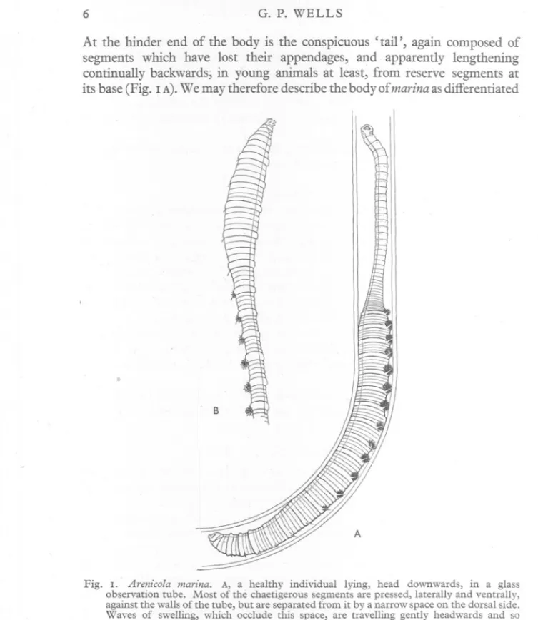
(6) G. P. WELLS. 6. At the hinder end of the body is the conspicuous' tail', again composed of segments which have lost their appendages, and apparently lengthening continually backwards; in young animals at least, from reserve segments at its base (Fig. I A).We may therefore describe the body of marina as differentiated. B. A. Fig. I. Arenico/a marina. A, a healthy individual lying, head downwards, in a glass observation tube. Most of the chaetigerous segments are pressed, laterally and ventrally, against the walls of the tube, but are separated from it by a narrow space on the dorsal side. Waves of swelling, which occlude this space, are travelling gently headwards and so driving water through the tube; one such wave is passing the 5th and 6th gills. B, the 'clubbed' attitude, which often appears in moribund specimens.. into three very distinct regions-an achaetous anterior region, or 'head', a chaetigerous middle region, or 'trunk', and an achaetous posterior region or 'tail'. The middle region shows various subsidiary differentiations, with.
(7) ANATOMY. OF ARENICOLA. 7. regard to the distribution of gills, nephridia, etc., among its component segments. The body of claparedii resembles, in all the above respects, that of marina. In ecaudata, the differentiation between' head' and' trunk' occurs as in the other species, but the specialized third region appears, at first sight, to be lacking. The worm has a great number of segments and the parapodia and gills continue as far as, or nearly as far as, the hinder extremity. There are, however, indications of a differentiation corresponding to, though less profound than, that between' trunk' and 'tail' in the other species (p. 37). The above method of describing the regional organization of Arenicola differs from that generally adopted in the literature. Audouin & Milne Edwards (1834) wrote of marina as 'compose de trois portions assez distinctes: l'une anterieure, ordinairement renflee et ne portant pas de branchies, une moyenne, etroite et branchifere,' et une posterieure, apode'. The anterior region, in this account, includes the' head' and the first six chaetigerous segments. The majority of subsequent writers, down to the present day, have adopted this method of subdividing the worm. Ashworth (19°4) instructs his students to 'note the shape of the worm; its division into an anterior abranchiate chaetigerous portion, middle branchiate chaetigerous region and posterior achaetous and abranchiate tail'. Fauvel (1927) describes marina thus: 'Region anterieure a 6 segments uncinigeres 'abranches. Region abdominale a 13 setigeres, branchiferes. Region caudale achete et abranche de longueur variable, fragile.' The gills have also been used to characterize the regions of ecaudata. 'Le corps', wrote Fauvel (1899a) of this species, 'se divise en deux parties: 1° la region anterieure ou thoracique qui comprend Ie prostomium, Ie segment buccal, un segment post-buccal achete et15 ou 16 segments pourvus de parapodes et de tores uncinigeres mais abranches; 2° la region abdominale dont tous les segments portent des parapodes et des branchies sauf parfois les 1 a 7 derniers qui sont abranches mais toujours setigeres.' It seems to the writer, that the method of subdividing the body with reference to the gills is rather misleading. Of the various divergent specializations which differentiate the segments of Arenicola from each other, some are extremely constant and fixed in position, not only from individual to individual but also from species to species. Such, for example, are the persistence of septa i, iii and iv of the middle region as, the well-known 'diaphragms', or the elaboration of the vessels of the (vanished) septum vii to form the ventricles, closely applied to the gut and separating its oesophageal from its gastric part. Others, on the other hand, fluctuate; for example, the nephridia are always restricted to a limited number of segments, but the number and position along the body of the segments concerned varies, not only from species to species but also to some extent from individual to individual. When seeking for criteria of regional differentiation, one should clearly choose characters of the former kind; yet the distribution of the gills, which so many.
(8) 8. G. P. WELLS. authorities use, is of the latter. In the' caudate' species, i.e. in all those with an achaetous 'tail', the most anterior gill is typically on segment vii, and the division between abranchiate and branchiate regions therefore coincides with the position of the hearts and boundary between oesophagus and stomach. In the so-called' ecaudate' species (ecaudata and grubii), the most anterior gill lies several segments farther back, though the cardiac and enteric differentiations remain at the same level as before. Even in the caudate species, including marina, 'the first gill is almost invariably small, and in a considerable percentage of examples, is reduced to minute proportions or is absent' (Ashworth, 1912). The same is true of ecaudata. Evidently, the boundary between abranchiate and branchiate regions is by no means a rigidly fixed one, and when more constant intersegmental differentiations are available, there is no justification for subdividing the body on the basis of the distribution of the gills. The division into' head', 'trunk' and 'tail', described above, is undoubtedly the most profound of the local specializations exhibited by Arenicola, and is merely obscured by throwing together the 'head' and the first few' trunk' segments into an 'anterior abranchiate chaetigerous portion' . In conclusion, a word may be said about the 'renflee' and 'etroite' of Audouin & Milne Edwards' description, cited above. One sometimes finds lugworms in which the first half a dozen or so segments are distended and the rest of the trunk is narrowed by contraction of the circular muscles. The worm as a whole is therefore club-shaped (Fig. I B). One never sees this attitude in vigorous, healthy worms, but only in dead or moribund specimens. It is very common in worms which have been badly collected, for example, in a hot, overcrowded jar. I believe its assumption to be an irreversible process, and a sign of approaching death; if any of the worms in my stock tanks exhibit it, they are thrown away before they die and foul the water. The point would be hardly worth mentioning, were it not that' clubbed' worms have appeared very often in the literature. The' renflee' and 'etroite' in Audouin & Milne Edwards's description, together with their PI. 8, fig. 8, show unmistakably that their worms were in the 'clubbed' attitude. Grube (1851) also refers to the anterior part of the body as 'mehr oder minder aufgebUiht'. More recently, a beautifully drawn figure of a 'clubbed' worm was published by Cunningham & Ramage (1888) and reproduced in the wellknown Handbuch of Kiikenthal-Krumbach (Hempelmann, 1934, p. 191). A striking example is to be found in the Cambridge Natural History (Benham, 1896, p. 333). In healthy worms, however, or in those prepared by the Mg-formalin method, no trace of' clubbing' can be seen. When we bear in mind, first, that the part of the body which distends in this abnormal attitude is also the pre-cardiac or 'anterior abranchiate' part, and, secondly, that many of the early authorities based their descriptions on specimens thus distorted, we may infer that the 'clubbed' attitude has some responsibility,. ..
(9) ANATOMY. OF ARENICOLA. 9. historically, for the idea that the boundary between abranchiate and branchiate regions represents a differentiation of major importance. THE BODY WALL AND ApPENDAGES. The account of the body wall follows the division of the body as a whole into three regions, as explained in the last section. The general plan of the body wall, common to all the regions, will be described first. THE GENERAL. STRUCTURE. OF THE BODY WALL. The body wall is divided externally into annuli, separated by interannular grooves. The position of the nerve cord is marked externally by a conspicuous, pale ventral groove, in marina and claparedii but not in ecaudata. The body wall consists of the following layers: (i) epidermis, (ii) subepidermal connective tissue, (iii) circular muscle, (iv) a layer of intermuscular connective tissue, which is brought out prominently by the aniline blue of Mallory's triple stain, (v) longitudinal muscle, (vi) coelomic epithelium. A series of oblique muscles, of the usual polychaete type, is generally present (Fig. 2). The body wall is ricWy vascularized, and one can generally.see in sections that the blood vessels lie in tubular spaces; these are extensions of the coelome, penetrating into the body wall. The circular muscle layer is interrupted by a radial partition of connective tissue at each interannular groove (Fig. 5 C-E, p. 17). According to Lillie (1905), the circular muscles develop very much later than the longitudinal in A. cristata Stimpson-a species very similar to marina. If one takes a piece of body wall from a distended specimen, clears it, spreads it flat on a slide, and then examines it between crossed polaroids, rotating the slide relative to the plane of polarization, one finds that the whole of the musculature at any given point on the body wall, both circular and longitudinal, blacks out in the same position. This means that the musculature of the general body surface consists only of these two series of fibres, whose molecules are orientated truly at right angles to each other. There can be no diagonal or spirally running fibres, as occur, for example, in several Oligo chaeta. The fibres of the longitudinal layer are grouped, in Imarina and claparedii, into a great number of longitudinal columns~ each covered by coelomic epithelium. These columns branch and anastomose with their neighbours, and form a conspicuous and characteristic background when one dissects either of these species (Fig. 3 A-C). In ecaudata, on the other hand, the body wall presents a smooth appearance internally; the longitudinal layer is covered over by a continuous peritoneal sheet which is only occasionally perforated or grooved to accommodate a blood vessel (Fig. 3 D). In all species, certain lines of separation can be traced in the longitudinal layer, which are important anatomical landmarks. They are usually evident in. ~.
(10) G. P. WELLS. 10. dissections, and are especiallyobvious in distended material. They appear, in ecaudata,as longitudinal clefts in the layer, and in marina and claparediias deep grooves between adjacent muscle columns, across which anastomoses seldom or never occur. The lines are (Fig. 2): (i) the ventral line, in which the nerve cord (n.c.) lies; (ii) the nephridial lines (neph.l.), at the level of the nephridiopores; (ill) the notopodiallines, (notop.l.), through which the inner ends of the notopodia protrude into the body cavity; and (iv) the dorsal line dors.l. c;rc.m.. ep.&c.t. stom.. notop./ong.v. in notop./.. neph. neph./ong.v. in neph.l.. obf.m.. fot.neur.v.. n.C.. Fig. 2. Arenicola marina. Transverse section of an ordinary annulus, drawn from a section slightly anterior to the eighth chaetigerous annulus. For explanation of the lettering on the figures, see list on p. 44.. (dors.l.), into which the dorsal mesentery, where present, is inserted. The ventral, notopodial and dorsal lines are always present, and may perhaps divide the longitudinal musculature into functionally distinct fields. The nephridial lines are more variable; they are evident along the whole length of ecaudata, and in the trunk, but not the tail, of marina. In claparedii, they can be made out only in the immediate neighbourhood of the nephridia. The ventral nerve cord is rounded in section and without segmental enlargements. It gives off a pair of interannular nerves in each interannular groove; these nerves run round the body to unite dorsally and so form a hoop enclosing.
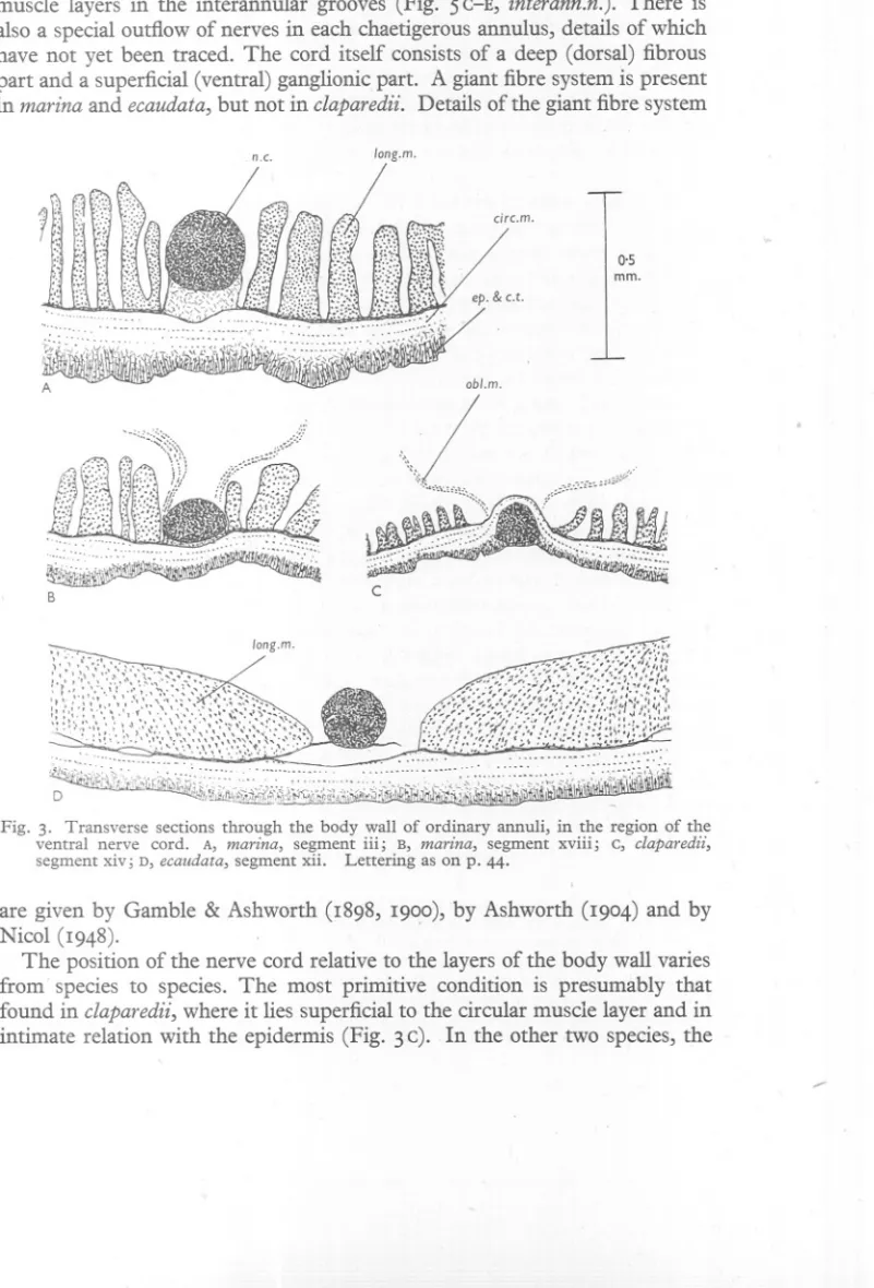
(11) ANATOMY. OF ARENICOLA. II. the body. They lie in the connective tissue partitions which divide the circular muscle layers in the interannular grooves (Fig. 5 C-E, interann.n.). There is also a special outflow of nerves in each chaetigerous annulus, details of which have not yet been traced. The cord itself consists of a deep (dorsal) fibrous part and a superficial (ventral) ganglionic part. A giant fibre system is present in marina and ecaudata, but not in claparedii. Details of the giant fibre system n.c.. long.m.. 0,5 mm.. obl.m.. \(\,. c. B. :>. !!t1[~,~~~~1~':. ,::::,: ..:. D. . . ~h~~i;I.t,,,,~:{,[,":/';j'i\J.I. ,. .. ~if~J~~j!}~!W:~~~~~, ,,,,,,,'''Y',. \-. Fig. 3. Transverse sections through the body wall of ordinary annuli, in the region of the ventral nerve cord. A, marina, segment iii; B, marina, segment xviii; c, claparedii, segment xiv; D, ecaudata, segment xii. Lettering as on p. 44.. are given by Gamble & Ashworth (I898, I900), by Ashworth (I904) and by Nicol (I948). The position of the nerve cord relative to the layers of the body wall varies from' species to species. The most primitive condition is presumably that found in claparedii, where it lies superficial to the circular muscle layer and in intimate relation with the epidermis (Fig. 3 c). In the other two species, the ......
(12) 12. G. P. WELLS. cord lies deep to the circular muscle layer. 1 In marina, it is separated from the circular layer by a pad of connective tissue, staining conspicuously blue with the aniline blue in Mallory's triple stain, and thicker in the anterior segments of the body than farther back (Fig. 3 A,B). In ecaudata, this pad is absent, and an extensive coelomic space separates the cord from the circular muscle (Fig. 3D). The cord is held in position by a membranous sheet running laterally to the longitudinal muscle layer, and perforated in many places to allow blood vessels to pass. The peripheral nerves reach the body wall by way of this membranous sheet. The oblique muscles have the form of flat, translucent strips. They run from the sides of the nerve cord to the notopodialline, where their fibres enter, and join, the circular muscle layer. Particularly thin, thread-like members of the oblique muscle series are atta<;hed to tl,1einner ends of most of the notopodia. . Oblique muscles are absent from the heads of all species, and (except for those attached to the notopodia) from the first 3 trunk segments of marina and claparedii anq the first 16 or 17 of ecaudata. Thereafter they continue to the hinder extremity of the body. There is of course no jointed skeleton, and the main skeletal function is presumably performed by the body fluid. The pressure in the coelome has been measured in marina under various conditions by Chapman & Newell (1947); they find that it increases with the level of activity of the worms and, in actively burrowing individuals, may exceed 3° cm. of sea water. We may suppose that in active animals, both layers are in a state of sustained tonic contraction, and that the changes of form are produced by local increases, or local decreases, in the tensions of the two layers. The following considerations suggest that the circular layer plays the greater part in the postural function of maintaining internal pressure. Suppose a cylindrical worm whose length is great and the thickness of whose body wall is small compared with its radius r em. Let the pressure in its body fluid be P g, wt.jcm.2, and let it be entirely due to the tensions in the circular and longitudinal layers, which, as with surface tension, we call So, SL g. wt.jcm. Now imagine a plane dividing the worm transversely. If no change of shape is taking place, the hydrostatic pressure acting over the area of section is balanced by the longitudinal muscle tension round the circumference, and we have 7Tr2P=27T1'SL or SL=!Pr. (i) 1 Ashworth (1904) writes of marina: 'In some specimens the cord in the tail and in the last chaetigerous segment lies only just beluw the epidermis.' I find, on examining serial sections of the tails of seven worms, that the cord lies deep to the circular muscles in all but one. In the single exception, the cord seems in some sections to lie as in claparedii while in others it is embraced by the circular muscle, the latter running both superficial and deep to it. This is much the smallest worm of the seven (tail diameter 1'1 mID.) so the appearances may represent a migration during development from the primitive to the final position. This specimen has the cord deep to the circular muscle in the chaetigerous segments..
(13) ANATOMY. OF ARENICOLA. 13. Now imagine the worm divided by a longitudinal plane passing through its long axis. This time, the pressure is balanced by the circular muscles, and, if the length is I cm., we have. Combining (i) and (ii). 2rlP=21So or So=Pr.. (ii). So = 2SL'. (iii). How far do these considerations apply to a real Arenicola? In the first place, the muscle layers have a measurable thickness; this is, however, not great compared with the radius and its effect will be to apply a small correction to the quotient 2 in equation (iii). Secondly, the oblique muscles may have a postural function; they are, however, very thin, and absent from a fairly considerable stretch of the body in ecaudata; and we may note, in passing, that septa and mesenteries are lacking over most of the trunk in all species. Thirdly, the body fluid is not the only skeleton. Isolated branchiate segments of marina, lying in a watch-glass of sea water, often undergo regularly rhythmical changes of shape, not perhaps very extensive but perfectly visible and corresponding in timing with the waves that traverse the body, when water is being pumped through the tube. This observation shows that there is a certain amount of elasticity in the body wall itself, though it seems probable that this factor plays only a minor role in the intact worm. Finally, there is the skeletal function of the surrounding mud. The movements of marina in glass tubes have been studied by various authors (Just, 1924; van Dam, 1937, 1938; Wells, 1944, 1945), and will be treated in a later section. For the present, we need only note that there appears to be an inverse relation between the degree of activity of the worm and the proportion of its surface which makes contact with the tube. At one extreme, it may be completely at rest: in this posture, the body is short and thick and presses against the tube with its whole surface; thus the relationships discussed above obviously do not apply. At the other, it is creeping actively forwards or backwards: the body is extremely elongated and at all points away from the tube, except that waves of swelling run along it, grip the tube, and so act as fixed points; here, as the waves follow each other fairly rapidly, the trouble lies in the assumption that no change of shape is taking place. Evidently, equation (iii) is not to be taken as precise. It may, nevertheless, point in the right direction. Lugworms kept in glass tubes often show a regular alternation of rest and rhythmic activity. The resting worms are short and thick, and the onset of an activity outburst is accompanied by lengthening and narrowing of the body (Wells, 1949a). Spontaneous activity is also associated with an increase in internal pressure (Chapman & Newell, 1947). The phasic responses to stimulation are suggestive in' this connexion. A nocuous stimulus may produce sudden shortening of the head or tail, or curving of the body (Just, 1924)-results evidently due, in the main, to ,. '.
(14) 14. G. P. WELLS. longitudinal muscle contraction. If a worm is creeping into a tube, and an attempt is made to pull it out backwards, it expands its front end very abruptly to grip the tube, a movement which suggests circular muscle inhibition. The data as a whole suggest that, in an active worm, the circular muscles are fairly highly contracted and the longitudinals less so; the phasic acts, whether reflexly or spontaneously produced, are mainly, at least, in the sense of longitudinal contraction and circular inhibition. Fox (r 949) has pointed outthatthe muscles of Arenicola contain haemoglobin, but does not discuss whether there is any inequality of distribution between the muscle layers. For the other pigments of the body wall, the works of Fauvel (r899b) and Lignac (r945) may be consulted. The epidermis is richly provided with unicellular gland cells. The skin secretes mucus, which is used, in marina, to impregnate the wall of the burrow and keep it firm (Osler, r826; Linke, r939). The mucus of ecaudata is particularly copious, as anyone who has handled the living worm is aware. If specimens of marina are kept in the laboratory, the water comes to contain' belts' of greyish mucus, of about the same diameter as the worms; these belts are sometimes seen round their bodies; they appear to be passed slowly headwards and to be a means of getting rid of such unwanted residues as the breakdown products of old chaetae. When handled, the lugworm produces a fluorescent, greenish yellow secretion, which stains the fingers and is also rather irritating.l THE MIDDLE REGION. (' TRUNK '). As the middle region of the body is in many ways the least specialized of the three, it will be taken first. Arenicola marina The trunk of this species consists of r9 segments (in exceptional individuals, 20). They exhibit a certain amount of structural and functional divergence. Each segment includes several annuli, of which one, the chaetigerous annulus, is larger than the others and bears the parapodia and gills. The chaetigerous annuli are shown white in Fig. 4. The boundaries between segments are given internally by septa, or, where the septa have disappeared, by the septal blood vessels. The septal planes correspond approximately, though not quite exactly, to the second groove behind each chaetigerous annulus: in other words, the penultimate annulus of each segment is chaetigerous. Typically, any two chaetigerous annuli are separated by 4 ordinary annuli, so that the number of annuli per segment is 5. In the first 3 segments, however, the number of animli is reduced. The first has 2, the second 3 and the third 4, except that in a 'laminarian variety', described by Gamble & Ashworth (r898), the third has 3. The fourth, and all subsequent trunk 1 The worm as a whole has a characteristic fragrance, especially when laid open. After he had worked on it for some years, it produced strong allergic symptoms in the writer (catarrh, asthma); this was put right by a course of injections.. L.
(15) ANATOMY. OF 4RJjNICOLA. 15. segments, have 5 each. The rule, that the chaetigerous annulus is penultimate, holds for the anterior segments in spite of this reduction. Ashworth gives no attention to the possible systematic usefulness of these numbers. It is true of all Arenicola species, that the majority of the trunk segments have 5 annuli, and that some degree of reduction occurs at the front h.l.. neurop.. neph.p. g.. A. marina. A. cfaparedii. A. ecaudata Fig. 4. Lateral views of the front ends of.the three species, to. show the annulation and the characters of the chaetigerous annuli and appendages (white). The proboscis is withdrawn in the marina and extended in the -other two. Lettering as on p. 44.. end. The extent of this reduction is not always the same, and I find that each of the species now under examination has a fairly constant and characteristic annulation formula, which holds for all the range of material which I have been able to examine (p. 2).. The most convenient way of describing the reduction is to use serial Roman figures for the chaetigerous annuli, and to put between them, in Arabic.
(16) G. P. WELLS. 16. figures, the numbers of intervening ordinary annuli. Thus the typical formula ... ... . for marina is: 1.2.11.3 .111.4. IV.4. v. While the 'laminarian variety' is: i.2. ii.2. iii.4. iV.4. v The structure of the ordinary annuli has already been sufficiently described. We turn now to the distinctive features of the chaetigerous annuli. These are: (i) the neuropodia, (ii) the notopodia, (iii) the parapodial girdles, (iv) the gills, and (v) the nephridia and'nephridiopores. The earlier authorities were mainly interested in the characters supposed to be of systematic importance-the detailed form of the chaetae, and the mode of branching of the gill. Full information about these points is to be found in the works of Gamble & Ashworth (1900) and Ashworth (1912). The musculature and functional topography of the appendages were described, for marina only, by myself (Wells, 1944). The main results for that species will now be summarized. A neuropodium consists essentially of a single dorsi-ventral row of chaetae, each with a sharply inclined rostrum projecting from the body surface and a gently curved shaft embedded in the body wall (Fig. 5A, B). Each chaeta lies in its own epithelial follicle. The whole row of chaetae and follicles may be termed the neuropodial plate. New chaetae and follicles are continually being formed in a highly basophil formative region (Fig. 5A, form.) at the ventral end of the plate, and old ones are destroyed at the dorsal end, where their products accumulate as greenish masses (dest.). These are somehow expelled from time to time. There therefore appears to be a continual dorsalwards procession of chaetae and their follicles along the neuropodial plate. To see the musculature of the I!europodium, longitudinal sections, cut at right angles to the body surface, should be studied (Fig. 5C). The inner end of the neuropodium projects into a cavity, cut off from the general coelOl;neby the longitudinal muscle layer. Retractor muscles run from the longitudinal layer to the outer edge of the neuropodial plate (ret.m.neurop.), and protractors run anteriorly and posteriorly from its inner edge to the neighbouring body wall (prot.m.neurop.. ).. The neuropodia of the more anterior segments are very short dorsi-ventrally, but they lengthen from segment to segment until, from segment x or xi backwards, they extend from a point slightly above the nephridial line to the side of the nerve cord (Fig. 4). I know of no published description of the movements of the neuropodial chaetae. They could obviously be protracted to some extent by the protractors, and withdrawn again by the retractors, if the substance of the annulus is sufficiently flexible. I am, however, inclined to guess that another movement is more significant. Simultaneous contraction of the posterior protractors and.
(17) ANATOMY. OF ARENICOLA. 17. anterior retractors would tend to incline the chaeta as a whole, so that its inner end would move backwards relative to its outer; at the same time, owing to the form of the chaeta and the position in which it normally lies in the neuropodial plate, it would rotate so that the rostrum points forwards (Fig. 5 A,B). Similarly, the anterior protractors and posterior retractors could turn the rostra backwards. The usefulness of this movement, in facilitating the ratchet or gripping function of the neuropodia when the worm is creeping along the tube, needs hardly to be stressed. notop.b.. notop.t.. ep.&c.t.. circ.m.. long.m.. h.f.. m.h.. prot.m.neurop.. h.f.. interonn.n.. c. ret.m.neurop.. dest.~. D pomp.c.. A. m.parop.c.. B E. Fig. 5. Arenicola marina. Diagrams of the parapodia. A, the essential structUre of a notopodium (above) and a neuropodium (below). B, a neuropodial chaeta. c, longitudinal section through one of the hinder neuropodia, at right angles to the body surface. Two ordinary annuli are also included. D, sagittal section through one of the first three chaetigerous annuli, near the mid-dorsal line. E, the muscles of a notopodium, exposed by a longitudinal cut through the body wall passing through its base. Lettering as on p. 44.. A notopodium can be regarded as a modified neuropodium. In external view, a dorsi-ventrally flattened tip (Fig. 5 A,E, notop.t.) can be distilJ.guished from a thin-walled, evaginable base (notop.b.). The chaetae are very long and emerge from the tip in two closely applied, parallel rows. To derive a notopodium from a neuropodium, the plate must be supposed to have duplicated itself into two plates lying close side by side like two leaves of a book, and then the inner jOURN. MAR. BIOL. ASSOC.vol. XXIX, I95°. 2.
(18) 18. G. P. WELLS. corners of the double plate to have been folded and rolled forwards, giving the inner end of the whole a somewhat scroll-like appearance (Fig. 5A). The resulting structure is complicated and not easy to visualize. Full details of the notopodium and its musculature were given elsewhere (Wells, 1944). The inner end of the notopodium protrudes through the longitudinal muscle layer into the general body cavity. The protractor muscles radiate from this end to the surrounding body wall (Fig. 5 E,prot.m.notop.). Most of these run anteriorly and posteriorly, but one or two are inserted on the body wall dorsal to the notopodium, close to its base, while others spread ventrally as far as the nephridial line (Fig. 6). A thin strand of muscle generally runs from the inner end of the notopodium to the side of the nerve cord, and this has often been described as the retractor of the notopodium. It is evidently a member of the oblique muscle series; and, as it is quite often absent, we cannot suppose that it plays an essential role in the movements of the podium. (Fig. 5 E, obl.m.notop.). The true retractors resemble those of the neuropodium; they run from the longitudinal muscle layer to the notopodial tip (Fig. 5 E, ret.m.notop.). There are also thin sheets of muscle sheathing the notopodial plates, and running between their inner ends. . The notopodia further resemble the neuropodia in that they are smallest in the more anterior segments, and increase gradually in size posteriorly. A notopodium is connected to the general body wall only by the muscles and the thin-walled, ip.vaginable base. It must therefore be held in position by its muscles, contracting tonically against each other. If a living worm is watched under a binocular Inicroscope, two types of movement of the notopodium can very readily be seen. The first is retraction and protraction, i.e. invagination or evagination of the base, and the second is the direction of the tip in an anterior or a posterior direction. We may conjecture that invagination is brought about by the retractors, with the possible assistance of the oblique muscle of the notopodium, evagination by the protractors, anterior pointing by the anterior retractors and posterior protractors, and backward pointing by the posterior retractors and anterior protractors. A high pressure of the body fluid will tend towards evagination. From the arrangement of the muscles, it would also be possible to swing the tip dorsally and ventrally, and perhaps to rotate the podium on its own long axis, but I am not aware that such movements have been witnessed or described. The parapodial girdle is a name now proposed to cover a group of special features of the chaetigerous annulus to which I drew attention elsewhere (Wells, 1944). These features are best seen in a sagittal section through one of the first three segments, somewhere near the dorsal line (Fig. 5 D). The chaetigerous annulus stands out at once in such a section, because it contains a conspicuous cavity, the 'parapodial canal' (parap.c.). This canal runs right round the annulus, and is separated from the general body cavity by the.
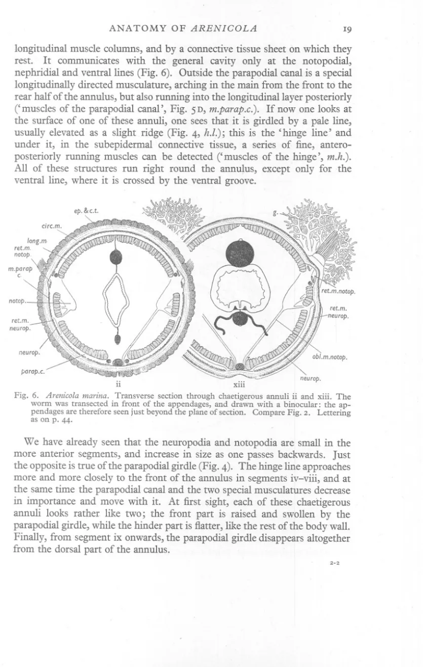
(19) ANATOMY. OF IlRENICOLA. 19. longitudinal muscle columns, and by a connective tissue sheet on which they rest. It communicates with the general cavity only at the notopodial, nephridial and ventral lines (Fig. 6). Outside the parapodial canal is a special longitudinally directed musculature, arching in the main from the front to the rear half of the annulus, but also running into the longitudinal layer posteriorly (' muscles of the parapodial canal', Fig. 5 D, m.parap.c.). If now one looks at the surface of one of these annuli, one sees that it is girdled by a pale line, usually elevated as a slight ridge (Fig. 4, h.l.); this is the' hinge line' and under it, in the subepidermal connective tissue, a series of fine, anteroposteriorly running muscles can be detected ('muscles of the hinge', m.h.). All of these structures run right round the annuJus, except only for the ventral line, where it is crossed by the ventral groove.. notop.. parap.c. ii. xiii. Fig. 6. Arenicola marina. Transverse section through chaetigerous annuli ii and xiii. The worm was transected in front of the appendages, and drawn with a binocular: the appendages are therefore seen just beyond the plane of section. Compare Fig. 2. Lettering as on p. 44.. We have already seen that the neuropodia and notopodia are small in the more anterior segments, and increase in size as one passes backwards. Just the opposite is true of the parapodial girdle (Fig. 4). The hinge line approaches more and more closely to the front of the annulus in segments iv-viii, and at the same time the parapodial canal and the two special musculatures decrease in importance and move with it. At first sight, each of these chaetigerous annuli looks rather. like two; the front part is raised and swollen by the parapodial girdle, while the hinder part is flatter, like the rest of the body wall. Finally, from segment ix onwards, the parapodial girdle disappears altogether from the dorsal part of the annulus. 2-2.
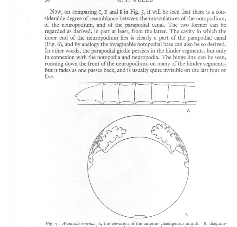
(20) 20. G. P. WELLS. Now, on comparing c, D and E in Fig. 5, it will be seen that there is a considerable degree of resemblance between the musculatures of the notopodium, of the neuropodium, and of the parapodial canal. The two former can be regarded as derived, in part at least, from the latter. The cavity in which the inner end of the neuropodium lies is clearly a part of the parapodial canal (Fig. 6), and by analogy the invaginable notopodial base can also be so derived. In other words, the parapodial girdle persists in the hinder segments, but only in connexion with the notopodia and neuropodia. The hinge line can be seen, running down the front of the neuropodium, on many of the hinder segments, but it fades as one passes back, and is usually quite invisible on the last four or five.. 2=:[Ccccr A. B Fig. 7. Arenicola marina. A, the elevation of the anterior chaetigerous annuli. B, diagrammatic section of a worm circulating water through a tube, as in Fig. I A.. There is no doubt at all about the importance of the parapodial girdle of the more anterior segments as a motor apparatus. If a marina is put into a large funnel of sea water, with the lower end of the stem closed with rubber tubing and a clamp, the worm usually burrows down into the stem, and the movements of the more anterior chaetigerous annuli can then be watched. At one moment they lie flat; then they are suddenly raised, and apparently distended, into the form of sharp, backwardly directed flanges; then they drop again (Fig. 7 A). The whole apparatus seems to act as a single unit, i.e. all of the' parapodialized'.
(21) ANATOMY. OF ARENICOLA. 2I. annuli, and the whole periphery of any one of them, rising and falling together, though the movements are most evident in the first three segments, in which the responsible structures are best developed. The movements are used in burrowing, to grip the sand and help to draw the worm in, and also, ~pparently, for certain other purposes, such as the drawing of surface sand down into the head end of the burrow (Wells, 1944). The gills are hollow, branched, contractile outgrowths of the body wall, borne by the chaetigerous annuli just behind the notopodia, and present on every segment from vii onwards (Fig. 4). The most anterior gill is smaller than the rest' and may be lacking altogether on one or both sides. For details of the mode of branching of the gills, the works of Ashworth (19°4, 1912) may be consulted. The sphinctered nephridiopores open just behind, and very slightly below,. the upper ends of the neuropodia, on segments iv to ix inclusive. According to Goodrich (1946) the 'nephridium' of Arenicola is a nephromixium and includes an ectodermal component which should logically be described with the rest of the body wall; it will, however, be included with the internal anatomy, which the writer hopes to describe in a later paper. We turn now from the various components of which the body wall of the trunk consists, to the plan of the region as a whole. It can be divided, on structural grounds, into the following three sections (Fig. 4): (i) Segments i, ii, ill. Gills absent. Neuropodia and notopodia small and apparently unimportant. Parapodial girdles massively developed round the whole circumference of the chaetigerous annuli. (ii) Segments iv to viii. Graded, transitional. (ill) Segments ix onwards. Gills present. Neuropodia and notopodia well developed. Parapodial girdle absent, except in connexion with the neuropodia and notopodia. Now this structural differentiation is very nicely paralleled by a physiological one. The movements of marina were first studied in detail by Just (1924), who pointed out that the first three or four segments (the boundary is not absolutely sharp) stand in functional contrast to the rest. His observations have been confirmed and extended by others (van Dam, 1937, 1938; Wells, 1944, 1945). In burrowing, in forwards or backwards creeping, and in the driving of water through the burrow, waves of swelling travel along the trunk. These waves may go in either direction and their form varies with the particular type of movement that is being carried out. At all times, however, they concern the hinder 15 or 16 segments, and (though the amount of worm they involve varies to some extent with the type and vigour of the movement) they are seldom, if ever, shown by the first three. Proboscis activity, on the other hand, is brought about by the integrated action of the proboscis itself, of the body wall of the head, and of the body wall of the first three trunk segments..
(22) 22. G. P. WELLS. If a worm is watched quietly pumping water through a glass tube (Fig. I), the .ventro-Iateral surfaces may be noticed, over most of the trunk, to be pressed tight against the tube; a space remains, however, between the dorsal surface and the tube, and the gills spread out into this space. Waves of swelling travel along the dorsal surface, occluding the space and so driving water through the tube; the gills contract as the waves approach and expand again as soon as they pass. A rather conjectural cross-section of a worm in the act of irrigation, based on watching worms in glass tubes from the side, is drawn in Fig. 7B. The problem at once arises, of how the close pressing of the flanks against the tube is achieved. The only possible answer, I think, is by means of the notopodial protractors. It will be seen from the drawing, that the contact could be maintained if the notopodia were pressing outwards and upwards. Meanwhile~the head and first few segments (which are not concerned in the irrigation waves, and in which the notopodia are poorly developed) arch away from the side of the tube (Fig. I). In active creeping, as Just (1924) described, the notopodia exert a ratchet action, being directed backwards for. headward locomotion, and forwards for tailward locomotion. We may fairly safely guess that the neuropodial rostra playa similar role. When swimming, the worm travels tail first, with lateral waves of great amplitude travelling headwards along the body, while the llotopodia are directed headwards and held close to the body surface. The podia are never used as oars or paddles, or even as punt poles; they are bracing and anti-slip devices. It seems, then, that the neuropodia and notopodia are adapted to assist in those wave movements which are th~ concern of the hinder segments, and it is in those segments that they are best developed. The first few trunk segments are specialized, partly to help in the extrusion of the proboscis by driving their contained body fluid forwards (Just, 1924) and partly for the flanging movement described above. The great development of the parapodial girdles which the latter function involves appears to carry with it a reduction in the size of the neuropodia and notopodia, and the disappearance of the gills. It is indeed striking, in a view of the whole worm"that the gills appear just as the parapodial girdles leave the dorsal faces of the chaetigerous annuli (Fig. 4). Arenicola claparedii. The general organization of the body wall of the trunk is the same in the other two species as in marina; it is therefore only necessary to note divergences. An immediately obvious feature of claparedii is the enormous development of the first three chaetigerous annuli (Fig. 4). In the living worm, the movements of these annuli are like those of marina in type, but very much more powerful and impressive. The amltomical features which distinguish these annuli are correspondingly well developed. At the same time, this expansion has entailed.
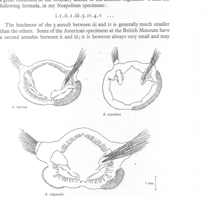
(23) ANATOMY. OF ARENICOLA. a great reduction in the ordinary annuli of the anterior segments. following formula, in my Neapolitan specimens:. i.I.ii.I.iii.3.iv.4.v. 23. I find the. .... The hindmost of the 3 annuli between iii and iv is generallymuch smaller than the others. Some of the American specimensat the British Museum have a second annulus between ii and iii; it is however alwaysvery small and may. \ /I. marina A. ecaudata. 1 mm-I A. c/aparedii. Fig. 8. Drawings, all to the same scale, of chaetigerous annulus xvi in each of the three species. Drawn from cleared specimens of the isolated annuli. The full extent of the chaetae is shown on the right, the projecting portions only on the left.. not extend round the whole circumference of the worm. In others, chaetigerous annulus iii hangs forwards in such a way that a minute ring might be present but hidden. We seem to be concerned here with an annulus in process of disappearance; and the variations described below ror ecaudata support the idea that it is always immediately in front of the ch~etigerous annuli that reduction occurs. ..
(24) 24. G. P. WELLS. In a posterior direction the parapodial girdle is seen to leave the dorsal surface after segment vi-so, although the girdle is better developed than in marina, it embraces rather fewer segments. The notopodia are more massive in claparedii than in marina, and so are the individual notopodial chaetae. The neuropodia are short in the more anterior segments and lengthen posteriorly, but they never approach the ventral line as closely as in marina (Fig. 8). Even in the hinder segments, the ventral ends of the neuropodia are separated by a distance about equal to the length of a neuropodium. The parapodial girdle extends from the lower end of the neuropodium to the si~e of the ventral groove, even in those hinder segments in which it has disappeared from the dorsal surface. Nephridiopores are present on segments v to ix inclusive. Arenicola ecaudata In this species, the reduction of the ordinary annuli of the more anterior segments is least marked, the formula being:. i.3 .ii.4.iii.4.iv.4.. v. .... In many specimens, all of the annuli are prominently developed, and the truth of the above formula is obvious. In others, however, there is a tendency to reduce the ordinary annuli which lie immediately in front of the chaetigerous annuli, and when this occurs it affects all of the first three or four segments. The reduced annuli may in extreme cases be so small, and so overhung by the following chaetigerous annuli, as to be invisible on surface inspection. The formula then appears to be: i.2 .ii. 3 .iii. 3 .iv .4. v. .... Nevertheless, in. all examples examined by myself, a sagittal cut with a razor blade reveals the hidden rings, and shows that the formula is in fact as previously stated. The parapodial girdles of this species are rather poorly developed, as the low degree of reduction of the annuli perhaps suggests. The hinge lines lie near the front margins of chaetigerous annuli i-iii, instead of bisecting them as in the other two species. The various components of the parapodial girdle are all present, but they are small, and all localized in the front part of the annulus. In a posterior direction these structures get gradually less and less well defined, until they can no longer be made out on the dorsal surface, as from about segment xiv. In other words, although less well developed than in the other species, they concern about twice as many segments. Now the most anterior gill, in ecaudata, is typically on segment xvi; as in marina, this gill is generally small and sometimes absent. The whole arrangement evidently confirms the idea, already suggested by the other two species, that j:he gills and the parapodial girdles tend to exclude each other from the dorsal surface..
(25) ANATOMY. OF LiRENICOLA. 25. The notopodia are relatively smaller, and more dorsally placed, than in the other species (Fig. 8). The neuropodia are exceedingly long, extending from the side of the nerve cord to a point well above the nephridial line. In the more anterior segments, far from being short (as in marina or claparedii), the neuropodia of ecaudata are best developed, and reach from the ventral line to the bases of the notopodia (Fig. 4). Nephridiopores are present on segments v to xvii. THE ANTERIOR REGION (' HEAD'). The' head' is the rougWy conical region extending forwards from the anterior margin of the first chaetigerous annulus (Fig. 4). Its segmentation is largely obscured, in the adult, by its profound functional modifications. It consists of the prostomium and a second large portion which, to quote Ashworth (1912), in most adult specimens of Arenicola, is divided by encircling grooves into three or four (or more) rings. There are good reasons for stating that this is composed of the peristomium and a body segment which is without chaetae in the adult. In post-larval stages of A. marina and ecaudata, the region between the prostomium and the first ordinary chaetiferous segment is subdivided by a groove into two parts. The anterior and usually rather smaller portion is undoubtedly the peristomium; it never bears chaetae, but the paired statocysts may be seen near its anterior margin. The posterior of the two parts is, in the post-larval stages which the writer has examined, achaetous, but a chaeta has been observed in this segment, in either A. marina or A. ecaudata, by Professors Ehlers, Benham, Mesnil and Fauvel, a fact which demonstrates that this is a true segment. Evidence confirmatory of this interpretation is afforded by the atrangement of the giant nerve cells. In later post -larval stages in which the annulation is making its appearance, the peristomium and the segment in question become subdivided into secondary rings.. . . The composition of this region is probably constant throughout the family.. Except for the important study of the statocysts by EWers (1892), previous writers on this region have mainly confined themselves to cursory accounts of its external features, and discussions of its segmental homology. Many interesting features, especially the musculature, have received little or no attention. The following account omits the proboscis, and certain special structures (e.g. the retractor muscle) associated therewith. I hope to describe them, with the internal anatomy in general, at a later date. Arenicola marina Apart from the absence of parapodia, the most noteworthy specializations of the body wall of the anterior region may be grouped under the following headings: (i) the mouth, (ii) the prostomium and nuchal pouch, (Hi) the central nervous system, (iv) the metastomial muscle, and (v) the statocysts. . At the mouth, which is terminal, the layers of the body wall continue on to the eversible proboscis. The latter organ is pardy extruded in Figs. 9 and ro..
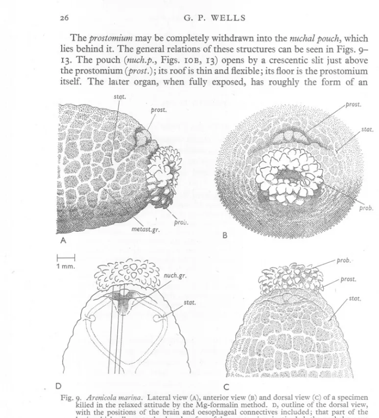
(26) G. P. WELLS. 26. The prostomium may be completely withdrawn into the nuchal pouch, which lies behind it. The general relations of these structures can be seen in Figs. 913. The pouch (nuch.p., Figs. lOB, 13) opens by a crescentic slit just above the prostomium (prost.); its roof is thin and flexible; its floor is the prostomium itself. The latter organ, when fully exposed, has roughly the form of an stqt.. B. A. I. i. 1 mm.. /1. "2~cO. prob.". -, -1",. :::J:):J. <;C-C.C C.O\);). -:s. CCL.C(,~()~ ((" '-' ~v ,,;;:.,.n-r~ '""1<JJ0j. nuch.gr.. ~. D. c. Fig. 9. Arenicola marina. Lateral view (A), anterior view (B) and dorsal view (c) of a specimen killed in the relaxed attitude by the Mg-formalin method. D, outline of the dorsal view, with the positions of the brain and oesophageal connectives included; that part of the brain which adheres to the dorsal surface of the prostomium is stippled; the nuchal groove is shown as a dashed line; the ruled lines give the positions of the sections in Fig. 12. Lettering as on p. 44.. isosceles triangle with its apex directed backwards; its dorsal face is impressed by a shallow Y-shaped groove (Fig. lOA). The worm must, however, be dissected if the whole prostomium is to be seen. In preserved material, it is always more or less completely overlapped by the dorsal lip of the nuchal pouch, so that one sees only its anterior margin; this is trilobed, owing to the.
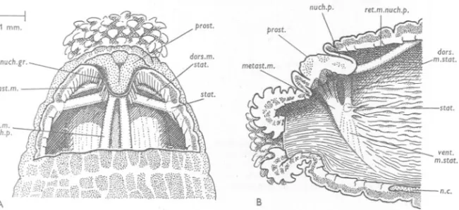
(27) ANATOMY. OF ARENICOLA. 27. fact that the arms of the dorsal, .y-shaped groove continue down the anterior face of the prostomium. Between the prostomium and the mouth is a strip of body wall, which Ashworth (1912) terms the 'upper lip'. The nuchal pouch as a whole may be regarded as a blind in-pushing of the epidermis and subjacent connective tissue, passing through the circular and longitudinal muscle layers. In sections, a large amount of transversely running muscle can be seen immediately ventral to the prostomium. Its position and course suggest that it is circular muscle; in fact, however, as will be shown below, it is made up of certain specialized muscles, derived, largely at least, from the longitudinal layer (the metastomial muscle, and the dorsal muscle of 1---1 1 mm.. auch.p. prost.. stat. ret.m. auch.p.. a.c. A. B. Fig. 10. Arenicola marina. A,dorsal dissection of the animal of Fig. 9; part of the body wall and the thin roof of the nuchal pouch have been removed. The front part of the dorsal vessel (stippled) can be seen between the retractors of the nuchal pouch. B, lateral dissection of another worm to show the coelomic aspect of the body-wall muscles of the right side; the animal has been bisected in the median plane and the gut and blood vessels have been removed. The ventral muscle of the statocyst is seen as a series of fine strands (dotted lines) running ventrally across the face of the metastomial muscle. Lettering as on p. 44.. the statocyst). The columns of the longitudinal layer pass by the sides of the nuchal pouch, and those immediately adjacent to it send slips which are inserted on its walls and help in its retraction. The chief part in retraction, however, is played by a paired muscle, the retractor of the nuchal pouch, clearly derived from the longitudinal layer, and running from the hind end of the pouch to a point on the body wall, about half way between the prostomium and the first chaetigerous annulus (Figs. 10 and 13, ret.m.nuch.p.). My impression is that retraction of the nuchal pouch never occurs as an isolated act, but only when the anterior end as a whole is shortened and thickened, as part of the general movement. There are no protractor muscles. Extrusion of the proboscis is presumably due to the pressure of the body fluid, and occurs, I believe, whenever the head as a whole lengthens and narrows, and at no other time. Fig. I I, drawn from preserved specimens, illustrates.
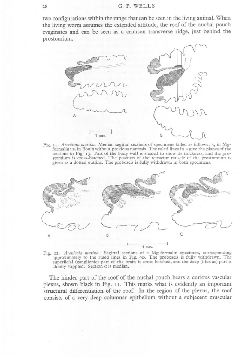
(28) G. P. WELLS. 28. two configurations within the range that can be seen in the living animal. When the living wOrm assumes the extended attitude, the roof of the nuchal pouch evaginates and can be seen as a crimson transverse ridge, just behind the prostomium.. e:: A. I. 1 mm.. I. c;::. Fig. I I. Arenicola marina. Median sagittal sections of specimens killed as follows: A, in Mgformalin; B, in Bouin without previous narcosis. The ruled lines in B give the planes of the sections in Fig. 13. Part of the body wall is shaded to show its thickness, and the prostomium is cross-hatched. The position of the retractor muscle of the prostomium is given as a dotted outline. The proboscis is fully withdrawn in both specimens.. --. A. c. B 1 mm.. Fig. 12. Arenicola marina. Sagittal sections of a Mg-formalin specimen, corresponding approximately to the ruled lines in Fig. 9D. The proboscis is fully withdrawn. The superficial (ganglionic) part of the brain is cross-hatched, and the deep (fibrous) part is closely stippled. Section c is median.. The hinder part of the roof of the nuchal pouch bears a curious vascular plexus, shown black in Fig. 11. This marks what is evidently an important structural differentiation of the roof. In the region of the plexus, the roof consists of a very deep columnar epithelium without a subjacent muscular.
(29) ANATOMY. OF ARENICOLA. 29. layer. Elsewhere, it has a shallower epithelium and a. thin circular muscle layer, continuous with the circular muscle layer of the general body wall. Now in the other two species, as we shall see, the prostomium cannot be retracted, and its hinder margin is marked by a nuchal groove, whose epithelium is very deep, ciliated and presumably sensory. This evidently corresponds to the hinder, vascular part of the roof in marina. The non-vascular part, present only in marina, is simply attenuated body wall, and it is this which converts the whole region into a pouch and makes retraction of the prostomium possible. 1 mm.. stat.. metast.m.. A. B. c. Fig. 13. Arenicola marina. Three transverse sections, corresponding approximately to the ruled lines in "Fig. I I B, of a specimen killed in Bouin. The sections are equidistant and 0'2 mm. apart; the most anterior is on the left. The dotted circle gives the position of the gut. Lettering as on p. 44.. The general plan of the central nervous system of the head is shown in Fig. 9 D. The ventral nerve cord continues forwards for a short distance on to the head; it then divides to give rise to the connectives, which run obliquely upwards and forwards to the prostomium. Just as the course of the ventral cord is marked externally by the ventral groove, so that of the connectives is marked by the more or less evident metastomial grooves (Fig. 9A, metast.gr.). The connectives resemble the ventral cord in consisting of a superficial ganglionic and a deep fibrous part, and in lying deep to the circular muscle . layer (Fig. 13, conn.). When they reach the lateral corners of the prostomium, where the nuchal pouch pushes through the circular layer, the ganglionic part of the connectives enters into intimate relation with the epidermis. The nerves now expand somewhat and, remaining in close contact with the epidermis, pass up the front face of the prostomium to reach its dorsal side. Here they run backwards for a short distance to' meet each other in the median plane. From their point of union, a pair of massive nerves runs back to the nuchal groove. That part of the central nervous system which lies in the prostomium,.
(30) 3°. G. P. WELLS. in intimaterelationwith the epidermis,is generallytermed the brain. Its form can be seen in Figs. 9D and 12. For the fine structure of the brain, see Gamble & Ashworth (1900). The shallow grooves, which divide the anterior margin of the prostomium into three lobes and trace a Y on its dorsal face, give the line along which the brain adheres to the epidermis. The body wall is therefore thick under these grooves. At the sides of the prostomium, and in its median anterior lobe, the body wall is thin and the coelome comes near the surface (Fig. 12). Immediately dorsal to the connectives, over the more ventral part of their course, lies the largest special muscle in the body wall of the worm. Thisthe metastomial muscle-originates as a union of contributions from all the. A. ~=tr[3tI~. -J.. ~. (~. ( (; (, ('{~p.~. c. ./. Fig. 14. Arenicola marina. Sketches to illustrate the process of reversing in the tube (see text).. longitudinal muscle columns ventral to the notopodialline. It runs forwards and upwards, following the general course of the connectives, until it reaches the level of the statocysts;. it then leaves the inner face of the longitudinal layer and crosses the body cavity, just below the brain, to continue into its fellow of the opposite side (Figs. 10, 13, metast.m.). This muscle was first described by Ehlers (1892), who said that it was inserted on to the prostomium. It is briefly mentioned by Gamble & Ashworth (1898) and by Ashworth (1904), who regard it as a retractor of the prostomium. Its true anatomical relationships, however, show that it cannot play that role. Its actual function is quite different, and very important. The lugworm sometimes reverses itself, either in. its own burrow or in glass observation tubes, by narrowing its body and then burrowing (with continual extrusions of the proboscis, and uprisings of the parapodial girdles) along its own ventral surface. In this way it can turn in a tube which, at other times, it seems comfortably to fill (Fig. I4c). The problem now arises, how does this performance start? Living Worms, when watched,! can occasionally be seen to assume a remarkable 1 To study the movements of the proboscis and head, it is convenient to tie the whole worm with fine string about at the level of chaetigerous annulus v, to cut away everything behind the ligature, and to put the isolated front end in sea water under a binocular microscope. The preparations stay active for hours, and frequently show the turning attitude described above..
(31) ANATOMY. OF ARENICOLA. 3r. attitude, in which the dorsal wall of the head and of the first two or three segments is greatly distended, while the ventral wall shows extreme longitudinal contraction (Fig. 14A). The prostomium and mouth are thereby come to be directed backwards: the attitude can be roughly imitated by 'burying one's chin in one's chest'. Now the assumption of this attitude always heralds an outburst of proboscis activity. The first extrusion begins while the worm is still in the attitude, and the proboscis therefore emerges in a tailward direction. As this extrusion completes itself, the whole anterior end assumes a more usual configuration. If the worm is lying in a dish of sea water, this results in a forward swing of the proboscis (Fig. 14B); but if the worm is in a tube, the forward swing cannot occur, and subsequent extrusions, made in the usual manner, will serve to pull the head farther and farther along the worm's ventral surface (Fig. 14C). The metastomial muscle runs like a sling over the mouth, and, if it contracts simultaneously with the more ventral longitudinal columns from which it arises, will play an important and perhaps essential role in the assumption of the attitude of Fig. 14A. The statocysts,1 whose form was beautifully described by Ehlers (1892), are a pair of blind in-pushings of the epidermal layer of the body wall, which pass through'the circular muscle layer (Figs. 9, 10, 13, stat.). Their openings are slit-like and dorsi-ventrally elongated. Each leads into a tube with a lumen of the same form, and which opens forwards at its deep end into a spherical bulb. The whole organ is therefore rather retort-shaped. . It contains various foreign objects, such as quartz grains, fragments of spicules and diatom shells, etc., covered with more or less well-marked layers of a 'chitinoid' secretion (Ashworth, 1904). The statocyst is provided with a rather complicated musculature, whose functions are obscure. The muscles are: (i) the dorsal muscle of the statocyst (Fig. lOB, dors.m.stat.), which runs dorsally and medially; its more anterior fibres continue into the corresponding muscle of the opposite side, while its hinder ones are inserted into the posterior end of the nuchal pouch, just below its retractor; (ii) a number of slips of muscle running back to the longitudinal columns adjacent to the statocyst; (iii) the. ventral muscle of the statocyst (Figs. 10, 13, vent.m.stat.), which crosses the metastomial muscle, as a thin sheet or as a series of fine strands, to the tissue round the connectives. On the whole, it seems likely that the dorsal muscle of the statocyst is derived, as the metastomial muscle is, from the longitudinal layer ; the anatomical relations of the ventral muscle, on the other hand, are consistent with its being a member of the oblique muscle series. 1 The statocysts lie at the level of the notopodia; as they are invaginations producing a chitinoid secretion, and as the notopodial chaetae have sensory nerve endings round their bases (Retzius, quoted by Ehlers, 1892), it is tempting to think of them as the peristomial notopodia. The idea was discussed at length by Ehlers (r892), who decided against it, on the ground that certain polychaetes of other families have statocysts, neuropodia and notopodia in the same segment..
(32) G. P. WELLS. 32. Accordingto vonBuddenbrock (1912, 1913), thestatocysts of Arenicolaare used to guide the worm when burrowing down 'into the mud. ot.gr.. laU.prost.. prob.. B. A. metost.gr.. 1 mm.. Dc. Fig. 15. Arenicola claparedii. Lateral view (A), anterior view (B) and dorsal view (c) of a Naples specimen killed by the Mg-formalin method. D, outline of the dorsal view with the brain and connectives given as in Fig. 9D; the ruled lines give the positions of the transverse sections in Fig. 17. Lettering as on p. 44.. Arenicola claparedii The prostomium of claparedii differs in two important respects from that of marina. In the first place, it is prolonged laterally into two large, vertical flaps, the lateral lobes (Fig. 15, lat.l.prost.). In the second place, it is not retractile; nuchal pouch and retractor muscle are lacking (Fig. I6D). The nuchal groove lies, as in marina, round the sides and hind-end of the prostomium (Figs. 15, 17, nuch.gr.). It will be seen, on comparing Figs. 9 and 15, that the prostomium is relatively larger in the latter. This results, partly at.
(33) -,. ~., \j. !). J. ). 'at.l.prost.. ~ .. ot.gr. at.gr.. ~~. /atL;~~. ( .. !:J. (. '). [)/ c. ). ~ C. ,). ). ~J. B A,. f. 1 mm.. 0. I. Fig. 16. Arenicola claparedii. American specimens, killed by means unknown to the writer. A, B, dorsal and lateral views of the same specimen.. c, sagittal section of the same, cut along the ruled line in A, to show how the lateral wing of the prostomium (cross-hatched) overhangs the otic groove. D, median sagittal section of another specimen, for comparison with Fig. 11. lat.l.prost.. lat./.prost.. nuch.gr.. C. A. nuch.gr. laU.prost.. 0. B. ". 1 mm.. Fig. 17. Arenicola claparedii. Naples specimens, killed in Mg-formalin. A, horizontal section through the nuchal groove and'the superficial part of the brain. B, horizontal section through the otic grooves and the deep part of the brain. c, D, transverse sections, corresponding approximately to the ruled lines in Fig. 15D. Conventions as in Fig. 12. Lettering as on p. 44. JOURN. MAR. BIOL. ASSOC.vol XXIX, 1950. ~---+--.
Figure


+7
Related documents
Nanotechnology helps agricultural sciences and reduce environmental pollution by production of pesticides and chemical fertilizers by using the Nano particles and
Mean pattern of study parameters with cardiac dysfunction and their correlation are shown in Table 4.The cardiac manifestations in both subgroups of study
Discusión Los efectos de la orientación topográfica, el tipo de superficie y las variaciones temporales de la demanda atmosférica sobre la evolución del agua del suelo y su trasvase
Table 10 shows the correlation of the different domains of sexual function of the BSFI, Table 11 shows the correlation of the different domains of sexual function
Using data from the United States National Longitudinal Study of Adolescent Health (n=2299 females and 2197 males, respectively), we utilise logit-link latent class analyses to
Environmental impact has a direct relationship with the importance that the company attaches to social actions and transparency, and an inverse relationship with
In this article I examine the potential feeling of time travel – historical immersion – in the World War II games Medal of Honor: Underground, Medal of Honor: Frontline,
21) Vaidya jadavaji trikamji acharya Charak samhita of agnivesha elaborated by charaka and drudhabala with ayurved dipika com- mentary by chakrapanidatta chikitsasthana
![9 (2 Methoxybenzylidene) 6 (2 methoxyphenyl) 4,8 diphenyl 2 oxa 3,7 diazaspiro[4 4]nona 3,7 dien 1 one](data:image/gif;base64,R0lGODlhAQABAIAAAP///wAAACH5BAEAAAAALAAAAAABAAEAAAICRAEAOw==)