CRYSTAL STRUCTURE OF
(1R,2S)-1,2-BIS(4-
CHLOROPHENYL)-3,8-DIMETHOXYACENAPHTHENE-1,2-DIOL: TETRAMERIC STRING OF FOUR CONFORMERS
CONNECTED BY CLASSICAL HYDROGEN BONDS AND
MOLECULAR ACCUMULATION ALIGNMENT BY LINKING OF THE
TETRAMERS WITH THE AID OF NON-CLASSICAL HYDROGEN BONDS
Takahiro Mido,
[a]Hiroaki Iitsuka,
[a]Takeshi Yokoyama,
[a]Genta Takahara,
[a]Kazuki Ogata,
[a]Noriyuki Yonezawa,
and Akiko Okamoto
[a]*Keywords: S-shaped tetramer composed of four conformers, pseudo-centrosymmetricity, classical hydrogen bonds, non-classical hydrogen bonds
Crystal structure of (1R,2S)-1,2-bis(4-chlorophenyl)-3,8-dimethoxyacenaphthene-1,2-diol, C26H20O4Cl2, is reported and discussed from the
viewpoints of characteristics in spatial organization, i.e., single molecular structures of four conformers, tetrameric aggregates, and higher ordered structures, with clarification of a classical and two non-classical hydrogen bonding interactions as the structure determining interactions. The title compound crystallizes with four independent molecules (conformers G, B, R, and Y) in the asymmetric unit. Furthermore, each independent molecule displays a meso configuration, with one 4-chlorophenyl group R and the other S. The four molecules are related by inversion center in the asymmetric unit of P-1 space group, exhibiting the number of molecules is eight, Z = 8.
Single molecular structure of each conformer shows that the two benzene rings are bonded with large dihedral angles against the naphthalene plane and the two phenyl rings are oriented in the same direction with respect to the naphthalene ring plane (syn-orientation). The four conformers are classified into two groups according to overlapping feature of phenyl rings, i.e., conformers G and Y have larger slippage of the phenyl rings than conformers B and R. In the molecular packing, four conformers Y, B, R, and G are connected by classical O–H…O(H) hydrogen bonds in head-to-head fashion forming S-shaped tetramer. Tetramers composed of four conformers are stacked into columnar structure along a-axis through non-classical C–H…Cl hydrogen bonds between conformers G. The columns are linked into a sheet structure by non-classical C–H…Cl hydrogen bonds between conformers G and Y along ab-diagonal. The waved sheets are interlocked by two types of non-classical C–H… hydrogen bonds forming the stripe structure along c-axis, i.e., non-classical C–H… hydrogen bonds between conformers R and Y, and those between conformers B and G.
* Corresponding Authors Fax: +81-42-388-7291
E-Mail: [email protected]
[a] Department of Organic and Polymer Materials Chemistry, Tokyo University of Agriculture and Technology, 2-24-16 Naka-machi, Koganei, Tokyo 184–8588, Japan
Introduction
Understanding of the nature of non-covalent bonding interactions is of great value in chemistry. As the representative non-covalent bonding interactions, classical hydrogen bonds and …stacking interactions have been regarded to play a decisive role in crystal structural motif and have been investigated in detail for a long time.1-6
Accumulation of X-ray crystal structure data has highlighted the importance of next weaker non-covalent bonding interaction, such as non-classical hydrogen bonds where C– H group acts as hydrogen donors, which is especially emphasized in crystal engineering and supramolecular architecture.7-10 On the other hand, the investigation of
weak non-covalent bonding interactions is obliged to be little accounted due to technical limitation in analysis. Contribution of non-classical hydrogen bonds is generally hidden by classical hydrogen bonds and … stacking interactions in organic crystals.
The authors envisioned that non-coplanarly accumulated aromatic rings molecules are suitable frameworks for analysing weak non-covalent bonding interactions, because the largely congested molecular circumstances presumably disturb formation of … stacking interactions. The authors’ recent work has focused on peri-aroylnaphthalene compounds and the homologous/analogous substances.11-20
According to the X-ray crystal structural analyses of ninety peri-aroylnaphthalene compounds, the two aroyl groups are non-coplanarly situated to the naphthalene ring and ordinary oriented in an opposite direction (anti-orientation). The molecular packing of peri-aroylnaphthalene compounds are mainly stabilized by cooperation of several kinds of weak non-covalent-bonding interactions, i.e., four kinds of non-classical hydrogen bonds, (sp2)C–H···O=C hydrogen bond,
(sp3)C–H···O=C hydrogen bond, (sp3)C–H···OR hydrogen
bond, and C–H···π hydrogen-bonding interaction, and π···π stacking interaction are observed in decreasing order of frequency.13 As a natural extension of a part of the authors’
structural study of sterically crowded 1,8-disubstituted naphthalene compounds, the corresponding reduced derivatives of 1,2-diarylated acenaphthene-1,2-diol compounds are undertaken. Two phenyl rings are connected to sp2 carbon of carbonyl group in peri-aroylnaphthalene
compounds, whereas two phenyl rings in 1,2-diarylated acenaphthene-1,2-diol are directly connected to sp3 carbons
opportunity to reveal the hitherto unknown interactions that determine the structure of aromatic rings accumulated molecules in crystalline state.
Herein, the authors report the crystal structure of (1R,2S )- 1,2-bis(4-chlorophenyl)-3,8-dimethoxyacenaphthene-1,2-di-ol,21 and discuss correlation among structural features of
molecular spatial organization, non-covalent bonding interactions, and molecular packing structure.
Experimental
Materials and methods
All reagents were of commercial quality and were used as received. Solvents were dried and purified using standard procedures.22 Synthetic methods and spectral data for the
precursor, 1,8-bis(4-chlorobenzoyl)-2,7-dimethoxynaph-thalene, have been reported in literature.11, 12, 20
Measurements
1H NMR spectra were recorded on a JEOL JNM-AL300
spectrometer (300 MHz). Chemical shifts are expressed in ppm relative to internal standard of Me4Si (δ 0.00). 13C
NMR spectra were recorded on a JEOL JNM-AL300 spectrometer (75 MHz). Chemical shifts are expressed in ppm relative to internal standard of CDCl3 (δ 77.0). IR
spectra were recorded on a JASCO FT/IR-4100 spectrometer (KBr tablet). High-resolution FAB mass spectra were recorded on a JEOL MStation (MS700) ion trap mass spectrometer in positive ion mode.
X-ray crystallography
For the crystal structure determination, the single-crystal of title compound was used for data collection on a four-circle Rigaku RAXIS RAPID diffractometer (equipped with a two-dimensional area IP detector). The graphite-mono-chromated Cu Kα radiation (λ = 1.54187 Å) was used for data collection. The lattice parameters were determined by the least-squares methods on the basis of all reflections with F2>2σ (F2). Crystal data, data collection and structure refinement details are summarized in Table 1. All H atoms could be located in difference Fourier maps, but were subsequently refined in optimized positions as riding atoms, with C–H = 0.95 (aromatic) and 0.98 (methyl) and with Uiso(H) = 1.2 Ueq(C). For data collection: PROCESS-AUTO23; cell refinement: PROCESS-AUTO23; data
reduction: CrystalStructure24; program(s) used to solve
structure: SIR200425; program(s) used to refine structure: SHELXL9726; molecular graphics: ORTEPIII27. The
hydrogen bond geometries of title compound are listed in Table 2. Molecular structures of four conformers with the atom-labelling scheme are displayed in Figure 1.
Synthesis of (1R,2S)-1,2-bis(4-chlorophenyl)-3,8-dimethoxyace-naphthene-1,2-diol
To a 10 mL two-necked round-bottomed flask, 1,8-bis(4-chlorobenzoyl)-2,7-dimethoxynaphthalene (87 mg, 0.20
mmol), zinc (93 mg, 1.2 mmol), zinc chloride (27 mg, 0.20 mmol) and NMP (0.40 mL) were stirred at 373 K under nitrogen atmosphere. After stirring for 2 h, the reaction mixture was poured into water (30 mL). The resulting aqueous solution was extracted with ethyl acetate (20 mL × 3). The combined organic extracts were washed with water (20 mL × 3) and brine successively. The organic layer thus obtained was dried over anhydrous MgSO4. The solvent
was removed under reduced pressure to give a cake. Then the cake was dissolved in chloroform (2.0 mL) and the solution was added drop-wisely to hexane (200 mL) for reprecipitation. The precipitates were collected by suction filtration (isolated yield 65 %). Colourless platelet single crystals suitable for X-ray diffraction were obtained by crystallization from methanol (52% yield).
1H NMR δ (300 MHz, CDCl
3) : 3.77 (6H, s), 4.37 (2H, s),
6.55–6.85 (4H, br), 6.90 (4H, d, J = 9.0 Hz), 7.22 (2H, d, J = 9.0 Hz), 7.85 (2H, d, J = 9.0 Hz) ppm; 13C NMR δ (75
MHz,CDCl3) : 56.062, 88.320, 114.06, 121.70, 125.11,
126.97, 127.74, 128.06, 132.19, 140.10, 140.87, 153.60 ppm; IR (KBr) : 3420 (–OH), 1627, 1503 (Ar, naphthalene), 1263, 1047 (C–O–C), 979, 822 (C–Cl) cm-1. HRMS (m/z):
[M+Na]+ calcd for C
26H20Cl2O4Na 489.0636, found
489.0677, m.p. = 485–487 K.
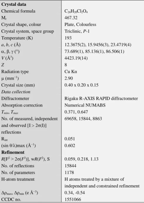
Table 1. Crystallographic data and structure refinement parameters of title compound.
Computer programs: PROCESS-AUTO (Rigaku, 1998), PROCESS-AUTO (Rigaku, 1998, CrystalStructure (Rigaku, 2007), SIR2004
(Burla et al., 2007), SHELXL97 (Sheldrick, 2008), ORTEPIII
(Burnett & Johnson, 1996).
Crystal data
Chemical formula C26H20Cl2O4
Mr 467.32
Crystal shape, colour Plate, Colourless Crystal system, space group Triclinic, P-1
Temperature (K) 193
a, b, c (Å) 12.3675(2), 15.9456(3), 23.4719(4)
, β, (°) 73.689(1), 85.136(1), 86.506(1)
V (Å3) 4423.19(14)
Z 8
Radiation type Cu Kα
µ (mm−1) 2.90
Crystal size (mm) 0.40 x 0.20 x 0.15
Data collection
Diffractometer Rigaku R-AXIS RAPID diffractometer
Absorption correction Numerical NUMABS
Tmin, Tmax 0.371, 0.647
No. of measured, independent and observed [I > 2σ(I)] reflections
69658, 15844, 8863
Rint 0.051
(sin θ/λ)max (Å−1) 0.602
Refinement
R[F2 > 2σ(F2)], wR(F2), S 0.059, 0.218, 1.13
No. of reflections 15844
No. of parameters 1178
H-atom treatment H atoms treated by a mixture of
independent and constrained refinement Δρmax, Δρmin (e Å−3) 0.34, -0.54
Figure 1. Molecular structures of (1R,2S )-1,2-bis(4-chlorophenyl)-3,8-dimethoxyacenaphthene-1,2-diol, with the atom-labelling scheme and displacement ellipsoids drawn at the 50% probability level: conformers G, B, R and Y are arranged at the top.
Table 2. Hydrogen-bond geometry (Å, ˚).
Cg2 = C1–C2–C3–C4–C10–C9 ring; Cg3 = C5–C10 ring; Cg30 = C79–C80–C81–C82–C88–C89 ring; Cg29 = C83–C88 ring; Cg21 = C57–C62 ring.
Results and discussion
The crystal of title compound has eight molecules of four types of conformers in an asymmetric unit. The conformers are labelled G (green), B (blue), R (red), and Y (yellow) as shown in Figure 2. Each conformer shows apparently the same topology of structure, i.e., phenyl rings in each conformer are oriented in the same direction with respect to the naphthalene ring plane (syn-orientation). These four conformers are grouped into two groups according to extent of overlap of phenyl ring planes, i.e., conformers G and Y have larger slippage of the phenyl ring planes than conformers B and R.
D―H…A D―H H…A D…A D―H…A
C30―H30···Cg2 0.95 2.99 3.735(5) 136
C51―H51···Cg3 0.95 2.92 3.685(5) 139
C56―H56···Cg30 0.95 2.88 3.660(4) 140
C77–H77...Cg29 0.95 2.93 3.772(4) 142
C83―H83···Cg21 0.95 2.91 3.705(5) 142
C12–H12B…Cl1 0.98 2.80 3.560(8) 135
C23–H23...Cl8 0.95 2.82 3.469(4) 127
O3―H3A···O12 0.84 1.91 2.702(4) 157
O11―H11···O7 0.84 1.88 2.708(4) 170
O8―H8···O16 0.84 1.85 2.677(4) 170
Figure 2. Four conformers classified into two groups by slippage of two phenyl rings: conformers G and Y, and conformers B and R
The respective dihedral angles between the phenyl rings in the four conformers are 36.4 (2)˚ for conformer G, 36.75 (19)˚ for conformer B, 39.26(19)° for conformer Y, and 41.13(19)˚ for conformer R. Dihedral angles between two phenyl rings and the naphthalene ring are 71.71(16) and 76.25(16)˚ for conformer B, 72.04(15) and 72.03(15)° for conformer R, 75.05(17) and 78.89(17)° for conformer Y, and 83.45(19) and 69.79(19)˚ for conformer G. Bond lengths of bridged C–C moiety in four conformers are longer than typical (sp3)C–C(sp3) bond. The bond lengths
are classified into two groups, i.e., 1.628 Å for conformer Y and 1.634 Å for conformer G, and 1.641 Å for conformer R and 1.642 Å for conformer B. The respective dihedral angles between naphthalene rings and five-membered rings are larger in order of 1.36(17)˚ for conformer B, 1.66(16)˚ for conformer R, 3.66(17)° for conformer Y, and 4.3(2)˚ for conformer G.
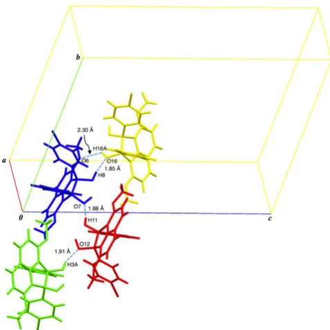
Figure 3. S-shaped tetramer composed four conformers Y, B, R and G.
Table 3. Single molecular structure data of conformers.
Dihedral angles between phenyl rings
{G}36.4 (2)˚
{B}36.75 (19)˚
{Y}39.26 (19)˚
{R}41.13 (19)˚
Dihedral angles between phenyl ring and naphthalene
{B}71.71(16), 76.25(16)˚
{R}72.04(15), 72.03(15)˚
{Y}75.05(17), 78.89(17)˚
{G}83.45(19), 69.79(19)˚
Bond lengths of bridged C–C bonds
{Y}1.628 Å
{G}1.634 Å
{R}1.641 Å
{B}1.642 Å
Dihedral angles between five-membered ring and naphthalene
{B}1.36(17)˚
{R}1.66(16)˚
{Y}3.66(17)˚
{G}4.3(2)˚
Torsion angles formed by bridged C–C–C–C bonds
{B}3.9(3)˚
{R}3.9(3)˚
{Y}11.4(3)˚
{G}12.8(4)˚
The torsion angles made by three bonds containing two carbons at 1- and 2-positions of the acenaphthene unit are 3.9(3)˚ for conformer B [C27–C39–C40–C34], 3.9(3)˚ for conformer R [C53–C65–C66–C60], 11.4(3)˚ for conformer Y [C79–C91–C92–C86], and 12.8(4)˚ for conformer G [C1– C13–C14–C8].
The structural data described above are summarized as Table 3. These single molecular structure data indicate that slippage of phenyl rings is related with distortion of five-membered ring moiety of the acenaphthene core.
Conformer B is linked with conformer R by classical O– H…OH hydrogen bond [{R}O–H…OH{B} hydrogen bonds (O11–H11…O7 = 1.88 Å)]. Conformers R and G are connected to each other by classical O–H…OH hydrogen bond [{G}O–H…OH{R} hydrogen bonds (O3–H3A…O12 = 1.91 Å)]. Whilst there is no classical O–H…O(H)
hydrogen bonds between conformers G and Y. Tetramers composed of four conformers are stacked into columnar structure along a-axis through non-classical C–H…Cl hydrogen bonds between conformers G [{G}(methoxy)C– H…Cl{G} (C12–H12B…Cl1 = 2.80 Å)] (Figure 4).
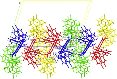
Figure 4. Waved sheet structure of tetramers formed by two types of non-classical C–H…Cl hydrogen bonds (dashed pink lines).
The columns are linked into a sheet structure by non-classical C–H…Cl hydrogen bonds between conformers G and Y along ab-diagonal [{G}(benzene)C–H…Cl{Y} (C23–H23…Cl8 = 2.82 Å)] (Figure 4). The waved sheets are interlocked by two types of non-classical C–H… hydrogen bonds forming stripe structure along c-axis, i.e., non-classical C–H… hydrogen bonds between conformers Y and R, and non-classical C–H… hydrogen bonds between conformers G and B [{R}(naphthalene)C– H···π(naphthalene){Y} (C56–H56···Cg30 = 2.88 Å; Cg30 = C79–C80–C81–C82–C88–C89 ring), {Y}(naphthalene)C– H···π(naphthalene){R} (C83―H83···Cg21 = 2.91 Å; Cg21
= C57–C62 ring) and {R}(benzene)C–
H…π(naphthalene){Y} (C77–H77…Cg29 = 2.93 Å; Cg29 = C83–C88 ring); {B}(benzene)C–H···π(naphthalene){G} (C51–H51···Cg3 = 2.92 Å; Cg3 = C5–C10 ring) and {B}(benzene)C–H···π(naphthalene){G} (C30–H30···Cg2 = 2.99 Å; Cg2 = C1–C2–C3–C4–C10–C9 ring)] (Figure 5).
As described in the preceding Results part, the four conformers have common topology that two aryl groups are non-coplanarly situated to the acenaphthene unit and oriented in the same direction against the acenaphthene ring, i.e., syn-conformation. Moreover, the four conformers are naturally divided into two pairs (conformers G and Y, and conformers B and R) from the viewpoint of the spatial organizations of the single molecular structures. The two conformers of the same pair have almost the same structural motif. Effective non-covalent bonding interactions extracted in the accumulation structure of conformers are taken into account the standpoint of crystal structure determining factors. First, classical O–H…O(H) hydrogen bonds are observed between conformers R and B, between conformers R and G, and between conformers B and Y. Secondary, non-classical C–H…Cl hydrogen bond between two molecules of conformer G along a-axis and that between conformers G and Y along ab-diagonal are observed. Three types of non-classical C–H… hydrogen bonds between conformers R and Y and two types of C– H… ones between conformers B and G are also observed. These effective hydrogen bonds are arranged in order of strength, i.e., classical O–H…O(H) hydrogen bonds, non-classical C–H…Cl hydrogen bonds, and non-non-classical C– H… hydrogen bonds. Based on the interpretation of preferential account of the strongest interaction, four conformers are linked into Y–B–R–G tetramer mother skeleton through classical hydrogen bonds. The central conformers B and R have essentially same spatial organization, and they are located with a pseudo-centrosymmetric center. The circumstances are essentially same for the alignment of conformers Y and G. Since four conformers have syn-conformation as common topology, each conformer has no centrosymmetric center in the molecule. In a natural consequence, they exhibit centrosymmetric center between the paired conformers. On the other hand, effective interactions between tetramers are unsymmetrically formed, especially for the interaction participated by conformers G and Y. Conformer G is linked with conformer G in the adjacent tetramer by non-classical C–H…Cl hydrogen bond along a-axis, and connected to conformer Y in neighboring tetramer by non-classical C– H…Cl hydrogen bond along ab-diagonal. Conformer R forms three types of non-classical C–H… hydrogen bonds with conformers Y in another tetramers, and conformer B
forms two types of non-classical C–H… hydrogen bonds with conformers G in another adjacent tetramers. The non-classical C–H…Cl hydrogen bonds two-dimensionally arrange the tetramers parallel to ab-plane, and weaker non-classical C–H… hydrogen bonds interlock the waved sheets forming the stripe structure along c-axis.
The molecular packing of title compound can be interpreted as follows: Four molecules related by centrosymmetric center are accumulated to form tetramer structure by stronger interactions of classical hydrogen bonding, which plays the prior function for determination of mother unit of the crystal. The tetramer discriminates the molecules into essentially two types of conformers according to the position in the tetramer, that is, the centered two molecules and the terminal ones. Consequently, the tetramer disproportionates to two kinds of conformers. The two kinds of conformers seem to be distinguished as four kinds of conformers by minute differences. On the basis of the existence of pseudo-centrosymmetricity in tetramer unit, a number of weak non-covalent bonding interactions contribute the arrangement of tetramer units forming second-ordered accumulation structure and the third-ordered one according to the strength of individual interaction. Naturally, the most stabilized accumulation crystalline structure needs to be perturbed. As a result, the crystal is composed of two types of conformers, each of which has two conformers having substantially same spatial organization with small differences.
Conclusion
both outer side resulting in formation of S-character like tetramer string bound by classical O-H…O(H) hydrogen bondings. The tetramers are connected with neighboured tetramer with two kinds of non-classical C–H…Cl hydrogen bondings at the terminal conformer molecules leading the waving planar aggregate on the plane parallel with a-axis. Furthermore, the planar aggregates are engaged with each other to be stacked along the c-axis. One terminal conformer molecule of a tetramer makes non-classical C– H…hydrogen bonding with a central conformer molecule of the adjacent tetramer to make the intertetramer connection. As described above, three kinds of non-covalent interaction of far different strength among the molecules of title compound in crystal are recognized to play governing factors to construct the higher ordered molecular accumulation structure by alignment of the molecules with different strength according to the spatial direction.
Though title compound has meso-form, syn-oriented structure prohibits to have centrosymmetric center in the inner side of molecule. In addition, two hydroxy groups are positioned at one side and the two phenyl groups are situated at the opposite side against acenaphthene plane. Such alignment makes the both side of acenaphthene ring largely different chemical environment. As a natural consequence, pseudo-centrosymmtrical dimeric aggregate formed by strong classical O-H…O(H) hydrogen bonding plays a role of coagulation core to be connected with two molecules at the both outer side by also strong classical O-H…OH hydrogen bonding, resulting in formation of tetramer molecular structure of pseudo-centrosymmetricity. The pseudo-centrosymmetric tetramers thus formed are coagulated to each other by rather weak interaction of non-classical hydrogen bonding in centrosymmetric fashion to yield highly ordered molecular aggregate structure. In the higher ordered structure, minimization of crystal energy might be achieved by adjustment the number and position of the non-classical hydrogen bondings to give rather small difference in the atomic alignment. Accordingly, this realizes the differentiation of molecules as four kinds of conformers belonging two types of spatial organization pattern.
Conclusively, the construction of crystal structure of title compound is recognized based on molecular motif of pseudo-centrosymmetric tetramer aggregation of molecules having largely differentiated aromatic faces connected by classical O-H…O(H) hydrogen bonding, where the tetramers accumulate with minimization of crystal energy by non-classical hydrogen bondings with perturbation of spatial organization of component molecules.
Acknowledgements
The authors would express their gratitude to Professor Keiichi Noguchi, Instrumentation Analysis Center, Tokyo University of Agriculture and Technology, for his technical advice. This work was partially supported by the Ogasawara Foundation for the Promotion of Science & Engineering, Tokyo, Japan.
References
1Desiraju, G. R. Acc. Chem. Res. 2002, 35, 565.
https://doi.org/10.1021/ar010054t
2Macháček, V., Bertolasi, V., Šimůnek, P., Svobodová, M.,
Svoboda, J. and Cernošková, E., Cryst. Growth Des., 2010,
10(1), 85. https://doi.org/10.1021/cg900295n
3Janiak, C., J. Chem. Soc., Dalton Trans. 2000, 3885.
https://doi.org/10.1039/b003010o
4Lehn, J.-M., Supramolecular Chemistry: Concepts and Perspectives; VCH: New York, 1995. https://doi.org/10.1002/3527607439
5Atwood, J. L., Davies, J. E. D., MacNicol, D. D. and Vogtle, F., In Comprehensive Supramolecular Chemistry; Pergamon, Oxford, UK, 1996; Vols. 1-11.
6Aakeröy, C. B. and Seddon, K. R., Chem. Soc. Rev., 1993, 397.
https://doi.org/10.1039/CS9932200397
7Desiraju, G. R., Crystal Engineering: The Design of Organic Solids; Elsevier: Amsterdam, 1989.
8Desiraju, G. R., Vittal, J. J. and Ramanan, A., Crystal Engineering. A Textbook, World Scientific Publishing, Singapore, 2011. https://doi.org/10.1142/8060
9Fourmigue, M. and Batail, P., Chem. Rev., 2004, 104, 5379.
https://doi.org/10.1021/cr030645s
10Zhong, Y-R., Cao, M-Li., Mo, H-J and Ye B-H., Cryst. Growth Des.,2008,8(7), 2282–2290.
ttps://doi.org/10.1021/cg700980v
11Okamoto, A. and Yonezawa, N., Chem. Lett.,2009, 38, 914–915.
https://doi.org/10.1246/cl.2009.914
12Okamoto, A., Mitsui, R. and Yonezawa, N., Chem. Lett., 2011, 40, 1283–1284. https://doi.org/10.1246/cl.2011.1283
13Okamoto, A. and Yonezawa, N., J. Synth. Org. Chem. Jpn., 2015, 73(4), 339–360.
https://doi.org/10.5059/yukigoseikyokaishi.73.339
14Takahara, G., Sakamoto, R., Ogata, K., Ohisa, S., Yokoyama, T.,
Yonezawa, N. and Okamoto, A., Eur. Chem. Bull., 2017,
6(1), 31-37. https://doi.org/10.17628/ecb.2017.6.31-37
15Okamoto, A., Tsumuki, T., Sasagawa, K., Siqingaowa and
Yonezawa, N. Eur. Chem. Bull. 2016, 5(6), 211-220. https://doi.org/10.17628/ecb.2017.5.211-220
16Okamoto, A., Muto, T., Siqingaowa, Takahara, G. and Yonezawa,
N., Eur. J. Chem., 2017, 8(1), 33-41.
https://doi.org/10.5155/eurjchem.8.1.33-41.1529
17Ogata, K., Nagasawa, A., Yonezawa, N. and Okamoto, A., Eur. J. Chem.,2017, 8(1), 20-24.
https://doi.org/10.5155/eurjchem.8.1.20-24.1530
18Siqingaowa, Tsumuki, T., Ogata, K., Yonezawa, N. and Okamoto,
A., Acta Cryst. 2016, E72, 1819-1823. https://doi.org/10.1107/s2056989016018077
19Okamoto, A., Nagasawa, A. and Yonezawa, N. Eur. J. Chem. 2014, 3(3), 263-268.
20Nakaema, Okamoto, A., Noguchi, K. and Yonezawa, N., Acta Cryst., 2007, E63, o4120.
https://doi.org/10.1107/s1600536807045114
21CCDC-1551066 contains the supplementary crystallographic data
for this paper. These data can be obtained free of charge from The Cambridge Crystallographic Data Centre via www.ccdc.cam.ac.uk/data_request/cif
22Armarego, W. L. F.; Chai, C. L. L. Purification of Laboratory Chemicals, Seventh edition, 2013, Elsevier Inc., Oxford.
23Rigaku (1998). PROCESS-AUTO. Rigaku Corporation, Tokyo,
24Rigaku (2010). CrystalStructure. Rigaku Corporation, Tokyo,
Japan.
25Burla, M. C., Caliandro, R., Camalli, M., Carrozzini, B.,
Cascarano, G. L., De Caro, L., Giacovazzo, C., Polidori, G., Siliqi, D. and Spagna, R., J. Appl. Cryst., 2007, 40, 609–613.
https://doi.org/10.1107/S0021889807010941
26Sheldrick, G. M., Acta Cryst., 2008, A64, 112–122.
https://doi.org/10.1107/S0108767307043930
27Burnett, M. N. and Johnson, C. K. (1996). ORTEPIII. Report
ORNL-6895. Oak Ridge National Laboratory, Tennessee, USA.