WEAK INTERMOLECULAR INTERACTION AND LATTICE
ENERGY ANALYSIS OF SOME BENZOTHIAZOLE
DERIVATIVES
Ratika Sharma
[a]and Rajni Kant
[a]*
Keywords: crystallography, benzothiazole, lattice energy, intermolecular interactions, hydrogen bonding.
A series of four chemically-similar-looking benzothiazole structures have been analyzed for their crystallographic comparison, weak intermolecular interaction analysis and lattice energy calculations.All the crystal structures are planar. However, the hydrazinyl group is slightly deviated with respect to the benzothiazole moiety in each structure. The N-H...N hydrogen bond plays an important role in the stabilization of the crystal packing in these derivatives. The related interactions of the type N-H...π, C-H...S and N....N act as an important linkage in most of the molecular pairs.A computational method has been used for the quantification of intermolecular interactions, as it allows partitioning of total interaction energy into corresponding coulombic, polarization, dispersion and repulsion contribution which facilitates a better understanding of the nature of intermolecular interactions contributing towards the crystal packing. The dispersive energy, however, lends significant contribution to the stabilization of these structures, thus making the combined nature of interaction energy in all the four molecules as predominately dispersive.
*Corresponding Author
[a] X-ray Crystallography Laboratory, Department of Physics and Electronics, University of Jammu, Jammu Tawi -180006, India.
Tel/Fax [O]: +91 191 243 2051 E-Mail: [email protected]
Introduction
Benzothiazoles, a class of sulfur and nitrogen containing heterocyclic compounds, have received considerable attention in recent years because of their medicinal and pesticidal importance. They possess considerable activity, including potent inhibition of human immune deficiency virus type 1 (HIV-1) replication by HIV-1 protease inhibition,1 antitumor,2 anthelmintic,3 analgesic and
anti-inflammatory,4 antimalarial,5 antifungal, anticandidal
activities6 and various activities related to the central
nervous system.7 As a part of our ongoing research work
on the preparation of X-ray diffraction quality single crystals and their X-ray structure analysis, we have taken up four chemically-similar-looking compounds for their comparative crystallographic analysis, weak interaction analysis and the energy contributions. The Crystallographic Information File (CIF) for each compound was obtained through the licensed CSD access. All important molecular motifs which provide maximum stabilization to the crystal structure were extracted and the nature and energy of these pairs was determined using PIXEL8.
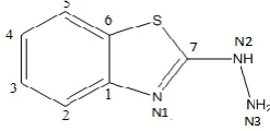
Figure 1. Chemical structure of benzothiazole moiety with atom numbering schemes
Structural features of benzothiazole structures
A representative view of benzothiazole moiety indicating the atomic numbering scheme is shown in Fig.1. The chemical name, molecular code and chemical structure for each compound is presented in Table 1.
Table 1. Chemical name, coding scheme and chemical Structure of the compounds
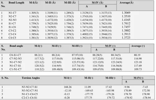
The precise crystallographic data for each compound is presented in Table 2. Most of the geometrical parameters, i.e., bond lengths and bond angles are in agreement with the literature values13-15. Some important bond lengths, bond
angles and torsion angles are presented in the Table 3. All the crystal structures are planar, except the hydrazinyl group which is slightly deviated with respect to the benzothiazole moiety.
Chemical Name Code Chemical Structure
2-Hydrazinyl- benzo[d]thiazole9
M-I
6-Chloro-5-fluoro-2-hydrazinyl
benzo[d]thiazole10
M-II
2-Hydrazinylmethyl- benzo[d]thiazole11
M-III
5-Chloro-2-hydrazinyl- benzo[d]thiazole12
M-IV
N S
NH NH2
N s
NH NH2
Cl F
N S
NH NH2
CH3
N S
NH NH2
Table 2. Precise crystallographic data of four benzothiazole derivatives
Table 3. Selected bond lengths, angles and torsion angles
S. No.
Bond angle M-I(◦) M-II (◦) M-III (◦) M-IV (◦) Average (◦) A B
1 C6-S1-C7 88.2(1) 88.2(4) 87.97(10) 88.28(5) 88.34(5) 88.19
2 C7-N2-N3 117.7(2) 117.01(8) 115.08(15) 117.22(8) 117.51(8) 116.90
3 N1-C7-N2 123.1(2) 122.9(9) 123.57(18) 123.12(9) 123.24(9) 123.18
4 N1-C7-S1 120.5(2) 116.9(9) 117.74(15) 116.75(8) 116.56(8) 117.69
5 C1-N1-C7 110.2(2) 109.48(8) 109.43(16) 109.67(9) 109.88(8) 109.73
S. No. Torsion Angles M-I(◦) M-II(◦) M-III(◦) M-IV(◦) A B
1 N3-N2-C7-S1 168.26 11.09 17.42 -9.96 -7.43
2 N3-N2-C7-N1 -12.10 -169.63 -165.98 170.89 172.50
3 N1-C1-C6-C5 -0.13 -177.75 -179.26 178.70 178.90
4 C2-C1-C6-S1 -0.28 177.75 179.72 -178.92 -178.94
The torsion around N2-C7 bond is insignificant. The hydrazinyl group located at the thiazole moiety is oriented in a synperiplanar (cis) or antiperiplanar (trans) conformation. In order to throw some more light on the molecular structure of benzothiazole derivatives, especially with regard to the role of hydrogen bonding, and the lattice energy calculations, the present work has been contemplated.
Hydrogen bonding analysis
Hydrogen bonding plays a key role in molecular recognition and crystal engineering. For this reason, crystal-packing studies are essential to understand the laws governing the intra- and inter-molecular H-bonding in a molecular crystal.
Data M-I M-II M-III M-IV
Formula C7H7N3S C7H5ClFN3S C8H9N3S C7H6ClN3S
No. of molecules per unit cell, Z 4 4 2 8
Crystal System Monoclinic Monoclinic Monoclinic Orthorhombic
Space group P21/n P21/c P21 Pca21
Cell parameters: a, Å
b, Å c, Å α, ◦ β, ◦
◦
10.839(5) 507552(5) 12.961(5) 90.00(5), 110.00(5) 90.00(5)
11.129(6) 5.664(3) 13.342(7) 90.00 108.55 90.00
3.893(2) 7.312(4) 14.137(8) 90.00 93.42(1) 90.00
13.023(13) 5.770(6) 21.708(2) 90.00 90.00 90.00
R-factor 0.037 0.024 0.028 0.019
S. No.
Bond Length M-I(Å) M-II (Å) M-III (Å) M-IV (Å) Average(Å) A B
1 N1-C7 1.305(3) 1.3109(11) 1.289(2) 1.3129(13) 1.3137(13) 1.3069
2 N2-C7 1.341(3) 1.3483(11) 1.375(3) 1.3743(10) 1.3437(10) 1.3510
3 N2-N3 1.413(3) 1.4172(10) 1.420(2) 1.4154(10) 1.4173(10) 1.4165
4 S1-C7 1.759(3) 1.7625(10) 1.756(2) 1.7639(10) 1.7621(10) 1.7612
5 S1-C6 1.748(2) 1.7429(9) 1.746(2) 1.7471(10) 1.7445(10) 1.7457
6 C3-C2 1.380(3) 1.3910(13) 1.389(3) 1.3877(15) 1.3935(14) 1.3882
7 C3-C4 1.385(4) 1.3977(13) 1.379(3) 1.4002(15) 1.3946(15) 1.3913
Table 4. Geometry of intermolecular hydrogen bonding in M-I – M-IV
The usual convention for the representation of the hydrogen bond is D-H…A, where D is the donor and A is the acceptor.
In hydrogen bonds, the distance between the hydrogen and the acceptor atom is shorter than the sum of their van der Waals radii16. Weak hydrogen interactions like C-H...π and have been recognized to play an important role in crystal structures of small biological molecules and protein structures17-19.
Table 5. Hydrogen bonding data
All the crystal structures are stabilized by N-H...N hydrogen bonding except for the molecule I which includes weak N-H...π interactions as well. The geometry of intra-and intermolecular hydrogen bonds is presented in Table 4. All the interaction parameters [H...A, D...A and θ] have
been compared with the literature values18 [Table 5] and
following observations have been made:
The average values of H...N distance in molecule I-IV are 2.29, 2.26, 2.28 and 2.20 Å, respectively. Thus making these interactions fall under the category of strong to weak interactions with over all range comes out to be (2.04-2.54). A weak N-H...π interaction has also been identified in molecule-I that plays a crucial role in the stabilization of its crystal structure.
The average values of N-H...N distance in molecules I- IV are 3.09, 3.03, 4.51, 3.01 Å, respectively, thereby making such interactions fall under the category of strong to weak interactions. The angular range [θ(D-H...A)] of 135º -176º also supports this observation.
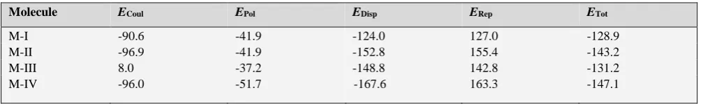
Energy calculations and discussion
In order to dwell more on the role of hydrogen bonding in small molecular assemblies of this kind, we have computed lattice energies of all the compounds by using the Coulomb-London-Pauli (CLP) model of intermolecular coulombic, polarization, dispersion and repulsion energies20, known as
the PIXEL method. Two output files are generated after the end of the calculation. The first (.pri file) consists of the total lattice energies partitioned into their coulombic, polarization, dispersion and repulsion contributions (Table 6). The second (mlc file) consists of molecule–molecule interaction energy along with the symmetry elements which relate to the molecules.
Table 6. Lattice energy from CLP (in kJ mol-1)
Molecule D-H…A D-H, Å H…A, Å D…A, Å θ[D-H…A, ◦]
M-I N-H…N
N-H…N N-H…N 0.90 0.82 2.44 2.04 2.54 2.54 2.93 3.23 3.34 175.0 144.0 167.0
M-II N-H…N
N-H…N 0.89 0.92 2.30 2.21 2.99 3.07 135.0 156.0
M-III N-H…N
N-H…N 0.81 0.85 2.13 2.44 2.94 3.13 176.9 139.5
M-IV N-H…N
N-H…N N-H…N 0.89 0.89 0.83 2.03 2.05 2.53 2.90 2.95 3.17 170.5 175.3 135.6
Properties Very strong
Strong Weak Molecule. I-IV
D…A range, Å
2.2-2.5 2.5-3.2 3.0-4.0 2.90-3.34
H…A range, Å
1.2-1.5 1.5-2.2 2.0-3.0 2.04-2.54
θ( D-H…A) range, (◦)
175-180 130-180 90-180 135-176
Effect on crystal packing
Strong Distinctive Variable Variable
Molecule ECoul EPol EDisp ERep ETot
M-I -90.6 -41.9 -124.0 127.0 -128.9
M-II -96.9 -41.9 -152.8 155.4 -143.2
M-III 8.0 -37.2 -148.8 142.8 -131.2
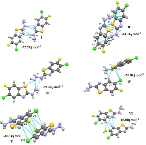
The interaction energy of selected molecular pairs (from the .mlc file), extracted from the crystal packing along with the involved intermolecular interactions are listed in Table 7, with the total energies being partitioned into their coulombic, polarization, dispersion and repulsion contributions. The molecular pairs are arranged in decreasing order of their stabilizing energies. As it is shown in the Table 6 that energies of molecule M-I and M-IV and that of II and III are comparable and the molecules M-II and M-M-III are more stable than M-I and M-IV. The combined nature of the interaction is predominately in all the four molecules is dispersive. The geometrical restrictions placed on the intermolecular H-bonds present in the selected molecular pairs are the sum of the Vander Waals radii + 0.3 Å and the directionality is greater than 110º. A precise description of each molecule with regard to its energy contribution and molecular pair formation is presented:
(M-I) 2-
hydrazinyl benzo[d]thiazoleAll the molecular pairs (I-IV) extracted from the crystal packing are shown in the Fig.2 having an interaction energy of -72.7 kJ mol-1. The most stabilizing molecular pair shows
the presence of N-H…N hydrogen bonding which leads to the formation of R2
2(8)21 graph-set motif. The second most
prominent interaction in this case is three centered bifurcated N-H...C hydrogen bonding with N1- H21 act as a donar for both the acceptor elements C1 and C7.
Figure 2. M-I: Molecular pair formations with their interaction energy contributions
The second most stabilized molecular pair in the crystal structure, formed via C-H…Cg-I molecular interactions (Cg-I is Centeroid of ring C1/C2/C3/C4/C5/C6), has a contribution of -50.3 kJ mol-1 (50% contribution to
stabilization from the dispersion energy) to the stabilization of the packing. These molecular stacks are interlinked via weak N-H…S, S-H…N bonding (motif III,-23.0 kJ mol-1)
and C-H…S and C2-N3 (motif IV, -15.0 kJ mol-1)
(M-II) 6-Chloro-5-fluoro-2-hydrazinyl benzo[d]thiazole
All the molecular pairs (I-VI) extracted after the PIXEL calculation are represented in Fig.3 along with their interaction energies. In this molecule the hydrogen atoms at the position 3 and 4 are replaced by Chlorine and Fluorine element.
Figure 3. M-II: Molecular pairs along with their interaction energies
Here motif I is the most stabilized pair with an interaction energy of –72.2 kJ mol-1 in the crystal packing and consist
of interaction involving H21 atom in three hydrogen bonds [N-H…C (involving H21 with C1 and C7) and N1-H21…N2] leads to the bifurcated hydrogen bonding. The nature of the interaction is governed by the couloumbic and repulsive energy with their maximum contribution to total stabilization energy. The motif II is the second most stabilized pair with interaction energy -44.1kJ mol-1
involving weak N-H…Cg-I(Cg-I is Centeroid of ring C1/C2/ C3/C4/C5/C6) N…N and N-H…S interactions. In rest of the motifs of this molecule the nature of the stabilized energy is predominately dispersive as this part of the energy contributes more than 70% to the total interaction energy except motif-IV (to which its contribution is around 40%), with energy interaction ranges from -18.0 to -24.8 kJ mol-1.
In these motifs the structure is stabilized by weak C-H…C, C-H…S, C…C, C…F, N…C and C...Cl interaction.
(M-III)
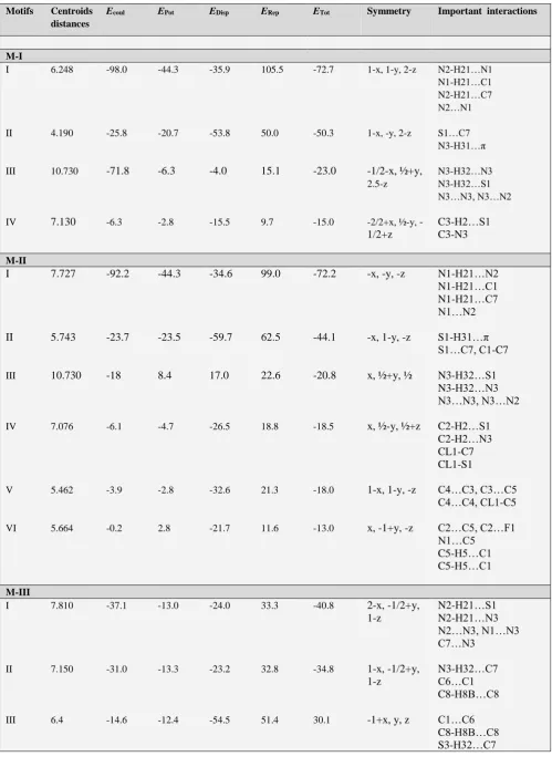
2-hydrazinylmethyl benzo[d]thiazoleTable 7. PIXEL interaction energies (I. E.) kJ mol-1 between molecular pairs related by a symmetry operation and the associated molecular interaction in the crystal
Motifs Centroids distances
Ecoul EPot EDisp ERep ETot Symmetry Important interactions
M-I
I 6.248 -98.0 -44.3 -35.9 105.5 -72.7 1-x, 1-y, 2-z N2-H21…N1
N1-H21…C1 N2-H21…C7 N2…N1
II 4.190 -25.8 -20.7 -53.8 50.0 -50.3 1-x, -y, 2-z S1…C7
N3-H31…π
III 10.730 -71.8 -6.3 -4.0 15.1 -23.0 -1/2-x, ½+y,
2.5-z
N3-H32…N3 N3-H32…S1 N3…N3, N3…N2
IV 7.130 -6.3 -2.8 -15.5 9.7 -15.0 -2/2+x, ½-y,
-1/2+z
C3-H2…S1 C3-N3
M-II
I 7.727 -92.2 -44.3 -34.6 99.0 -72.2 -x, -y, -z N1-H21…N2
N1-H21…C1 N1-H21…C7 N1…N2
II 5.743 -23.7 -23.5 -59.7 62.5 -44.1 -x, 1-y, -z S1-H31…π
S1…C7, C1-C7
III 10.730 -18 8.4 17.0 22.6 -20.8 x, ½+y, ½ N3-H32…S1
N3-H32…N3 N3…N3, N3…N2
IV 7.076 -6.1 -4.7 -26.5 18.8 -18.5 x, ½-y, ½+z C2-H2…S1
C2-H2…N3 CL1-C7 CL1-S1
V 5.462 -3.9 -2.8 -32.6 21.3 -18.0 1-x, 1-y, -z C4…C3, C3…C5
C4…C4, CL1-C5
VI 5.664 -0.2 2.8 -21.7 11.6 -13.0 x, -1+y, -z C2…C5, C2…F1
N1…C5 C5-H5…C1 C5-H5…C1
M-III
I 7.810 -37.1 -13.0 -24.0 33.3 -40.8 2-x, -1/2+y,
1-z
N2-H21…S1 N2-H21…N3 N2…N3, N1…N3 C7…N3
II 7.150 -31.0 -13.3 -23.2 32.8 -34.8 1-x, -1/2+y,
1-z
N3-H32…C7 C6…C1 C8-H8B…C8
III 6.4 -14.6 -12.4 -54.5 51.4 30.1 -1+x, y, z C1…C6
M-IV [A...B]
I 7.754 -95.9 -46.0 -34.9 104.8 -71.9 ½+x, 2-y, z N2A-H21A…NIB
N2B-H21B…N1A N2A-H21A…C7B N2B-H21B…C7A N1A-N2B, N2A…N1B
II 5.448 -26.8 -22.2 -57.2 59.5 -46.6 ½+x, 1-y, z N3B-H32B… π
N3A-H32A… π S1A…C7B, C7A-S1B
III 9.946 -21.2 -8.6 -18.0 24.2 -23.6 x, y, z N3B-H2B…S1A
N3B-H2B…N3A N2B-N3A
IV 10.037 -16.7 -7.0 -16.7 18.9 -21.6 x, 1+y, z CL1B… π
C1A, C4A C5A, C6A
V 6.443 -3.2 -3.1 -24.8 16.2 -14.9 x,
1-y1/2+z
CL1A…C1B
CL1A…C6B C3A-H3A…CL1B C3A-H3A…C4B
VI 7.101 -3.5 -1.8 -14.3 6.5 -13.1 ½-x, y, ½+z CL1A…H3B
M-IV [A....A]
I 7.085 -7.4 -4.3 -19.2 14.2 -16.7 -1/2+x, 1-y,
z
C2A…H2A-N3A
N3A-H31A…C1A C2A-H2A…S1A S1A…C2A N3A…C2A
II 5.777 -2.5 -3.3 -22.8 13.4 -15.2 x, -1+y, 2-y CL1A…C2A
C2A…C5
M-IV [B…B]
I 6.966 -7.5 -4.5 -20.2 15.9 -16.2 -1/2+x, 1-y,
z
C2B…SIB
N3B…C2B C2N-H2B…S1B
II 5.777 -1.4 -2.8 -21.8 11.3 -14.7 x, y-1, z CL1B…C2B
C5B… C2A
In the first two motifs the maximum stabilization energy comes from the coulombic part where as the motif-III is stabilized by the dispersive energy as in this case it contributes more than 50% to the total stabilization energy. The motif-I is stabilized due to the presence of weak N-H…S and N-H…N (H21, N2, N3 & S1) interaction. It also involves the N1...N3, N2…N3 and N3…C7 interaction. The next two stabilized molecular pairs (II and III) involve C-C molecular stacking, however along with this interaction the motif-II shows the presence of N-H…C where as motif-III is shows the presence of N-H...N hydrogen bonding.
(M-IV)
5-chloro-2-hydrazinyl benzo[d]thiazoleFigure 4. M-III: Molecular pair formations with their interaction energy contributions
Figure 5a. M-IV: Molecular pair formations with their interaction energy contributions
(i) A-B interaction motifs
The two molecules in the asymmetric unit are packed with the involvement of dimeric interactions represented by the graph set motif R2
2(8) formed with a strong N-H…N and
weak N-H…C (involving N1B and C7B with bifurcated donar pair N2A-H21A) hydrogen bonds and this pair is the most stabilized pair in the crystal, energy being -71.9 kJ mol-1 and the principal stabilization of around 50%
corresponding to coulombic component. The second most stabilized molecular pair (motif II) in the crystal structure is stacked via N-H…π interaction involving N3-H32 and benzene ring of both the asymmetric unit A and B along with C...S interactions and has a contribution of -46.6 kJ mol-1 to the stabilization of the crystal packing. In motif-III
the two asymmetric units A and B are connected by bifurcated N3B-H32B…S1A and N3B-H32B…N3A
hydrogen bonding with N3B-H32B acts as a common donar to both the elements. This interaction has maximum contribution from coulombic and repulsive part with total stabilization energy of value -23.6 kJ mol-1. In rest of these
motifs the two symmetry units are linked through the weak bonding involving chlorine atom with C3B-CL1B…π in motif-IV[ where π is the centeroid of the benzene ring of molecule A in the asymmetric unit], bifurcated hydrogen bonding in motif-V [involves C3-H3… Cl1 in both the symmetric units A and B] represented by the graph set motif R4
2(8) and C3B-H3B…CL1A hydrogen bonding in the case
of motif VI.
(ii) A-A interaction motifs
The two important motifs (I, II) showing the interaction between the two similar molecules (A, A) in the asymmetric unit are extracted from crystal packing along with their stabilization energies are represented in the Fig. 5b. Both these interactions motifs are dispersive in nature as it contributes maximum to the total stabilization energies -16.7 and -15.2 kJ mol-1 in case of motif I and II respectively. The
motif I is stabilized with the presence of C2A-H2A…S1A, S1A…C2A and N3A…C2A and motif II shows the involvement of C3A-Cl1A…C2A and C5A…C2A for the stabilization of the molecular packing.
Figure 5b. M-IV: Molecular pair formations with their interaction energy contributions in A-A interaction
Figure 5b. M-IV: Molecular pair formations with their interaction energy contributions in B-B interaction
(iii) B-B interaction motifs
the motif I and II respectively in A-A interaction and results in the generation of similar packing motifs. In this case the energies are predominately dispersive in nature with the total energy values -16.2 and -14.7 kJ mol-1 in motif I and II
which are quite similar as we have seen in above described A-A interactions.
Conclusions
An analysis of the energetics of the neighbouring molecular pairs of four thiazole derivatives shows the presence of different intermolecular interactions participating in the crystal packing. The total interaction energy among different molecular pairs has been divided into corresponding coulombic, polarization, dispersion and repulsion contribution which facilitates a better understanding of the nature of intermolecular interactions contributing towards the crystal packing. The dispersive energy contributes maximum to the stabilization of the structure in all the four molecules. Hence the combined nature of the interaction energy in all the four molecules is predominately dispersive. It has been found that N-H...N hydrogen bonds plays an important role in the stabilization of the crystal packing. Along with these hydrogen bonds, N-H...π, C-H...S and N....N interaction has also become the important linkage in most of the molecular pairs. It is of interest to extend this evaluation of energies of molecular pairs in other thiazole derivatives which will enable us to have better understanding of weak intermolecular interactions.
Acknowledgement
One of the authors (Rajni Kant) acknowledges the Department of Science & Technology for single crystal X-ray Diffractometer as a National Facility under Project No. SR/S2/CMP-47/2003 and DST Project No: EMR/2014/000467. RS acknowledges University Grant Commission (UGC) for the award of NET-JRF scholarship under ref. No. 23/12/2012 (ii) EU-V.
References
1Liu, X. F., Liu, X.-H., Acta Cryst.,2011, E67, o202.
2Liu, X. H., Tan, C. X., Weng, J. Q., Phosphorus Sulfur Silicon
Relat. Elem.,2011a, 186, 552-557.
3Liu, X. H., Tan, C. X., Weng, J. Q., Phosphorus Sulfur Silicon
Relat. Elem.,2011b, 186, 558-557.
4Yaseen, A., Haitham, A. S., Houssain, A. S., Najim, A., Z.
Naturforsch. Teil B., 2006, 62 523–528.
5Kini, S., Swain, S. P., Gandhi, A. M., Indian J. Pharm. Sci., 2007,
69, 46–50.
6Munirajasekhar, D., Himaja, M., Sunil, V. M., Int. Res. J.
Pharm.,2011, 2, 114–117.
7Gurupadayya, B. M., Gopal, M., Padmashali, B., Manohara, Y. N., Indian J. Pharm. Sci.,2008, 70, 572–577.
8Gavezzotti, A., New J. Chem., 2011, 35, 1360-1368.
9Sharma, R., Prakash , S. N., Narayana, B., Gupta, V.K., Kant, R.,
Eur Chem. Bull.,2014, 3(4), 337-339.
10Hoong-Kun, F., Ching, K. Q, Sarojini, B. K., Mohan,B. J., Narayana, B., Acta Cryst,.,2012, E68, o2459.
11Xu-Feng, L., Xiao-Yong, Y., Shao-Liang, J., Acta Cryst., 2011,
E67, o1641.
12Hoong-Kun, F., Chin W. O., Sarojini., B. K., Mohan, B. J., Narayana,B., Acta Cryst., 2012, E68, o691–o692.
13Burke-Laing, M., Laing, M., Acta Cryst., 1976, B32, 3216- 3224.
14Allen, F. H., Kennard, O., Watson, D. G., Brammer, L., Orpen, A. G.,Taylor, R.., J. Chem. Soc. Perkin Trans.,1987, 2, S1– 19.
15Fun, H.-K., Arshad, S., Himaja, M., Munirajasekhar, D., Sarojini, B. K., Acta Cryst.,2011a, E67, o2412.
16Taylor, R., Kenard, O., J. Am. Chem. Soc., 1982, 104, 5063-5070.
17Hunter, C. A., Sanders, J. K. M., J. Am. Chem. Soc., 1990,112, 5525-5534.
18Desiraju, G. R., Steiner, T., The Weak Hydrogen Bond in
Structural Chemistry and Biology, 1999, Oxford University Press, Oxford.
19Calhorda, M. J., Chem. Commun..,2000, 801-809.
20Dunitz, J. D., Gavezzotti, A., Cryst. Growth Des., 2005,5, 2180-2189.
21Bernstein, J., Davis, R. E., Shimoni, L., Chang, N.L., Angew.
Chem. Int. Ed. Engl., 1995, 34, 1555–1573.