ARTIGO ORIGINAL
RESUMO
Introdução: A gangrena de Fournier é uma infeção polimicrobiana potencialmente fatal que afeta os tecidos moles do períneo com ponto de origem em patologias urológicas, coloretais ou cutâneas. Apesar de ser mais frequente no sexo masculino e em idosos, pode afetar ambos os géneros e qualquer idade. O abcesso perianal, a diabetes mellitus e a Escherichia coli são respetivamente a causa, a co-morbilidade e o micro-organismo mais frequentemente encontrados. Este estudo teve como objetivo descrever a experiência de um Serviço de Cirurgia Plástica e Queimados de um Hospital terciário no tratamento e reconstrução de defeitos perineais causados por gangrena de Fournier, disponibilizando detalhes sobre a sua demografia.
Material e Métodos: A amostra é constituída por todos os doentes internados no serviço de Cirurgia Plástica e Queimados com o diagnóstico de gangrena de Fournier. Os autores realizaram uma colheita e análise retrospetiva de dados clínicos e demográficos durante um período de 10 anos incluindo género, idade, tempo de internamento, causa, número de desbridamentos, fatores predispo-nentes, resultados microbiológicos de culturas de pus, técnicas reconstrutivas cirúrgicas e suas complicações, intervenções cirúrgicas adicionais e o resultado final.
Resultados: Foram identificados 15 doentes: 14 homens (93%) e uma mulher (7%); a idade média foi 66,9 anos (amplitude: 46 - 86); tempo médio de internamento foi 46,8 dias (amplitude: 20 - 71 dias) e o número médio de desbridamentos foi 3,3 (amplitude: 1 - 4). O fator predisponente mais frequente foi a diabetes mellitus, e as causas mais frequentes o abcesso perianal (n = 2) e o abcesso cutâneo (n = 2). Em oito (53,3%) doentes não foi identificada a causa da gangrena de Fournier. Foram utilizadas várias técnicas reconstrutivas e realizadas conco (33,3%) intervenções cirúrgicas adicionais (uma cistostomia, duas orquidectomias, duas ileostomias); seis doentes (40%) apresentaram complicações de técnicas reconstrutivas com resultado final adequado.
Discussão: O micro-organismo mais frequentemente isolado nas culturas de pus foi o Staphylococcus aureus, o que contrasta com a literatura onde a Escherichia coli é o agente mais frequentemente isolado. Foi identificado um número superior ao esperado de
Fournier’s Gangrene: 10-Year Experience of a Plastic
Surgery and Burns Department at a Tertiary Hospital
Gangrena de Fournier: 10 Anos de Experiência de um
Serviço de Cirurgia Plástica e Queimados num Hospital
Terciário
1. Department of General Surgery. Coimbra Hospital and University Centre. Coimbra. Portugal. 2. Department of Plastic Surgery and Burns. Coimbra Hospital and University Centre. Coimbra. Portugal.
Autor correspondente: João Mendes Louro. [email protected]
Recebido: 01 de julho de 2018 - Aceite: 10 de dezembro de 2018 | Copyright © Ordem dos Médicos 2019
João Mendes LOURO1, Miguel ALBANO1, João BALTAZAR2, Miguel VAZ2, Carla DIOGO2, Sara RAMOS2, Luís CABRAL2
Acta Med Port 2019 May;32(5):368-374 ▪ https://doi.org/10.20344/amp.11003
ABSTRACT
Introduction: Fournier gangrene is a polymicrobial life threatening infection of perineal subcutaneous soft tissues with its point of origin in urologic, colorectal or skin diseases. Although more frequent in elderly and men, it can affect all genders and age groups. Perianal abscess, diabetes mellitus and Escherichia coli are the most frequent cause, predisposing comorbidity, and microorganism found in tissue culture analysis respectively. The objective of this study was to describe the experience of a Plastic Surgery Department of a tertiary Hospital in reconstructing Fournier’s gangrene perineal defects and its detailed demography.
Material and Methods: The sample is composed of all patients with Fournier gangrene admitted in the Plastic Surgery and Burns Department. The authors retrospectively collected and analyzed demographic and clinical data during a period of 10 years including gender, age, length of stay, cause, number of debridements, predisposing factors, microbial culture results, surgical reconstructive techniques and its associated complications, additional surgical procedures and outcomes.
Results: Fifteen patients were identified: 14 males (93%) and one female (7%); mean age was 66.9 years (range: 46 - 86); mean, length of stay was 46.8 days (range: 20 - 71 days) and mean number of debridements was 3.3 (range: 1 - 4). The most frequent predis-posing factor was diabetes mellitus, the major cause was perianal (n = 2) and skin abscess (n = 2). Eight (53.3%) patients had no iden-tifiable source of Fournier gangrene. Various types of reconstructive techniques were employed; and 5 additional surgical interventions (33.3%) were undertaken (one cystostomy, two orchidectomy, two ileostomy); six patients (40%) presented reconstructive technique complications with adequate final outcome.
Discussion: In contrast with the literature, where Escherichia coli was the most frequently isolated agent, Staphylococcus aureus was the most frequent microorganism found in tissue biopsy/pus collection analysis. A higher than expected number of patients (n = 8) had no identifiable source of Fournier gangrene. This findings can be explained by the retrospective non-multicentre study limitation, with a potencial source of bias patients that were transferred from other hospitals in advanced stage, without point of origin of Fournier’s gangrene identified.
Conclusion: Early recognition and extensive necrotic tissue debridement, along with prompt and adequate antimicrobial treatment, are the mainstay of Fournier gangrene management, thus reducing morbidity and mortality in these patients. Surgical reconstruction chal-lenges derived from this condition should be addressed by specialized teams due to the risk of dysfunctional sequelae and conspicuous deformities. Taking in account the single-center and retrospective observational character of the present study, these premises require proper validation from a multicenter prospective study.
ARTIGO ORIGINAL doentes sem causa identificável (n = 8) de gangrena de Fournier. Estes achados podem ser explicados pelo facto de se tratar de um
estudo retrospetivo multicêntrico, com um potencial viés por existirem doentes que foram transferidos de outras institucões em estado avançado de doença, sem foco de origem de gangrena de Fournier identificado.
Conclusão: O precoce reconhecimento e extenso desbridamento do tecido necrosado, em conjunto com um adequado tratamento antibiótico, são os pilares do tratamento da gangrena de Fournier reduzindo assim a morbilidade e mortalidade destes doentes. Os desafios cirúrgicos reconstrutivos que advêm desta patologia devem ser abordados por uma equipa especializada, pelo risco de se-quelas funcionais e estéticas. Tendo em conta o carater observacional, retrospetivo e unicêntrico do presente estudo, estas premissas requerem uma validação adequada através de um estudo prospetivo e multicêntrico.
Palavras-chave: Cirurgia Plástica; Fasciite Necrosante; Gangrena de Fournier/cirurgia; Portugal; Procedimentos Cirúrgicos Recons-trutivos
INTRODUCTION
Fournier’s gangrene (FG) is a severe infection affect-ing subcutaneous soft tissues of the perineum, perianal and genital zones. It results from a breach in the integrity of the urethral or gastrointestinal mucosa, creating a rapidly pro-gressive and life threatening type of necrotizing fasciitis.1,2 Muscle cells are less frequently affected due to its rich vas-cularisation.3 This condition is more frequently diagnosed in males (10:1) and in the elderly,4,5 but may affect patients of all genders and ages. Despite a low prevalence (3 - 7/ 1 000 000)6, mortality rate can reach up to 40%.7
FG is usually a symbiotic polymicrobial infection caused by aerobic and anaerobic bacterial flora, arising from the low gastrointestinal (i.e. perianal/ischiorectal abscess) or genitourinary tracts, (i.e. urinary tract infection, traumatic catheterization) or from the skin (i.e. perineal skin abscess, allergic reactions).8 Immunosuppressed patients due to concomitant conditions like diabetes mellitus (DM), chronic ethanol abuse, malignancies, liver cirrhosis, HIV infection, organ transplantation or steroids use have a higher risk of developing the disease.4,7
Early recognition of clinical signs is imperative: the spread rate of the necrotizing fasciitis can be as high as 2 – 3 cm/ hour, rapidly progressing to the gluteal muscles, scrotum, penis, abdominal and thoracic wall.9-11 The clini-cal presentation is variable, but suspicion should be high when abrupt severe pain is present and associated with ec-chymosis, fever, and cutaneous anaesthesia; rarely, sub-cutaneous gas with crepitation may be present.12-14 Con-ventional X-rays may show subcutaneous gas, however, the best imaging modality is a computed tomography (CT) scan that allows a more precise assessment of the exten-sion and depth of the infection as well as the presence of abscesses.15,16
Early antibiotic administration and urgent surgical drain-age along with debridement of necrotic tissues are critical for sepsis control in FG. Debridements may create major defects presenting great reconstructive challenges.17 The purpose of this study is to analyse demographic, clinical and surgical data of all patients with FG admitted at the Plastic Surgery and Burns Department of a tertiary hospital, in Coimbra, Portugal, during a ten-year period and compare it to the available literature.
MATERIALS AND METHODS
The authors state that STROBE checklist was complet-ed.
Study population
Observational retrospective analysis of patients with Fournier’s gangrene admitted to the Plastic Surgery and Burns Department of Centro Hospitalar e Universitário de Coimbra (Hospital and University Centre of Coimbra - CHUC), Coimbra,Portugal, during a period of 10 years (from January 1st, 2007, to December 31st, 2016).
Demographic and clinical data were collected from med-ical records including age, gender, predisposing factors/ae-tiology, comorbidities, number and nature of surgical inter-ventions, culture findings, length of stay (LOS) and clinical outcome.
Retrospectively reviewing the medical records allowed us to only include in our cohort, patients with confirmed FG on histology; this process minimised selection bias.
Limitations of the study
Retrospective non-multicenter study.
Statistical analysis
All data were collected and analysed using Microsoft® Excel® for Mac 2011, version 14.7.1 (161129).
RESULTS
Fifteen patients with FG were identified: fourteen males (93%) and one female (7%). The mean age was 66.9 years (range: 46 - 86). The mean LOS was 46.8 days (range: 20 - 71 days). The mean number of debridements was 3.3 (range: 1 - 4). There were no fatalities in this group of pa-tients (Table 1).
The most common identified causes were perianal (n = 2: 13.3%) and skin abscesses (n = 2: 13.3%); other causes included bartholinitis (n = 1: 6.7%), perineal trauma (n = 1: 6.7%) and hernioplasty suture site infection (n = 1: 6.7%). In 8 patients (53.3%) there was no identifiable point of ori-gin of FG. DM was the most frequent comorbidity (n = 6: 40%); other predisposing factors were identified namely
Table 1 – Demographic and clinical data
n (% / range) Gender
Male
Female 14 (93%)1 (7%)
Age (range) 66.9 (46 - 86)
LOS (range) 46.8 (20 - 71)
ARTIGO ORIGINAL
monoclonal gammopathy of undetermined significance, chronic renal failure, alcoholic cirrhosis and colon cancer, with one case each (Table 2).
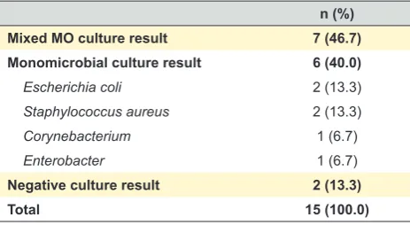
Cultures of pus were obtained in all 15 surgical wounds (Table 3): seven patients (46.7%) had mixed microorgan-isms (MO) (mixture of aerobic ± anaerobic ± fungus); 6 pa-tients (40%) had one bacterial isolation, and two papa-tients (13.3%) had negative microbiological results. The most frequent MO found was Staphylococcus aureus (n = 7: 46.7%), followed by Enterococcus faecalis (n = 5: 33.3%),
Escherichia coli (n = 3, 20%), Acinetobacter baumannii (n = 3, 20%) and Pseudomonas aeruginosa (n = 2, 13.3%); other bacteria were only present in one culture each: Strep-tococcus pyogenes, Enterococcus faecium, Enterococcus cloacae, Klebsiella pneumoniae, Streptococcus epidermid-is, Bacteroides fragilepidermid-is, Corynebacterium, Candida albicans and Aspergillus fumigatus. Fig. 1 shows all MO found in culture results. Monomicrobial bacteria culture identification was present in 6 patients: Escherichia coli (n = 2, 13.3%),
Staphylococcus aureus (n = 2, 13.3%), and Corynebacte-rium and Enterobacter cloacae isolated in one patient each. One patient had multidrug resistant Staphylococcus aureus. Anaerobic bacteria (Bacteroides fragilis) were identified in one patient (6.7%).
All patients were subjected to reconstructive surgical
procedures (Table 4). In 5 patients (33.3%) the only re-constructive surgery performed was a split thickness skin graft (STSG). Eight patients (53.3%) had one reconstructive surgery with flaps (one scrotal reconstruction with bilateral internal pudendal pedicled flaps; one scrotal reconstruction with contralateral rotational flap; one internal thigh bilateral fasciocutaneous transposition flaps; one vulvar reconstruc-tion with McGregor propeller flap; one pedicled anterolat-eral thigh (ALT) flap with tunnelling to the defect; one local sliding flap; one medial femoral circumflex artery perfora-tor fasciocutaneous flap; one internal thigh rotational flap). One patient (6.7%) needed two flap reconstructive surger-ies (scrotal reconstruction with bilobed internal thigh flaps and fasciocutaneous transposition flaps for perineal defects closure, and one patient (6.7%) had a full thickness skin graft for reconstruction of a degloved penis shaft. Deriva-tive ileostomy was performed in two patients (13.3%), and diverting cystostomy in one (6.7%). In addition to scrotal, vulvar and perineal reconstruction, two patients (13.3%) re-quired unilateral orchidectomy (Table 4).
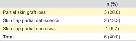
Regarding complications, three patients (20%) pre-sented partial skin graft loss, two patients (13.3%) suffered skin flap partial suture dehiscence and there was one case (6.7%) of partial necrosis of the skin flap (Table 5).
DISCUSSION
FG is characterized by a rapidly progressive poten-tially fatal necrotizing infection of the external genitalia and perineum that can spread along Buck’s fascia of the penis, Colle’s fascia of the perineum and extend to Scarpa’s fascia of the anterior abdominal wall, sometimes even reaching the thoracic wall. This necrosis is caused by vessel oblitera-tion related to polymicrobial proliferaoblitera-tion along the fascial plane, generating oedema, microthrombosis and hypoxia, favouring anaerobic bacteria overgrowth.18 In the present study, only one patient was confirmed to be infected with anaerobic bacteria (Bacteroides fragilis). Regardless, one should consider that false negatives for anaerobic bacteria
Table 3 – Culture results
n (%) Mixed MO culture result 7 (46.7) Monomicrobial culture result 6 (40.0)
Escherichia coli 2 (13.3)
Staphylococcus aureus 2 (13.3)
Corynebacterium 1 (6.7)
Enterobacter 1 (6.7)
Negative culture result 2 (13.3)
Total 15 (100.0)
Table 2 – Causes and predisposing factors
n (%) Causes
Idiopathic 8 (53.3)
Perianal abscess 2 (13.3)
Skin abscess 2 (13.3)
Bartholinitis 1 (6.7)
Perineal trauma 1 (6.7)
Hernioplasty suture site infection 1 (6.7)
Predisposing factors
Diabetes mellitus 6 (40.0)
No isolated predisposing factor identified 5 (33.3)
Monoclonal gammopathy of undetermined significance 1 (6.7)
Chronic renal failure 1 (6.7)
Alcoholic cirrhosis 1 (6.7)
ARTIGO ORIGINAL
in biopsy cultures are a frequent finding and do not exclude their presence. One can postulate that more anaerobic pathogens would have been identified if routinely searched for in appropriate tissue biopsies.
In this study, the most common cause of FG was per-ineal abscess: 4 patients (26.7%) - 2 perianal and 2 skin abscesses. Three patients (20%) had other causes (bar-tholinitis, perineal trauma, hernioplasty suture site infection) and more than half of the patients (n = 8: 53.3%) had no identifiable cause of FG. There was an unexpected inver-sion in statistics because idiopathic FG usually accounts
for 25% of the group and perineal abscess (perianal and skin abscess) around 50%.15,19,20 However, when the diag-nosis is delayed to later stages of the disease it becomes more difficult to identify the primary source. Some of the idi-opathic cases of FG might have originated from colorectal or skin sources even though the authors could not find such information in the clinical records.
This retrospective 10-year analysis found a rather low number of patients with FG – only 15 patients. A possi-ble explanation might be the fact that the study sample is composed only by patients admitted to Plastic Surgery and
Figure 1 – Microorganisms found in al culture results of patients with Fournier’s gangrene Staphylococcus aureusEnterococcus faecalis
Escherichia coli
Acinetobacter baumanniiPseudomonas aeruginosa
Aspergilus fumigatusBacteroides fragilis
Candida albicans Corynebacterium
Enterococcus faeciumEnterococcus cloacae Klebsiela cloacae
Klebsiela pneumoniae
Streptococcus epidermidisStreptococcus pyogenes 0
2 1 4 3 6 5 8 7 9 10
46.7%
33.3%
20.0% 20.0% 13.3%
6.7% 6.7% 6.7% 6.7% 6.7% 6.7% 6.7% 6.7% 6.7% 6.7%
Table 4 – Surgical procedures performed
n (%) Reconstructive surgical procedures
Split skin graft (as the only procedure performed) 5 (33.3)
Internal pudendal pedicled flap 2 (13.3)
Contralateral rotational flap 1 (6.7)
Internal thigh bilateral fasciocutaneous transposition flaps 1 (6.7)
McGregor propeller flap 1 (6.7)
Local sliding flaps 1 (6.7)
Medial femoral circumflex artery perforator fasciocutaneous flap 1 (6.7)
Internal thigh rotational flap 1 (6.7)
Bilobed internal thigh flaps 1 (6.7)
Full thickness graft 1 (6.7)
Additional surgical procedures
Derivative ileostomy 2 (13.3)
Diverting cystostomy 1 (6.7)
Orchidectomy 2 (13.3)
ARTIGO ORIGINAL
Burns Department. All patients were initially operated in the General Surgery or Urology Departments, and most of them were also treated there, which may underestimate the ac-tual number. In addition, some of these patients also had the first debridement in another institution. They were trans-ferred to CHUC only after stabilization and septic source control in order to have further debridement and reconstruc-tive surgery, and might have been registered as cutaneous ulcers of undetermined origin.
Commonly, wound cultures are polymicrobial with mixed aerobic and anaerobic bacteria. Our findings corroborate this data, as shown in Table 1. On the other hand, the most frequent bacteria usually found in the literature is Escheri-chia coli with a frequency of 43% - 80%20 which contrasts with our group where the most frequent one was Staphy-lococcus aureus (n = 7: 46.7%), followed by Enterococ-cus faecalis (n = 5: 33.3%), Escherichia coli (n = 3, 20%),
Acinetobacter baumannii (n = 3, 20%), Pseudomonas aer-uginosa (n = 2, 13.3%) and other bacteria only present in one culture each (Bacteroides fragilis, Corynebacterium, Enterococcus cloacae, Enterococcus faecium, Klebsiella pneumoniae, Streptococcus pyogenes, Streptococcus epi-dermidis). There were also two fungal specimens isolated, namely Aspergillus fumigatus and Candida albicans. It is the authors’ consideration that microbial cultures were not systematically obtained (pus collection versus tissue bi-opsy) or registered in clinical records of all patients. Fur-thermore, one can suggest another simple explanation for the high number of negative cultures obtained, and possible false negatives: the fact that many samples obtained were indeed sterile necrosis due to the widespread use of local (i.e. silver sulfadiazine) or systemic antibiotic therapy and the use of antiseptic solutions (i.e. chlorohexidine, iodopovi-done); other reasons may be contamination at the Burn Unit and/or the timing of sampling for culture analysis.
In this study, DM had an incidence of 40%, which is simi-lar to other series like Korkut et al with an incidence of 46% - 76.9% 21. Other predisposing factors with lower incidence were also found (alcoholic cirrhosis, chronic renal failure, colon cancer and monoclonal gammopathy of undeter-mined significance) and all of them might have contributed to the severity of the disease. Five patients had no isolated predisposing factor identified.
No fatalities were verified, which contrasts with the lit-erature with mortality rates reaching up to 40%.7 This can be due to early necrotic debridement, early antibiotic pre-scribing, and to the fact that the majority of our patients got ward beds at the Burn Unit, where complete aseptic care is provided, besides closer monitoring and antimicrobial
stewardship. This absence of casualties might also reflect a selection bias as patients with the worst outcomes were frequently first admitted in the Intensive Care Units and eventually die before they were fit to undergo reconstruc-tive surgery.
Some authors propose the use of prognostic factor out-come predictors: Laor et al22 created the Fournier Gangrene Severity Index (FGSI) that gathers clinical (fever, respira-tory rate, heart rate) and laborarespira-tory data (serum sodium, potassium, bicarbonate, creatinine, haematocrit and white blood cell count) allowing to predict patients’ prognosis and mortality: when the score > 9, the mortality can reach up to 75%, and if < 9 survival can be expected in 78% of the patients, but many authors like Tuncel et al23 argue that the index cannot be relied on to predict mortality alone; Frieder-ichs et al24 state that procalcitonin (PCT) ratio levels 1 or 2 days after surgical debridement may be a valuable indica-tor of infection source elimination, indicating that when PCT values rises, wider margin debridement is required. Kincius M et al25 evaluated the feasibility of predicting the outcome using the LRINEC (Laboratory Risk Indicator for Necrotizing Fasciitis) score using laboratory data (C-reactive protein, sodium, white blood cell count, hemoglobin, serum sodium, creatinine and glucose); they concluded that a score higher than 9 can be used as a high-value threshold predictor of death during the first evaluation of patients with FG. According to Taken et al,18 the management of FG can be divided in four main steps: rapid and aggressive debride-ment of necrotic tissue, resuscitation and hemodynamic/ fluid support, empirical broad spectrum parenteral antibi-otic therapy and sterile wound dressing. It is the opinion of the authors that reconstructive surgery should definitely be added as a fifth parameter, improving outcomes and quality of life.
In this series we found a mean of 3.33 necrotic tissue debridements per patient with a range between 1 and 4. When tissue vitality was doubtful, after the first wide ne-crosectomies, additional tissue debridements were under-taken, guided by the extension of the necrosis beyond the wound limits due to progression of the infectious process. A higher number of debridements may be related with worse prognosis and a higher mortality rate26 given the fact that the infection source is not completely eradicated after the first surgical procedure. Conversely, patients with less se-vere disease would require less surgical debridements. De-spite this popular belief, Laor et al22 state that the number of debridements is not related with worse prognosis. The fact that patients with less severe disease, in contrast with non-survivors, will have more time to be undergo multiple de-bridements, will allow infection eradication and tissue heal-ing with better outcomes. Other treatment’ adjuvants were sometimes employed but in a non-systematic way, like the use of vacuum therapy. When available, hyperbaric oxygen therapy (HOT) is treatment of great value, since it can op-timize infected tissue oxygenation, and has bacteriostatic and bactericide properties.27 However, none of the patients had access to HOT.
Table 5 – Complications after reconstructive surgery n (%)
Partial skin graft loss 3 (20.0)
Skin flap partial dehiscence 2 (13.3)
Skin flap partial necrosis 1 (6.7)
ARTIGO ORIGINAL Additional surgical interventions were undertaken in five
patients (33.3%): two ileostomies (13.3%), one cystostomy (6.7%) and two orchidectomies (13.3%). Surgical faecal di-version aims to reduce perineal wound contamination when there is anal sphincter involvement, faecal incontinence or continued wound contamination. Nevertheless, it increases morbimortality and health care costs. Paying attention to these facts, rectal sealing collecting systems, like Flexi-Seal Faecal Management System®, may be considered as the first faecal diversion of choice.28, 29 When a surgical fae-cal diversion is deemed necessary, the procedure elected is usually a colostomy. In the present series however, two ileostomies were performed because one patient had been subjected to left hemicolectomy and the other had a left colon cancer. Both patients had their ileostomy closed at a later date without complications. The incidence of surgi-cal faesurgi-cal diversion was 13.3%, which is similar to the lit-erature, which refers an incidence of 15% of colostomies performed.30 Urinary diversion is indicated when there is a large perineal wound after debridement or abscess with ei-ther urethral or extensive penile involvement. Some authors recommend urethral catheterization to achieve diversion,31 but others suggest the use of cystostomy when there is a large perineal involvement, with some series indicating that nearly half of the patients with FG receive a cystostomy for urinary diversion.32
The reconstructive option was directed to the specific patient’s defect. Literature information regarding the type of reconstructive technique is vast, addressing both cosmetic and functional results as well as complications, but some-times is somewhat difficult to interpret. Karian L et al33 con-cluded that given the fact that most patients with FG have serious comorbidities, the reconstructive surgical procedure should be the fastest and the least expensive because pa-tients are at high risk of developing complications requiring longer or multiple procedures; regarding testicular expo-sure, if it is less than 50% of the scrotal area, Karian L et al
advocate scrotal advancement flaps or healing by second-ary intention; for larger defects with perianal involvement, more complex procedures should be undertaken, including skin grafts or flap reconstruction, sometimes resorting to the use of microsurgical techniques.
Regarding operative complications, Chen et al34 report an 11% rate of split-thickness skin graft partial loss, which is half of our figures (20%). However, Carvalho et al35 report 18% of graft infection. Karian et al33 support the use of split thickness skin graft despite its possible complications (con-traction, graft loss), which are acceptable given the simplic-ity and low donor-site morbidsimplic-ity. In this sample there was a skin flap partial dehiscence and a skin flap partial necrosis:
the first was re-sutured and the second healed by second-ary intention and sterile dressings, having a suitable final result. It is known that flap reconstructive surgery is more complex and more prone to donor and receiver site com-plications such as seroma, hematoma, wound dehiscence, donor site scaring and partial or total flap loss. In addition to these complications the surgeon must be aware of cos-metic and functional problems that must be individually ad-dressed.
CONCLUSION
FG is a rapidly progressive necrotizing fasciitis of the perineum arising from urologic, colorectal or skin foci, and must be considered as a surgical emergency. It is more common in males and in the elderly, and DM is the most frequently associated comorbidity. Contaminating flora is of-ten polymicrobial, and the most common causative microor-ganism is Escherichia coli. The present study corroborates the indications for an early, aggressive, and wide debride-ment, plus adequate antimicrobial therapy as the mainstay of treatment.
In the authors’ opinion, surgical reconstruction chal-lenges derived from this condition should be addressed by specialised teams due to the risk of dysfunctional sequelae and conspicuous deformities. Since the present study has a single-centre and retrospective observational character, these premises require proper validation from a multicentre prospective study.
SOURCE OF BIAS
Patients transferred from other hospitals in advanced stage, without point of origin of Fournier’s gangrene identi-fied.
PROTECTION OF HUMANS AND ANIMALS
No ethical approval was required for retrospective stud-ies according to The National Legislation on Clinical Trials. The authors declare that the procedures followed the Hel-sinki Declaration of the World Medical Association.
DATA CONFIDENTIALITY
The authors declare having followed the protocols in use at their working center regarding patients’ data publica-tion.
CONFLICTS OF INTEREST None.
FUNDING SOURCES None.
REFERENCES
1. Laucks SS. Fournier’s gangrene. Surg Clin North Am. 1994;74:1339. 2. Stephens BJ, Lathrop JC, Rice WT, Gruenberg JC. “Fournier gangrene:
historic (1764-1978) versus contemporary (1979-1988) differences in aetiology and clinical importance”. AM Surg. 1993;59:149-54. 3. Gozal D, Ziser A, Avigdor S, Gruenberg JC. Necrotizing fasciitis. Arch
Surg. 1986;121:233.
ARTIGO ORIGINAL
5. Rodríguez Alonso A, Pérez García MD, Núñez López A, Ojea Calvo A, Alonso Rodrigo A, Rodríguez Iglesias B. Fournier’s Gangrene: anatomo-clinical features in adults and children. Therapy update. Actas Urol Esp. 2000,24:294-306.
6. Kasper DL, Braunwald E, Fauci AS, Hauser SL, Longo DL, Jameson JL. Harrison’s principle and practice of medicine. 16th ed. New York:
McGraw-Hill; 2005.
7. Sorensen MD, Krieger JN, Rivara FP, Broghammer JA, Klein MB, Mack CD, et al: Fournier’s gangrene: population based epidemiology and outcomes. J Urol. 2009;181;2120–6.
8. Ullah S, Khan M, Asad U, Jan M. Fournier’s gangrene: a dreadful disease. Surgeon. 2009;7,138-42.
9. Paty R, Smith A. Gangrene and Fournier’s gangrene. Urol Clin North Am. 1992;19:149-62.
10. Uppot R, Levy H, Patel P. Case 54: Fournier gangrene. Radiology. 2003;226:115–7.
11. Saijo S, Kuramoto Y, Yoshinari M, Tagami H. Extremely extended Fournier’s gangrene. Dermatologica. 1990;3:228-32.
12. Mandell GL, Douglas RG, Bennett, JE, Dolin R, Blaser MJ: Cellulitis and subcutaneous tissue infections. In: Principles and practice of infectious diseases. 6th ed. Philadelphia: Churchill Livingstone; 2005.
13. Sudarsky LA, Laschinger JC, Coppa GF, Spencer FC. Improved results from a standardized approach in treating patients with necrotizing fasciitis. Ann Surg. 1987;206:661-5.
14. Stevens DL, Bisno AL, Chambers HF, Dellinger EP, Goldstein EJ, Gorbach SL, et al. Practice guidelines for the diagnosis and management of skin and soft tissue infections: 2014 update by the infectious diseases society of America. Clin Infect Dis. 2014;59:e10-52.
15. Smith GL, Bunker CB, Dineen MD. Fournier’s gangrene. Br J Urol. 1998;81:347-55.
16. Wysoki, M, Santora T, Shah R, Friedman A. Necrotizing fasciitis: CT characteristics. Radiology. 1997;203:859–63.
17. Sarvestani AS, Zamiri M, Sabouri M. Prognostic factors for Fournier’s Gangrene: a 10-year experience in Southeastern Iran. Bull Emerg Trauma. 2013;1:116-22.
18. Taken K, Oncu MR, Ergun M, Eryilmaz R, Demir CY, Demir M, et al. Fournier’s gangrene: Causes, presentation and survival of sixty-five patients. Pak J Med Sci. 2016,32:746-50.
19. Vick R, Carson C. Fournier’s disease. Urol Clin North Am. 1999,26:841– 9.
20. Eke N. Fournier’s gangrene: a review of 1726 cases. Br J Surg. 2000;87:718–28.
21. Korkut M, Icoz G, Dayangac M, Akgun E, Yeniay L, Erdoğan O, et al. Outcome analysis in patients with Fournier’s gangrene: Report of 45 cases. Dis Colon Rectum. 2003;46:649-52.
22. Laor E, Palmer L, Tolia B, Reid RE, Winter HI. Outcome prediction in patients with Fournier’s gangrene. J Urol. 1995;154:89–92.
23. Tuncel A, Aydin O, Tekdogan U, Nalcacioglu V, Capar Y, Atan A. Fournier’s gangrene: three years of experience with 20 patients and validity of the Fournier’s gangrene severity index score. Eur Urol. 2006;50:838–43.
24. Friederichs J, Hutter M, Hierholzer C, Novotny A, Friess H, Bühren V, et al. Procalcitonin ratio as a predictor of successful surgical treatment of severe necrotizing soft tissue infections. Am J Surg. 2013;206:368-73. 25. Kincius M, Telksnys T, Trumbeckas D, Jievaltas M, Milonas D. Evaluation
of LRINEC Scale Feasibility for Predicting Outcomes of Fournier Gangrene. Surg Infect.2016;17:448-53.
26. Chawla SN, Gallop C, Mydlo JH. Fournier’s gangrene: an analysis of repeated surgical debridements. Eur Urol. 2003;43:572-5.
27. Rosa I, Guerreiro F. Hyperbaric oxygen therapy for the treatment of Fournier’s gangrene: a review of 34 cases. Acta Med Port. 2015;28:619-23.
28. Chennamsetty A, Khourdaji I, Burks F, Killinger KA. Contemporary diagnosis and management of Fournier’s gangrene. Ther Adv Urol. 2015;7:203–15.
29. Ozturk E, Sonmez Y, Yilmazlar T. What are the indications for a stoma in Fournier’s gangrene?. Colorectal Dis. 2011;13:1044–7.
30. Mallikarjuna MN, Vijayakumar A, Patil VS, Shivswamy BS. Fournier’s gangrene: current practices. ISRN Surg. 2012;2012:942437.
31. Yanar H, Taviloglu K, Ertekin C, Guloglu R, Zorba U, Cabioglu N, et al. Fournier’s gangrene: risk factors and strategies for management. World J Surg. 2006;30:1750–4.
32. Hollabaugh RS, Dmochowski RR, Hickerson WL, Cox CE. Fournier’s gangrene: therapeutic impact of hyperbaric oxygen. Plast Reconstr Surg. 1998;101:94–100.
33. Karian LS, Chung SY, Lee ES. Reconstruction of defects after Fournier gangrene: a systematic review. Eplasty. 2015;15:e18.
34. Chen SY, Fu JP, Chen TM, Chen SG. Reconstruction of scrotal and perineal defects in Fournier’s gangrene. J Plast Reconstr Aesthet Surg. 2011;64:528-34.