© 2020 by the Serbian Biological Society How to cite this article: Vasileva IA, Ivanova JG, Gigova LG. Selection of nitrogen 291 source affects the growth and metabolic enzyme activities of Chlorella vulgaris
(Beijerinck) strain R-06/2 (Chlorophyta). Arch Biol Sci. 2020;72(2):291-300.
Selection of nitrogen source affects the growth and metabolic enzyme activities of
Chlorella vulgaris
(Beijerinck) strain R-06/2 (Chlorophyta)
Ivanina A. Vasileva*, Juliana G. Ivanova and Liliana G. Gigova
Laboratory of Experimental Algology, Institute of Plant Physiology and Genetics, Bulgarian Academy of Sciences, Acad. G.
Bonchev Str., bldg. 21, 1113, Sofia, Bulgaria
*Corresponding author: [email protected]
Received: February 19, 2020; Revised: April 27, 2020; Accepted: May 6, 2020; Published online: May 13, 2020
Abstract: The choice of nitrogen source in a cultivation medium can specifically affect the physiology and biochemistry of microalgae. To increase the production of low-cost valuable biomass, the preferred nitrogen form for each alga should be determined. The aim of our study was to analyze the effects of different nitrogen sources and cultivation times on the growth, biochemical composition and the activities of glutamine synthetase, glutamate synthase, glutamate dehydrogenase, malate dehydrogenase, aspartate aminotransferase and proteases of Chlorella vulgaris R-06/2. Media supplemented with urea or ammonium nitrate provided similarly (p>0.05) high growth rates for a short cultivation time (4 days). The two nitrogen compounds applied simultaneously ensured better biomass yield but for prolonged cultivation. In the exponential growth phase, ammonium nitrate stimulated (p<0.05) protein production, whereas urea enhanced (p<0.05) the carbohydrate content in older cultures as compared to the other nitrogen sources. The activity of each of the studiedmetabolic enzymes
of C. vulgaris R-06/2 varied specifically depending on the nitrogen source and the growth phase, ensuring the maintenance
of efficient, balanced metabolism under all cultivation conditions. When using large-scale cultivation to produce biomass for various useful applications, the selection of nitrogen source should be based on algal metabolism.
Keywords: Chlorella sp.; growth rate; biochemical composition; nitrogen metabolism; in-gel enzyme activity
Abbreviations: nitrogen (N), carbon (C), dry weight (DW), polyacrylamide gel electrophoresis (PAGE), glutamine syn-thetase (GS), glutamate synthase (GOGAT), glutamate dehydrogenase (NAD-GDH), aspartate aminotransferase (AAT), malate dehydrogenase (NAD- MDH).
INTRODUCTION
The use of microalgae dates back thousands of years. In recent decades they have been rediscovered as an excellent source of a vast range of valuable compounds due to the strong demand for natural, safe, eco-friendly and renewable products. The cultivation of microalgae is a promising process for obtaining their proteins, which contain all essential amino acids, carbohydrates, lipids (especially essential fatty acids), pigments, vitamins, etc., that have different potential applications in animal or human nutrition, nutraceuticals, pharmaceuticals and cosmetics [1,2]. To produce large quantities of low-cost biomass through autotrophic cultivation of microalgae, it is important to achieve efficient usage of light [3] and nutrients [4]. Nitrogen (N) is considered one of the most critical macronutrients for cellular
lower energetic costs for its uptake and assimilation. In addition, ammonium both inhibits and represses the transport and assimilation of nitrate [8] and, in some green algae, of urea [7,9]. Thus, ammonium ap-pears to be the most favorable N form for the growth of algae. The variety of regulatory mechanisms for N assimilation depends on the species/strain; however, the N status of the cells and some environmental conditions allow specific preference of each alga to a particular N source. In addition to growth, the source of N in the nutrient medium can also specifically in-fluence the biochemical composition of algae [10,11]. Therefore, to increase the production of biomass and its valuable components for practical applications, it is important to select the most suitable N source for each biotechnologically promising strain.
Chlorella is a widely commercialized eukaryotic green alga used as a health food and animal feed in aquaculture, as well as in nutraceutical and pharma-ceutical industries. In addition, Chlorella is one of the microalgal genera widely used for greenhouse gas biomitigation and wastewater bioremediation [12]. Although Chlorella species are among the best studied algae, the response of their N-metabolizing enzymes to changes in the N source in the nutrient medium is well characterized only when ammonium is replaced by nitrate [13-16 and references therein].
Chlorella R-06/2, isolated from a geothermal spring (42°C) in the region of Rupite village (SW Bulgaria) and classified as Chlorella vulgaris Beijerinck 1890 (Chlorophyta) [17], was recently characterized in terms of its responses to environmental stressors [18], as well as the biological activities of its cellular com-ponents [19,20]. In this study we examined the effects of different sources of N, urea, ammonium nitrate, and a combination of both, on the physiology and metabolism of C. vulgaris R-06/2 during its growth. For this purpose, the growth rate, biomass production and biochemical composition as well as the isoenzyme patterns and activities of some enzymes that link N and carbon (C) metabolism were analyzed and compared.
MATERIALS AND METHODS Algal strain and cultivation conditions
C. vulgaris strain R-06/2, deposited in the collection of the Laboratory of Experimental Algology, Institute of Plant Physiology and Genetics, BAS, Sofia, Bul-garia, was used in this study.Non-axenic, monoalgal cultures were grown in a modified mineral nutrient medium composed of NH4NO3 (0.4 g L-1), CO(NH
2)2
(0.3 g L-1), MgSO
4.7H2O (0.494 g L-1), KH2PO4 (0.17
g L-1), NaHCO
3 (2.0 g L-1) and trace elements [21,22],
conditionally referred to as “standard medium”. To study the responses of the strain to different N sources, ammonium nitrate plus urea in the standard medium was substituted by only ammonium nitrate or only urea separately with equal final concentrations of N (20 mM). An initial culture density of 0.8 g L-1 dry
weight (DW) was used for all treatments. Experi-ments were carried out at 30±1.0°C and continuous lateral illumination with cool-white fluorescent lamps at a photon flux density of 132 μmol photonsm-2 s-1
measured at the surface of 200 mL flasks. A source of C was provided by bubbling sterile 2% CO2 (v/v) in air through the cultures. All experimental cultures were harvested after 96 h (during the exponential growth phase, as determined in preliminary experiments). Ammonium nitrate and urea cultures were harvested after 192 h and the ammonium nitrate plus urea culture after 480 h (at the early stationary phase). Cells were collected by centrifugation (5000 × g, 20 min), rinsed three times with distilled water, frozen, and stored at -70°C until further analysis.
Determination of biomass dry weight and specific growth rate
The growth of C. vulgaris R-06/2 was evaluated by the increment in algal biomass DW. The DW (g L-1) was
determined gravimetrically. Algal suspensions (3 × 5 mL each) were filtered through Whatman GF/C glass filters (Whatman International Ltd, Maidstone, UK), rinsed with tap water to eliminate salts and oven-dried at 105°C to a constant weight.
The specific growth rate was calculated using the following formula: μ=ln(mt2/mt1)/t2–t1 [23], where mt1 and mt2 represent the DW at the start of the experiment (t1) (t1=0) and on the 2nd or 4th days of cultivation (t
Biochemical composition analysis
Frozen fresh biomass was used for all biochemical analyses. Total protein content was measured as de-scribed [24], with bovine serum albumin (BSA) as standard. Total carbohydrates were quantified as glucose equivalents by the phenol-sulfuric acid method [25]. For determining the lipid content, the collectedalgal samples were repeatedly extracted with hot ethanol under reflux until the extract became colorless, and then re-extracted with chloroform. The extract was released from the chloroform at 40-45°C on a rotary vacuum evaporator and the amount of lipid was de-termined gravimetrically [26].
Preparation of cell extracts, polyacrylamide gel electrophoresis (PAGE) and in-gel enzyme activity staining
Cells, collected from 50 mL of each culture by cen-trifugation (5000 × g, 20 min), were resuspended in 15 mL of 60 mM Tris base with 0.1 mM EDTA (TE) buffer (pH 7) and mechanically homogenized at 4°C, using the vibration homogenizer VHG1 (Germany). The cell homogenate was centrifuged at 13000 × g for 15 min. The concentration of soluble proteins in the supernatant was determined by the method of Bradford [27], with BSA as a standard. Equal amounts (9 μg) of protein from cells grown under different conditions were subjected to discontinuous PAGE as described [28], but under non-denaturing, non-reducing condi-tions (in the absence of sodium dodecyl sulfate (SDS) and β-mercaptoethanol (βME). Electrophoretic sepa-ration was performed on 10% polyacrylamide gels at a constant current of 35 mA per gel for 3-4 h. Upon completion of the electrophoresis, separate gels were stained for the activities of glutamine synthetase (GS, EC 6.3.1.2) [29], glutamate synthase (NADH-GOGAT, EC 1.4.1.14) [30], glutamate dehydrogenase (NAD-GDH, EC 1.4.1.2) [31], malate dehydrogenase (NAD-MDH, EC 1.1.1.37) [32] and aspartate aminotransferase (AAT, EC 2.6.1.1) [33]. Substrate gel electrophoresis and detection of protease activities were performed as described [34]. Non-heated, non-reduced samples (9 μg protein each) were separated on gels, prepared by copolymerization of 10% acrylamide/0.1% gelatin (w⁄v) in the presence of 2% SDS. Electrophoresis was 30 min at 100 V, followed by 75 min at 150 V. After
electrophoresis, the proteins were renatured in 2.5% (v⁄v) Triton X-100 for 15 min, two times prior to in-cubation, overnight in 50 mM Tris-HCl, pH 8, 10 mM CaCl2 at 37°C. The gels were stained with 0.1% (w⁄v) Coomassie Brilliant Blue G250 in 30% ethanol and 10% (v⁄v) acetic acid. The reagents used for enzyme activity staining were purchased from Sigma (Sigma Inc., St. Louis, MO, USA).
Gel patterns were photographed immediately after staining using the UVItec gel documentation system (Cambridge, UK). Image analysis of the gels was performed on a PC using Gel-Pro32 Analyzer software (Media Cybernetics, Bethesda, MD USA). The activity of each isoenzyme (band) was recorded as integrated optical density (IOD), in arbitrary units. When an enzyme had multiple bands, the sum of their IOD values was considered as total enzyme activity for a specific experimental condition. For easier compari-son, the values in the figures are presented relative to the total enzymatic activity of the exponentially grown urea culture, the value of which was conditionally as-sumed to be 100%.
Statistical analysis
Experiments were conducted in triplicate. The data were presented as the means±standard deviation. The significance of differences between the treatments was evaluated by one-way analysis of variance (ANOVA) and Bonferroni’s post hoc test using GraphPAD InStat Software (San Diego, CA, USA). Values of p<0.05 were considered significant.
RESULTS
Comparison of growth on different N sources
On the 2nd and 4th days of cultivation, the DW and the
specific growth rate of C. vulgaris R-06/2 grown on ammonium nitrate or urea were significantly higher (p<0.05) than that of algae grown on ammonium nitrate plus urea (standard) medium (Table 1). There was no significant difference (p>0.05) in the DW and growth rate between the ammonium nitrate- and urea-cultured cells at both measurement points. On the 8th
ammonium nitrate culture, while the algae grown in urea had a lower value (p<0.05). It should be noted that at that time, the urea and ammonium nitrate cultures entered into the stationary growth phase, while in the standard medium the exponential growth of C. vulgaris R-06/2 continued until the 20th day when the biomass
DW was 9.9±0.1 g L-1.
Effect of N source and cultivation time on the biochemical composition
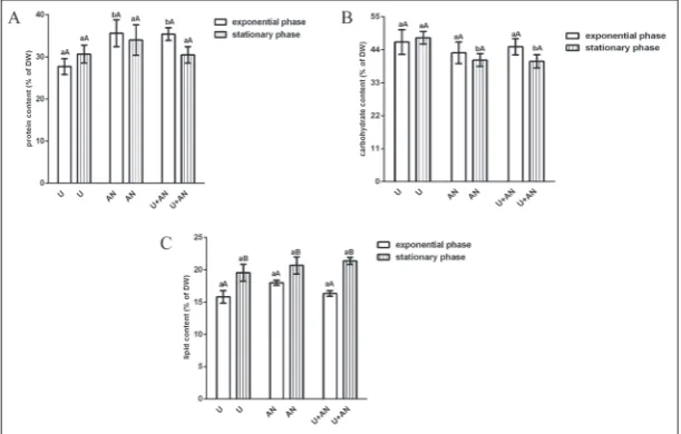
Variations in the content of the main biomass compo-nents of C. vulgaris R-06/2 depending on the N source in the medium and the cultivation time are presented on Fig. 1. The protein content ranged between 30-36% of DW in exponentially grown cells, and between
30.5-34% of DW in the stationary cultures (Fig. 1A). On the 4th day, the
cultures grown in standard medium and medium supplemented with am-monium nitrate showed significantly increased (p<0.05) protein synthesis (by 17 and 20%, respectively) com-pared to the urea culture. There were slight variations (p>0.05) in the pro-tein levels between the older cultures as well as between the stationary and exponential phase of each culture.
Carbohydrates were the most abundant component of C. vulgaris R-06/2 biomass. Their content, rang-ing from 41 to 47.6% DW, was not significantly affected by the N source in the culture medium or the growth time. A moderate but significant in-crease (p<0.05) in carbohydrates (by 11-14%) was only observed in the older culture grown in urea as com-pared to the other two cultures in the stationary phase (Fig. 1B).
The lipid content of the C. vul-garis R-06/2 biomass varied within the range 15.8-18% DW in the young cells and 19.6-21.4% in the older cells (Fig. 1C). The selection of N source had no significant impact on lipid synthesis. All cultures, however, showed a significant increase (by 16-25%, p<0.05) in their lipid content in the stationary as compared to the exponential phase.
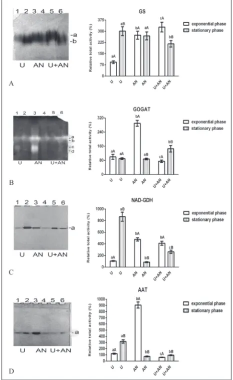
Isoenzyme pattern and activity of metabolic enzymes of C. vulgaris R-06/2 grown in a medium with different nitrogen sources
GS of C. vulgaris R-06/2 was represented by two diffu-sion bands designated as “a-b” and arranged according to the increase in their migration mobility (Fig. 2A, left). The relative total GS activity was the least pronounced in the urea culture and grown until the exponential phase (Fig. 2A, right). In the stationary phase it was increased by 220%. When algae were grown with am-monium nitrate, the enzyme activity was relatively high
Table 1. Effect of N source and cultivation time on the biomass dry weight and specific
growth rate of C. vulgaris R-06/2.
Nitrogen source DW t=2 µ t2=2 DW t=4 µ t2=4 DW t=8
Urea 3.0±0.1 a 0.64±0.02 a 4.5±0.1a 0.422±0.006a 3.6±0.2a Ammonium nitrate 3.0±0.1 a 0.64±0.02 a 4.7±0.1a 0.433±0.005a 4.5±0.2b Urea+ammonium
nitrate 2.1±0.1 b 0.46±0.02 b 3.5±0.1b 0.359±0.007b 4.9±0.2b
DW – biomass dry weight (g L-1); [µ] – specific growth rate; t – cultivation time (days). Means
in a column indicated by different letters are significantly different (p<0.05) between N sources for a specific cultivation time (ANOVA and Bonferroni’s post hoc test).
and did not show cultivation time-dependent changes. The highest GS activity was observed in the standard medium culture during the exponential phase, but with entry of the culture into the stationary phase, it decreased by about one-third.
Four isoforms of GOGAT (a-d) were observed on the gels after native PAGE and subsequent stain-ing (Fig. 2B). The ammonium nitrate-supplemented medium had a strong enhancing effect on relative total GOGAT activity in the exponential phase of growth of C. vulgaris R-06/2 (Fig. 2B). The total enzyme ac-tivity of algae grown in urea medium did not change significantly with cultivation time, but the activities of isoforms “c” and “d” were higher in the younger as compared to the older culture. With prolonged cultiva-tion of the alga in the standard medium, the activity increased approximately two-fold when compared to the exponential phase.
C. vulgaris R-06/2 showed one isoform (a) оf NAD-dependent GDH (Fig. 2C). The enzyme activity was the highest in the case of algae cultivated in urea medium to the stationary phase (Fig. 2C), whereas in the exponentially growing culture in the same medium, the band intensity was more than nine times weaker. In the ammonium nitrate and standard media, the NAD-GDH activity was higher in the samples obtained in the exponential than in the stationary phase of growth. Among the older cultures, the activity was lowest in the algae cultivated in ammonium nitrate medium.
One band of AAT activity was observed in C. vul-garis R-06/2 under all cultivation conditions examined (Fig. 2D). The enzyme activity was highest in the alga that was grown in ammonium nitrate-supplemented medium to the exponential phase (Fig. 2D). AAT functioned better in the urea medium compared to the standard medium during both growth phases, and in the stationary compared to the exponential phase in both media.
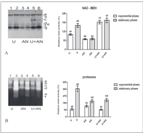
During the study of the isozyme profile of NAD-MDH of C. vulgaris R-06/2, the presence of seven iso-forms (a-g) was established (Fig. 3A). Isoform “c” was clearly visible only when using the standard medium, its activity being 4-fold higher in the exponential than in the stationary phase. Isoforms “g” and “a” were the most intensive (active) in all samples. The intensity of isoform “g” changed with the changes in cultivation
Fig. 2. Isoenzyme patterns (on the left) and changes in total
conditions and most strongly affected overall enzyme activity, while isoform “a” activity varied slightly. When using the urea medium, the intensity of band “g” and total NAD-MDH activity were by about 60% higher in the stationary as compared to the exponential phase (Fig. 3A). The relative total enzyme activities of algae culti-vated in ammonium nitrate medium were the weakest. The gelatin zymograms revealed the presence of six protease bands (a-f) in C. vulgaris R-06/2 (Fig. 3B). The relative total protease activity was significantly higher in the stationary than in the exponential phase of growth due to the increased intensity of bands “b”, “d” and “f”, regardless of the N source in the medium (Fig. 3B). Among the younger cultures, the ammonium nitrate-grown cells had the highest activity, whereas
the urea-grown cells were with the most active proteases among the older cultures.
DISCUSSION
The results of our investigation showed that the media supplemented with only urea or only ammonium nitrate provided faster growth of C. vulgaris R-06/2 and biomass accumulation for a short period of time (4 days), while the medium with both urea and ammonium nitrate ensured better biomass yield but at prolonged cultivation (more than 8 days). This pattern could be the consequence of the higher assimilation rate of the single N-containing compounds than a mixture of two. In contrast to what was observed for C. vulgaris R-06/2, the ammonium nitrate-containing modified Hoagland medium was found not to be growth-supportive for the green algae Chlorella pyrenoidosa and Scenedesmus obliquus and theeustigmatophyceaen alga Nannochloropsis oculata cultured for 10 days [35]. Chlorellasorokiniana did not show a preference for any of the N sources, displaying similar (p>0.05) specific growth rates with sodium nitrate, urea, ammonium carbonate and ammonium chloride, while Chlorella vulgaris had a clear preference for sodium nitrate over the other N sources [36]. In general, the efficiency of N utilization from different N sources for growth varies among algal species.
The biochemical profile of C. vulgaris R-06/2 was not strongly influenced by the N source of the medium and the cultivation time. There were no significant differences (p>0.05) between ammonium nitrate- and ammonium nitrate plus urea-grown cultures in carbo-hydrate, lipid and protein contents, expressed as % of DW, during both growth phases. Only the use of urea resulted in some significant changes (p<0.05) in the biochemical composition. The urea-grown younger culture had a lower protein content than the other two exponentially growing cultures, while in the stationary growth phase, urea stimulated carbohydrate accumu-lation when compared to the other used N sources.
Fig. 3. Isoenzyme patterns (on the left) and changes in total enzyme activity
(on the right) of C. vulgaris R-06/2 in response to N source in the medium and cultivation time. A – NAD-dependent malate dehydrogenase (NAD-MDH); B
The effect of the cultivation time was expressed as a significant increase (p<0.05) in lipid content with aging of the cultures, regardless of the N source. Similarly, Chlorella sp. BUM11008 accumulated more lipids during the stationary phase when compared to the exponential phase [37]. This common response can be related to a gradual depletion of N in the nutrient medium throughout algal growth.
The influence of different N sources [10,11] and, to a lesser extent, the simultaneous effect of cultivation time and the N source [38] on the biochemical profile, has been investigated in some other microalgae, with most studies focusing on lipid accumulation [38-40]. The literature examples, along with the data of the present study, demonstrated that the selection of N source affects the biochemical composition of the microalgae, providing evidence of strongly species-specific responses to the different N sources. As a com-mon trend, it can be noted that inefficient utilization of the applied N source decreases the availability of N in cells and leads to an increase in the production of carbohydrates or lipids or both, but slows down algal growth. Studies have shown that the N source itself can simultaneously stimulate algal growth and the productivity of lipids, carbohydrates and proteins [11], lipid productivity [39] or protein productivity (ammonium nitrate in our study).
In order to gain deeper insight into the metabolic responses of C. vulgaris R-06/2 to the change in N source, the alterations in the isoenzyme patterns and relative activities of five enzymes that link N and C (energy) metabolism (GS, GOGAT, NAD-GDH, AAT and NAD-MDH) were followed during the exponential and stationary phases of algal growth. Proteases were included in the studies as they contribute to the total intracellular amino acid pool. GS and GOGAT, as partners in the GS-GOGAT cycle, are key enzymes in the biosynthesis of glutamate [41], one of the central players in N metabolism, since it is capable of both receiving and donating to the N group [42]. NAD-dependent GDH catalyzes the deamination of glutamate into alpha ketoglutarate. This biochemical reaction provides an oxidizable carbon source used for energy production, as well as the reduced electron carrier NADH, thus linking amino acid metabolism to the tricarboxylic acid (TCA) cycle [6]. AAT catalyzes the interconversion of aspartate and alpha ketoglutarate
to glutamate and oxaloacetate. The amino group transfer from aspartate or glutamate to the respective keto acids is crucial in both amino acid biosynthesis and degradation. In our study, the activity of AAT was analyzed in a glutamate-producing direction. Using NAD as a cofactor, MDH catalyzes the conversion of malate to oxaloacetate [43]. This reaction is a part of many metabolic pathways, including the TCA cycle.
Employing the excellent capabilities of the meth-ods for separation of native proteins by PAGE and subsequent in-gel activity staining, the isoforms of GS, GOGAT, NAD-GDH, AAT, NAD-MDH and proteases of C. vulgaris R-06/2 were visualized. Being determined in crude cell extracts with equal total protein contents, the images on each gel also allowed comparison of the enzyme activities. It was found that the isoenzyme patterns of C. vulgaris R-06/2 did not change, but the intensity of some bands varied, depending on the N source in the culture medium, resulting in changes in relative total activity of the respective enzyme. Analysis of the results obtained also showed that there was no direct relationship between the activity of the metabolic enzymes in the medium with a defined N source and the growth rate and biomass accumulation in the same medium. For example, in cells cultured in ammonium nitrate-containing medium to the exponential phase, the relative total activities of glutamate-producing (GOGAT, AAT) and glutamate-catabolizing (NAD-GDH) enzymes were the highest and NAD-MDH activity was the lowest, with medium GS activity compared to the exponentially growing cultures in the urea and the standard media. The use of urea as a source of N instead of ammonium nitrate led to a decrease in activity of all studied enzymes, except for NAD-MDH. Despite the differences in the metabolic enzymes’ responses to urea vs ammonium nitrate, both N sources provided nearly equivalent growth rates and biomass dry weight (p>0.05). The regulation of metabolic enzymes in dependence of the N source during the stationary growth phase differed from that in the exponential phase. In the older urea culture of C. vulgaris R-06/2, for example, the activities of GS, NAD-GDH, AAT and NAD-MDH were significantly higher (p<0.05) than those in exponentially growing cells supplied with urea.
protease activity in these cells, suggesting a role for these enzymes in the reassimilation of the ammonium produced by protein degradation, as reported for Chla-mydomonas reinhardtii [44]. On the other hand, the observed high activity of NAD-GDH and especially of NAD-MDH in cultures supplied simultaneously with ammonium nitrate and urea during both growth phases, was probably associated with the greater energy requirements of these cells. As with the urea-grown C. vulgaris R-06/2, an increase in GDH-deaminating activity with the aging of cultures was shown in Chla-mydomonas reinhardtii as well [45].
There are a number of earlier studies on how mi-croalgae metabolic enzyme responses can vary with different N sources, but all relate to the replacement of nitrate in the nutrient medium with ammonium, or the replacement of ammonium with nitrate [13-16,45,46] and rarely with urea [47]. Chlorella and Chlamydomonas species are the model algae in these studies. Their GS, GOGAT, GDH and AAT are best characterized with respect to the enzyme kinetic properties, changes in the enzyme patterns and activity levels.
The activities of the studied enzymes of C. vulgaris R-06/2 changed in response to the change in the N source, as well as with the culture age, and these altera-tions can be related to the uptake rates and interacaltera-tions between the different chemical forms of N, the activity and capacity of nitrate, ammonium and urea trans-port systems, the activity of nitrate reductase, nitrite reductase and ATP-urea amidolyase, and the size of endogenous pools of ammonium and intermediary metabolites like glutamate and alpha ketoglutarate.
CONCLUSIONS
This study shows the tolerance of C. vulgaris R-06/2 to changes in the N source in the nutrient medium. The ability of the strain for flexible regulation in response to a change in N source as well as cultivation time en-sured maintenance of efficient functioning of metabolic enzymes in each of the studied cultivation conditions and ultimately resulted in good algal growth. When large-scale cultivation of C. vulgaris R-06/2 is used to produce biomass for various useful applications, urea would be the most appropriate choice in terms of economic profitability, as it is a cheaper N source than ammonium nitrate.
Funding: This work was co-funded by the Program for career
development of young scientists, BAS (project ДФНП-207) and the National Science Fund, Bulgaria (project КП-06-ОПР04/1).
Author contributions: Ivanina Vasileva participated in all phases
of the research and in writing the manuscript; Juliana Ivanova participated in the cultivation of the algae and performed the laboratory measurements; Liliana Gigova contributed to the drafting and writing of the manuscript. All authors reviewed and approved the final manuscript.
Conflict of interest disclosure: The authors declare no conflicts
of interest.
REFERENCES
1. Singh R, Parihar P, Singh M, Bajguz A, Kumar J, Singh S, Singh VP, Prasad SM. Uncovering potential applications of cyanobacteria and algal metabolites in biology, agriculture and medicine: current status and future prospects. Front Microbiol. 2017;8:515.
2. Sharma P, Sharma N. Industrial and biotechnological applica-tions of algae: A Review. J Adv Plant Biol. 2017;1(1):1-25. 3. Jethani H, Patel Pravin, Mudliar SN, Sarada R, Chauhan VS.
Growth and biochemical response of an indigenous oleagi-nous microalga Scenedesmus obtusus cultivated in outdoor open ponds. Indian J Exp Biol. 2019;57(1):40-9.
4. Markou G, Vandamme D, Muylaert K. Microalgal and cya-nobacterial cultivation: The supply of nutrients. Water Res. 2014;65:186-202.
5. Solomon CM, Glibert PM. Urease activity in five phytoplank-ton species. Aquat Microb Ecol. 2008;52:149-57.
6. Berges JA, Mulholland MR. Enzymes and N cycling. In: Capone DG, Bronk DA, Mullholland M, CarpenterEJ, edi-tors.Nitrogen in the marine environment.Amsterdam: Else-vier; 2008. p. 1361-420.
7. Molloy CJ, Syrett PJ. Interrelationships between uptake of urea and uptake of ammonium by microalgae. J Exp Mar Biol Ecol. 1988;118(2):85-95.
8. Sanz-Luque E, Chamizo-Ampudia A, Llamas A, Galvan A, Fernandez E. Understanding nitrate assimilation and its regu-lation in microalgae. Front Plant Sci. 2015;6:899.
9. Hodson RC, Williams SK, Davidson WR. Metabolic control of urea catabolism in Chlamydomonas reinhardi and Chlorella pyrenoidosa. J Bacteriol.1975;121(3):1022-35.
10. Effect of different nitrogen sources on growth and bio-chemical composition of the green microalgae Scenedesmus obliquus and Chlorella kessleri. Third International Confer-ence on Biological SciConfer-ences; 2004 Apr 24-29, Tanta, Egypt. Tanta (Egypt): [publisher unknown]; c.2004. 419p.
11. Lourenço SO, Barbarino E, Mancini-Filho J, Schinke KP, Aidar E. Effects of different nitrogen sources on the growth and biochemical profile of 10 marine microalgae in batch culture: an evaluation for aquaculture. Phycologia, 2002;41(2):158-68.
(Advances in Biochemical Engineering/Biotechnology; Vol. 153).
13. Ahmad I, Hellebust JA. Nitrogen metabolism of the marine microalgae Chlorella autotrophica. Plant Physiol. 1984;76(3):658-63.
14. Ahmad I, Hellebust JA. Partial characterization of enzymes of nitrogen metabolism in Chlorella autotrophica Shihira & Krauss. New Phytol. 1993;123:685-92.
15. Tischner R, Lorenzen H. Changes in the enzyme pattern in synchronous Chlorella sorokiniana caused by different nitro-gen sources. Z Pflanzenphysiol.1980;100:333-41.
16. Shatilov VR, Sofin AV, Kasatkina TI, Zabrodina TM, Vladi-mirova MG, Semenenko VE, Kretovich WL. Glutamate dehy-drogenase of unicellular green algae: effects of nitrate and ammonium in vivo. Plant Sci Lett. 1978;1(1):105-14. 17. Gärtner G, Uzunov B, Ingolic E, Kofler W, Gacheva G, Pilarski
P, Zagorchev L, Odjakova M, Stoyneva M. Мicroscopic inves-tigations (LM, TEM and SEM) and identification of Chlorella
isolate R-06/2 from extreme habitat in Bulgaria with a strong biological activity and resistance to environmental stress fac-tors. Biotechnol Biotechnol Equip. 2015;29(3):536-40. 18. Gacheva G, Pilarski P. The resistance of a new strain Chlorella
sp. R-06/2, isolated from an extreme habitat, to environmental stress factors. Gen Appl Plant Physiol. 2008;34(3-4):347-60. 19. Najdenski HM, Gigova LG, Iliev II, Pilarski PS, Lukavsky J,
Tsvetkova IV, Ninova MS, Kussovski VK. Antibacterial and antifungal activities of selected microalgae and cyanobacteria. Int J Food Sci Technol. 2013;48(7):1533-40.
20. Gigova GL, Toshkova RA, Gardeva EG, Gacheva GV, Ivanova NJ, Yossifova LS, Petkov GD. Growth inhibitory activity of selected microalgae and cyanobacteria towards human cervi-cal carcinoma cells (HeLa). J Pharm Res. 2011;4(12):4702-07. 21. Setlik I. Contamination of algal cultures by heterotrophic
microorganisms and its prevention. In: Lhotský O, Nečas J, editors. Annual Report of the Laboratory of Experimental Algology and Department of Applied Algology for the Year 1966. Třeboň, Czech Republic: Institute of Microbiology, ČSAV; 1967. p. 89-100.
22. Georgiev D, Dilov H, Avramova S. Buffered nutrient medium and intensive culture method of green microalgae. Hydrobiol. 1978;7:14-23. French.
23. Levasseur M, Thompson P, Harrison P. Physiological acclima-tion of marine phytoplankton to different nitrogen sources. J Phycol. 1993;29(5):587-95.
24. Lowry O, Rosenbrough N, Farr AZ, Randball RJ. Protein measurement with the Folin phenol reagent. J Biol Chem. 1951;193(1):265-75.
25. DuBois M, Gilles KA, Hamilton JK, Rebers PA, Smith F. Colo-rimetric method for determination of sugars and related sub-stances. Anal Chem. 1956;28(3):350-6.
26. Petkov G, Dilov H. On the composition of alcoholic extract of microalgae of the Scenedesmus Meyen. Hydrobiol. 1987;29:41-4.
27. Bradford MM. A rapid and sensitive method for the quan-tification of micrograms quantities of protein utilizing the principle of protein-dye binding. Anal Biochem. 1976;72(1-2):248-54.
28. Laemmli UK. Cleavage of structural proteins during the assem-bly of the head of bacteriophage T4. Nature. 1970;227:680-5.
29. Simonović AD, Gaddameedhi S, Anderson MD.In-gel pre-cipitation of enzymatically released phosphate. Anal Bio-chem.2004;334(2):312-7.
30. Matoh T, Ida S, Takahashi E. Isolation and characterization of NADH-glutamate synthase from pea (Pisum sativum L.). Plant Cell Physiol. 1980;21(8):1461-74.
31. Nash DT, Davies ME. Isoenzyme changes during the growth cycle of Paul’s scarlet rose cell suspensions. Phytochemistry. 1975;14(10):2113-8.
32. Honold GR, Farkas GL, Stahmann MA. The oxidationreduc-tion enzymes of wheat. I. A qualitative investigaoxidationreduc-tion of the dehydrogenases. Cereal Chem. 1966;43(5):517-28.
33. Griffith SM, Vance CP. Aspartate aminotransferase in alfalfa root nodules 1. Purification and partial characterization. Plant Physiol. 1989;90(4):1622-9.
34. Lodemel JB, Maehre HK, Winberg J-O, Olsen RL. Tissue dis-tribution, inhibition and activation of gelatinolytic activities in Atlantic cod (Gadus morhua). Comp Biochem Physiol B Biochem Mol Biol. 2004;137(3):363-71.
35. Bajwa K, Bishnoi NR, Kirrolia A, Sharma J, Gupta S. Com-parison of various growth media composition for physio-biochemical parameters of biodiesel producing microalgal species (Chlorococcum aquaticum, Scenedesmus obliquus, Nannochloropsis oculata and Chlorella pyrenoidosa). European J Biotechnol Biosci. 2017;5(6):27-31.
36. Podevin M, De Francisci D, Holdt SL, Angelidaki I. Effect of nitrogen source and acclimatization on specific growth rates of microalgae determined by a high-throughput in vivo microplate autofluorescence method. J Appl Phycol. 2015;27(4):1415-23.
37. Praveenkumar R, Shameera K, Mahalakshmi G, Akbarsha MA, Thajuddin N. Influence of nutrient deprivations on lipid accumulation in a dominant indigenous microalga Chlorella
sp., BUM11008: evaluation for biodiesel production. Biomass Bio-energ. 2012;37:60-6.
38. Kim G, Mujtaba G, Lee K. Effects of nitrogen sources on cell growth and biochemical composition of marine chlorophyte
Tetraselmis sp. for lipid production. Algae, 2016;31(3):257-66. 39. Agwa OK, Abu GO. Influence of various nitrogen sources on biomass and lipid production by Chlorella vulgaris. Br Bio-technol J, 2016;15(2):1-13.
40. Soni SM, Sankneniwar SS, Rasheed MA, Rao PLS, Hasan SZ. Effect of various nitrogen sources on microalgal growth and lipid content in Chlorella pyrenoidosa NCIM 2738 and ANK-1. Int J Curr Microbiol App Sci. 2017;6(8):3099-108. 41. Scanlan DJ, Post AF. Aspects of marine cyanobacterial
nitrogen physiology and connection to the nitrogen cycle. In: Capone DG, Bronk DA, Mullholland M, CarpenterEJ, editors.Nitrogen in the marine environment.Amsterdam: Elsevier; 2008. p. 1073-96.
42. Dagenais-Bellefeuille S, Morse D. Putting the N in dinoflagel-lates. Front Microbiol. 2013;4:369.
43. Minárik P, Tomášková N, Kollárová M, Antalík M. Malate dehydrogenases - structure and function. Gen Physiol Bio-phys, 2002;21(3):257-65.
edi-tors. Inorganic nitrogen metabolism. Berlin:Springer-Verlag; 1987. p. 132-6.
45. Moyano E, Cárdenas J, Muñoz-Blanco J. Involvement of NAD(P)+- glutamate dehydrogenase isoenzymes in carbon and nitrogen metabolism in Chlamydomonas reinhardtii. Physiol Plant.1995;94(4):553-9.
46. Muñoz-Blanco J, Cárdenas J. Changes in glutamate dehy-drogenase activity of Chlamydomonas reinhardili under different trophic and stress conditions. Plant Cell Environ. 1989;12(2):173-82.