CERVICAL PRECANCEROUS LESIONS IN HIV-POSITIVE WOMEN IN CAMEROON: PREVALENCE, PREDICTORS AND POTENTIAL IMPACT OF SCREENING
JULIUS ATASHILI
A dissertation submitted to the faculty of the University of North Carolina at Chapel Hill in partial fulfillment of the requirements for the degree of Doctor of Philosophy in the
Department of Epidemiology
Chapel Hill 2009
Approved by:
William C Miller, MD PhD MPH
Adaora A Adimora, MD MPH
Joseph Eron, MD
Jennifer S Smith, PhD MPH
iii ABSTRACT
JULIUS ATASHILI: Cervical precancerous lesions in HIV-positive women in Cameroon: prevalence, predictors and potential impact of screening
(Under the direction of William C. Miller, MD, PhD, MPH)
Cervical cancer is the most common cancer in women in low-income countries.
Although cervical cancer incidence and mortality is higher in HIV-positive women, resource
limitations restrict the implementation of systematic screening programs in these women.
The purpose of this dissertation was to explore the potential for targeted screening by
assessing the prevalence and clinical predictors of cervical squamous intra-epithelial lesions
(SIL) in HIV-positive women in Cameroon. Furthermore, we sought to explore the potential
impact of antiretroviral therapy and screening on mortality from cervical cancer.
We initially conducted a cross-sectional study of HIV-positive women in Cameroon. A
total of 282 women, aged 19 to 68 years with a median CD4 cell count of 179
cells/microliter, were enrolled. SIL were detected in 43.5% of the 276 women with
satisfactory samples: including high-grade SIL (HSIL) in 3.3%. None of the clinical factors
assessed significantly predicted the presence of any lesion. Among patients with CD4
counts less than 200 cells/microliter, the prevalence of SIL was higher in patients aged
26-59 years compared to younger women, while there was essentially no difference amongst
women with CD4 counts greater than 200 cells/microliter.
Using a Markov state-transition model of a cohort of HIV-positive women in
Cameroon, we examined the potential impact of scenarios including: no HAART and no
iv
age 35 (HS35). Compared to NHNS, lifetime cumulative cervical cancer mortality doubled
with HNS. It will require 202 women being screened at age 35 to prevent one cervical
cancer death amongst women on HAART.
The high prevalence of SIL in women initiating antiretroviral therapy in Cameroon
underscores the need for screening in this population. With neither age nor any other clinical
factor being a good predictor of SIL, alternative affordable screening options need to be
explored. Furthermore, the long-term evolution of SIL needs to be assessed in prospective
studies of these women. Screening has the potential of reducing cervical cancer mortality in
v
To my late dad, Mr Tita Sangbong Nicholas, and my mom, Mrs Siri Mary:
vi
ACKNOWLEDGEMENTS
This dissertation would not have been possible without a series of people/institutions:
I am very grateful to:
Dr William C Miller for a superb mentorship all the time I was at UNC.
Dr Adaora Adimora for giving me the opportunity to get to and through the program at UNC and her guidance with the dissertation work.
Dr Jennifer S Smith for all her support during my training and guidance with the dissertation.
Dr Joseph Eron for his patience, guidance and availability throughout the dissertation.
Dr Evan Myers for his patience, guidance and availability throughout the dissertation process.
Prof Peter M Ndumbe for giving me the inspiration and opportunity focus on epidemiology as a career and his continued support.
Drs Marcel Yotebieng, Prema Menezes, Larissa Braga, Linda Kalilani, Brian Pence, Padjama Patnaik, Maria Khan, Abigail Norris Turner, and all other colleagues who made this experience less painful.
Drs Jay Kaufman, Charles Poole, Frieda Behets, Annelies Van Rie, Sharon Weir, Victor Schoenbach, Irvine Hoffman, Marcia Hobbs, and all other UNC faculty for all their guidance.
vii
Kirsten Leysieffer, Kathy James, Cathy Emrick and all the staff at the infectious diseases department.
Dr Vani Vannappagari for her support.
All study participants and the following health care providers who assisted in study implementation: Drs Kinge Thompson, Akam Wilfred, Charles Kefie, Tayong Gladys, Sume Gerald and Etogo; Egbearong Ashu, Noella Njabanou, Brigitte Wandji, Martha Mesembe, Emilia Lyonga and Alexi.
George Ikomey and Allen Rinas for assistance with cytology and Rob Krysiak for assistance with material acquisition.
My entire family for enduring my absence.
I also acknowledge the following funding:
A Fogarty fellowship supported by NIH Fogarty grant DHHS/NIH/FIC 5 D43 TW01039-08 AIDS International Training and Research Program to the University of North Carolina at Chapel Hill.
Primary data collection for this dissertation was funded by a Developmental Award from the University of North Carolina at Chapel Hill’s, Center for AIDS Research (CFAR Grant Number NIH #9P30 AI 50410).
viii
TABLE OF CONTENTS
LIST OF TABLES ... xi
LIST OF FIGURES ... xii
LIST OF ABBREVIATIONS ... xiii
CHAPTER ONE: INTRODUCTION, SPECIFIC AIMS AND HYPOTHESES ... 1
1.1 Introduction ... 1
1.2 Specific Aim 1 ... 3
1.3 Specific Aim 2 ... 3
1.4 Specific Aim 3 ... 4
1.5 Overview ... 4
CHAPTER TWO: BACKGROUND AND SIGNIFICANCE ... 5
2.1 Cervical cancer ... 5
2.2 Cervical cancer and precancerous lesions in HIV ... 15
2.3 Need to screen for cervical cancer in HIV in resource-poor settings including Cameroon ... 20
2.4 Age and cervical cancer ... 22
2.5 Review of previous studies ... 26
CHAPTER THREE: RESEARCH DESIGN AND METHODS ... 49
3.1 Methods to determine the prevalence, severity and predictors of cervical squamous intraepithelial lesions in HIV-positive women on antiretroviral therapy in Cameroon. ... 49
3.2 Methods to describe the age trends in the prevalence of cervical squamous intraepithelial lesions in HIV-positive women on antiretroviral therapy in Cameroon. ... 55
ix
CHAPTER FOUR: CERVICAL SQUAMOUS INTRAEPITHELIAL LESIONS IN
WOMEN INITIATING ANTIRETROVIRAL THERAPY IN CAMEROON:
PREVALENCE AND PREDICTORS ... 70
4.1 ABSTRACT ... 70
4.2 INTRODUCTION ... 71
4.3 METHODS ... 72
4.4 RESULTS ... 76
4.5 DISCUSSION ... 79
4.6 REFERENCES ... 91
CHAPTER FIVE: AGE AND THE PREVALENCE OF CERVICAL SQUAMOUS INTRAEPITHELIAL LESIONS AMONG HIV-POSITIVE WOMEN IN CAMEROON ... 93
5.1 ABSTRACT ... 93
5.2 INTRODUCTION ... 94
5.3 METHODS ... 95
5.4 RESULTS ... 98
5.5 DISCUSSION ... 99
5.6 REFERENCES ... 105
CHAPTER SIX: POTENTIAL IMPACT OF ANTIRETROVIRAL THERAPY AND SCREENING ON CERVICAL CANCER MORTALITY IN HIV-POSITIVE WOMEN IN SUB-SAHARAN AFRICA ... 108
6.1 ABSTRACT ... 108
6.2 INTRODUCTION ... 109
6.3 METHODS ... 110
6.4 RESULTS ... 113
6.5 DISCUSSION ... 115
6.6 REFERENCES ... 126
CHAPTER SEVEN: CONCLUSION ... 128
APPENDICES ... 131
x
Appendix 2: Sample normal cervix high CD4 (>500/uL) subtree ... 133
Appendix 3: Sample HSIL moderate CD4 (200-500/uL) subtree ... 134
Appendix 4: Sample distant ICC low CD4 (<200/uL) subtree ... 135
Appendix 5: Study questionnaire ... 136
xi
LIST OF TABLES
Table 2.1: Terminologies/classification of cervical precancerous/cancerous lesions ... 9
Table 2.2: Summary of cervical cancer screening guidelines in the US ...15
Table 2.3: Summary epidemiology of HPV, cervical precancerous and cancerous lesions in HIV positive women in developing countries ...27
Table 2.4: Summary of studies of that addressed the impact of age on the prevalence of lesions, the progression of lesions, the prevalence of HPV or diagnostic work-up. ...33
Table 2.5: Summary of cost-effectiveness studies of the cervical cancer screening. ...40
Table 3.1: Plausible transitions (blank cells) between stages in the Markov model. ...68
Table 3.2 Baseline values ...69
Table 4.1: Socio-demographic and clinical characteristics in 282 women initiating HAART in Cameroon ...83
Table 4.2: Association of clinical predictors with prevalent cervical precancerous lesions in 282 women initiating HAART in Cameroon ...85
Table 4.3A: Definition of clinical risk scores developed and assessed for predicting the prevalence of cervical precancerous lesions in women initiating HAART in Cameroon ...87
Table 4.3B: Performance of potential clinical risk scores for targeting cervical screening in 282 women initiating HAART in Cameroon ...88
Table 5.1: Age-specific prevalence of cervical precancerous epithelial lesion in 276 women initiating HAART in Cameroon ... 102
Table 6.1: Baseline parameters used in modeling cervical cancer mortality in HIV positive women ... 118
Table 6.2: Projected cumulative (lifetime) cervical cancer mortality in HIV-positive women in Cameroon on HAART and or screened for cervical cancer ... 119
xii
LIST OF FIGURES
Figure 2.1: Age-specific incidence and mortality of cervical cancer ...25
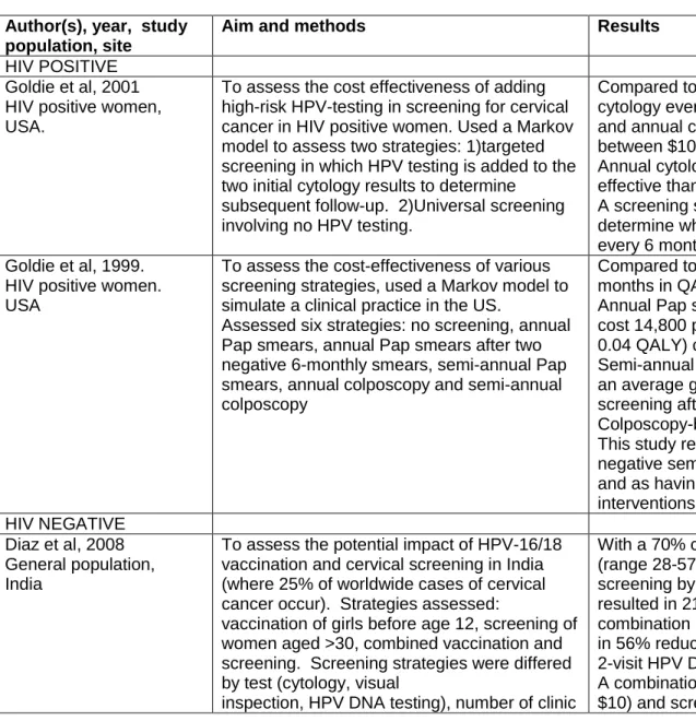
Figure 3.1: Precision of prevalence estimates based on a sample size of 276 women ...50
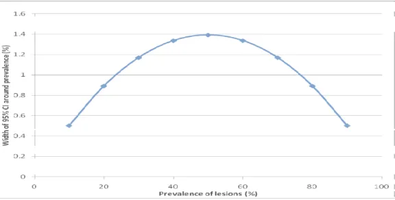
Figure 3.2: Directed acyclic graph (DAG) of the relationship between age, prevalent cervical lesions and other covariates. ...57
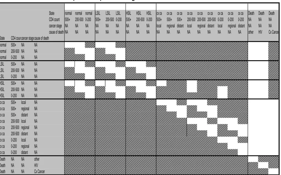
Figure 3.3: Summary of states in the Markov model ...64
Figure 4.1: Total weighted errors associated with screening for any precancerous lesion by the relative ‘cost’ of ‘false negative’ errors compared to ‘false positive’ errors. (A-C: Screening based on risk score with a cut-off targeting 25%, 50% and 75% of women for A,B and C respectively; D: Universal screening; E: No screening). ...89
Figure 4.2: Total weighted errors associated with screening for ASC-H/HSIL by the relative ‘cost’ of ‘false negative’ errors compared to ‘false positive’ errors. (A-C: Screening based on risk score with a cut-off targeting 25%, 50% and 75% of women for A,B and C respectively; D: Universal screening; E: No screening). . ...90
Figure 5.1: Trends in age-specific prevalence of precancerous lesions and ASC_H/HSIL in 276 women initiating HAART in Cameroon (estimates based on locally weighted regression models). ... 103
Figure 5.2: Sensitivity analysis of outcome misclassification on the observed prevalence difference between age groups (26-59 versus 18-25 and 60+ years) by CD4 count ... 104
Figure 6.1: Summary of states in the Markov model ... 121
Figure 6.2: Cumulative mortality from cervical cancer (A) or from all causes of death (B) by intervention in a cohort of HIV positive women getting infected at age 25. ... 121
Figure 6.3: Sensitivity of cumulative cervical cancer mortality to the relative effect of HAART in reducing HIV-mortality (A); the progression rate of precancerous lesions (B) and cervical cancer mortality rate(C). ... 121
xiii
LIST OF ABBREVIATIONS
AIDS Acquired Immune Deficiency Syndrome
ANOVA Analysis of variance
aOR Adjusted odds ratio
ASC-H ASCUS, cannot exclude a high grade lesion
ASCUS Atypical Squamous Cells of Unknown Significance
ATC AIDS treatment center
AUC Area under the curve
CD Cluster designate
CDC Center for Disease Control and prevention
CE Cost-effective
CEA Cost-effectiveness analysis
CI Confidence Interval
CIN Cervical Intraepithelial Neoplasia
CSCCD Center for the Study and Control of Communicable Diseases
DAG Directed acyclic graph
DNA Deoxyribonucleic acid
GDP Gross Domestic Product
HAART Highly Active Antiretroviral Therapy
HC Hybrid Capture
HIV Human Immunodeficiency Virus
HNS HAART and No screen
HPV Human Papillomavirus
HR Hazard ratio
xiv
HS35 HAART and Screen at age 35
HSHI HAART and Screen at HAART initiation
HSIL High-grade Squamous Intraepithelial Lesion
IARC International Agency for Research on Cancer
ICC Invasive Cervical Cancer
ICER Incremental cost-effectiveness ratio
LBC Liquid Based Cytology
LEEP Loop electrosurgical excision procedure
LOWESS Locally-weighted Sums of Squares methods
LRT Likelihood ratio test
LSIL Low-grade Squamous Intraepithelial Lesion
MLE Maximum Likelihood Estimation
NA Not applicable
NHNS No HAART and No Screen
NPV Negative predictive value
OR Odds ratio
PCR Polymerase Chain Reaction
PD Prevalence difference
PMTCT Prevention of Mother-to-Child Transmission of HIV
PPV Positive predictive value
QALY Quality-adjusted life years
RTI Reproductive Tract Infection
SCC Squamous Cell Carcinoma
SCJ Squamocolumnar Junction
xv
STI Sexually Transmitted Infection
UK United Kingdom
UNAIDS Joint United Nations Program on HIV/AIDS
US United States
USD United States Dollars
VIA Visual Inspection with Acetic acid
VIAM Magnified visual inspection with acetic acid
VILI Visual Inspection with Lugol’s Iodine
CHAPTER ONE: INTRODUCTION, SPECIFIC AIMS AND HYPOTHESES
1.1 Introduction
Cervical cancer is the most common cancer in women in low-income countries
[WHO, 2006] and the second most common cancer in women worldwide [Stewart and
Kleihues, 2003]. Compared to immuno-competent women, HIV-positive women have a
higher prevalence, incidence and progression rate of precancerous cervical lesions
[Palefsky, 2006]. By the end of 2007, women accounted for 50% of the estimated 33 million
people living with HIV worldwide, and close to 59% of the 22 million in sub-Saharan Africa
[UNAIDS, 2008]. With the recent increase in access to antiretroviral therapy these women
are expected to live longer thus potentially allowing sufficient time for cervical cancer to
develop. In addition to longer life expectancy, antiretroviral therapy is associated with a
reduction of competing causes of death, such as Kaposi sarcoma and tuberculosis, thus
potentially increasing the proportion of morbidity and mortality attributable to cervical cancer
[Franceschi and Jaffe, 2007]. Enhancing early detection and treatment of precancerous
lesions, through screening could reduce the burden of cervical cancer in these HIV-positive
women [Goldie et al, 2005; Franceschi and Jaffe, 2007].
Despite the relatively high association of HIV with precancerous and cancerous cervical
2
HAART in most developing countries (including Cameroon) does not include a systematic
screen for cervical cancer or precancerous lesions. We hypothesize that cervical cancer
goes undiagnosed and that early diagnosis of precancerous lesions by a screen at
HAART-initiation could be cost-effective in reducing overall morbidity and mortality rates in these
HIV-positive women. Targeted screening could potentially increase the cost-effectiveness of
screening in these resource-limited settings. However, for targeted screening to be effective
the factors associated with a higher prevalence and severity of lesions need to be identified.
Age has been a common consideration in the targeted screening for precancerous
lesions in the general population. In effect, current guidelines for screening the general
population of women in the United States (US) suggest screening commence no later than
at age 21 years, reducing the frequency of screening at age 30 and stopping screening at
age 65 (or 70 in some guidelines) [USPSTF, 2008; ACOG 2003, Saslow et al, 2002]. World
Health Organization (WHO) guidelines aimed primarily at resource-limited settings are less
stringent, recommending screening begin at age 30, need not be annual and need not be
done over the age of 65 [WHO, 2006]. These age considerations may not necessarily be
ideal for HIV-positive women in whom higher human papilloma virus (HPV) prevalence,
higher HPV persistence, and a faster progression of lesions could mean an earlier
occurrence and or a longer persistence of precancerous lesions. The optimal age for
screening in HIV positive individuals could differ substantially from those in the general
population and needs to be better described.
This dissertation sought to provide data aimed at improving the detection and
management of cervical cancer in HIV-positive women. In Cameroon, and other
resource-limited settings, the period of HAART initiation may be a critical time during which
HIV-positive women could be screened as these women already undergo a series of clinical and
laboratory assessment required for HAART. Age may also be an important consideration to
3
To assess the need and potential effect of a screening program in a resource-limited
setting like Cameroon, this dissertation had the following specific aims:
1.2 Specific Aim 1
To determine the prevalence, severity and predictors of cervical squamous intraepithelial lesions in HIV-positive women initiating antiretroviral therapy in Cameroon.
Rationale: Targeted screening may be a potential cost-effective alternative for the screening
of HIV-positive women in resource-limited settings. Determining clinical characteristics that
predict cervical lesions will help identify sub-populations which could be targeted for
screening and thus potentially increase the ratio of the number of cases detected per
screening test. We will develop a predictive model for prevalent cervical epithelial lesions in
women initiating HAART in Cameroon and assess the use of risk-scores to guide screening.
Hypothesis: We hypothesize that cervical precancerous lesions are prevalent in women
initiating HAART in Cameroon and that readily available socio-demographic and clinical
characteristics can be used to predict the presence of lesions. We further hypothesize that
these characteristics could be used to develop a score that could in turn be used to reliably
predict which women need to be screen.
1.3 Specific Aim 2
To describe the age trends in the prevalence of cervical squamous intraepithelial lesions in HIV-positive women on antiretroviral therapy in Cameroon.
Rationale: The optimal ages to begin and discontinue cervical cancer screening in HIV
positive women are unknown. By assessing the age-specific prevalence of lesions we would
estimate the minimum age at which lesions occur, the age with maximum occurrence and
4
useful in the development of age-targeted screening guidelines in HIV positive women in
Cameroon.
Hypothesis: We hypothesize that the prevalence of lesions is dependent on age and that
age alone could be used as a criterion for targeted screening.
1.4 Specific Aim 3
To quantify the potential effect of antiretroviral therapy and screening, on mortality from cervical cancer in HIV-positive women in Cameroon
Rationale: The long term effect of cervical cancer screening in HIV-positive women in
Cameroon is unknown. Mortality from cervical cancer and mortality from HIV can be
competing risks in the evolution of each other disease. The increased survival that is
expected to result from the increased access to HAART in Cameroon may also be
accompanied by an increase in the incidence and mortality due to cervical cancer. By
assessing the potential impact of modifiable factors such as antiretroviral therapy and
screening, we would quantify the potential gains that could be expected from these
interventions and help policymakers in the allocation of limited resources.
Hypothesis: We hypothesize that the cumulative mortality due to cervical cancer would
increase with antiretroviral therapy while screening would substantially reduce this mortality.
1.5 Overview
To achieve these aims we conducted two distinct studies: a cross-sectional
descriptive study of women in HIV clinics in Cameroon who were interviewed on
demographic and clinical characteristics and then screened for cervical precancerous
lesions; and a computer-simulated analysis of the potential outcomes in a group of
CHAPTER TWO: BACKGROUND AND SIGNIFICANCE
In this section we present the burden of cervical cancer, its pathogenesis and risk
factors, and the role of screening in reducing cervical cancer mortality in the general
population. We then summarize the literature on the peculiarities of cervical cancer in
HIV-infected women, the occurrence of precancerous and cancerous lesions by age and end by
reviewing published studies of the prevalence of precancerous lesions in HIV in developing
countries, and studies of the impact of age on cervical cancer.
2.1 Cervical cancer
2.1.1 Classification and epidemiology
The term ‘cervical cancer’ is generally used in reference to squamous cell carcinoma of the uterine cervix although other histological forms are plausible. The cervix is covered by two types of epithelia: a squamous cell epithelium usually limited to the
ectocervix and a columnar (glandular) epithelium usually limited to endocervix. The
pathogenesis of squamous cell carcinoma (SCC) involves the exposure and subsequent
squamous metaplasia of the squamocolumnar (‘transformation’) zone, the intersection of the
ecto- and endocervix. Other histological forms of cervical neoplasia include
6
adenosquamous carcinoma and small cell carcinoma amongst others [Silverberg and Loffe
2003]. SCC is however by far the most common form of cancer accounting for 80% of
primary cervical cancers [Waggoner, 2003]. Any unspecified use of ‘cancer of the cervix’ in
this document will be a reference to SCC.
Cervical cancer is the second cause of cancer in all-age women worldwide and the second cause of cancer death in sub-Saharan Africa. It is estimated that 500,000 new cases are diagnosed yearly with 85% of these in the developing world [Waggoner,
2003; Ferlay et al, 2004]. Cervical cancer is also estimated to cause over 270,000 deaths
annually with the majority of deaths occurring in developing countries. Worldwide these
represent incidence and mortality rates of 16.0 and 8.9 per 100,000 respectively. The
incidence and mortality rates in Africa (19 and 14.8 per 100,000 respectively) [WHO/ICO
2007] are higher than those observed in South-East Asia (15.9 and 8.4 per 100,000
respectively) and substantially higher than those observed in North America (9.0 and 3.6 per
100,000 respectively) [Ferlay et al, 2004]. These rank cervical cancer the first cause of
cancer incidence and mortality in women of all ages in Africa.
In Cameroon, cervical cancer is estimated to be the first cause of cancer and cancer mortality in women of all-ages (and the third cause of cancer mortality in women
aged 15-44 years, after breast cancer and Kaposi sarcoma) [WHO/ICO, 2007]. The
incidence and mortality rates of 22.6 and 18.2 per 100,000 women in Cameroon more than
double the rates observed in the US (9.0 and 3.2 per 100,000 women respectively) where
cervical cancer is the 13th most frequent cancer.
2.1.2 Pathogenesis
7
HPV is increasingly recognized as a necessary, albeit not a sufficient, cause of cervical cancer [Castellsague, 2008]. The etiologic role of HPV was first suggested when case-control studies worldwide, detected HPV DNA in 90-100% of adequately collected and
preserved cervical tissue from cases as opposed to 5-20% of cervical samples in controls
[Bosch et al, 2002]. These findings were confirmed in prospective studies in which women
with HPV-16 had higher incidence of advanced cervical intraepithelial neoplasia (CIN III)
[Schiffman and Castle, 2003; Munoz et al, 2006].
HPV are DNA viruses with more than 100 types identified based on the DNA diversity.
Anogenital tissues are infected by close to 40 HPV types. Based on epidemiologic data 13
types (types 16, 18, 31, 33, 35, 39, 45, 51, 52, 56, 58, 59) have been established to have
high oncogenic risk [Munoz et al, 2003; Munoz et al, 2006]; six other types (types 26, 53, 66,
68, 73, 82) are considered by some to probably have a high risk[Munoz et al, 2006]. Other
types are generally considered to be of low oncogenic risk.
HPV-16 and 18 are estimated to account for 70% of cervical cancers. There is
however a variability in the frequency and geographic distribution of the different oncogenic
types. While HPV 16, 31,51 and 53 were the most common types in low-grade lesions
worldwide, 16 appears more frequent in Europe (2-fold compared to Africa) while
HPV-18 appears more frequent in North America than in Europe and South/Central America
[Clifford et al, 2005]. Furthermore, a recent meta-analysis reports HPV 16/18 in 74-77% of
high-grade squamous intra-epithelial/invasive cancer cases in North America, Europe and
Australia versus a prevalence of 65-70% in Africa, Asia and South/Central America[Smith et
al, 2007].
The HPV virion has a circular genome enclosed in a capsid shell made of two
proteins: a major capside protein L1 and a minor protein L2 [Steben and Duarte-Franco,
8
the virus’ life cycle. The viral genome also codes for six early proteins (E1, E2, E4, E5, E6
and E7) that interact with host cell genomes and are involved in viral transcription,
replication and assembly[Munoz et al, 2006]. HPV infects squamous epithelial cells,
particularly in differentiating cells such as those of the skin or the squamocolumnar junction.
The complex interaction between viral (particularly E6/E7) proteins and host proteins may
result in changes the regulation of host cell differentiation and neoplasia.
HPV infection is not considered sufficient for the development of cervical cancer as
despite its high prevalence very few infections lead to cancer. The development of
precancerous and cancerous lesions is a function of persistent infection. In multiple studies the relative risk of high grade lesions in women with persistent HPV infection
compared to HPV negative women ranged from 1.3 (95%CI: 1.1, 1.5) to 813 (95%CI: 168.2,
3229.2) [Koshiol et al, 2008]. Persistence appears to be multi-factorial depending on viral
factors (type, viral load), exogenous factors and host factors.
HPV-persistence may lead to precancerous lesions. Multiple systems have been proposed to classify these precancerous lesions. The most frequently used Bethesda 2001
system classifies lesions based on cervical cytology as unsatisfactory, negative (normal),
atypical squamous cells of uncertain significance (ASCUS), atypical squamous cells
(ASC-H), low-grade squamous intraepithelial lesions (LSIL), high-grade SIL (HSIL), or invasive
cervical cancer[Solomon et al, 2002]. The correspondence between this system and other
9
Table 2.1: Terminologies/classification of cervical precancerous/cancerous lesions [source: WHO, 2006]
Cytological classification (screening)
Histological classification (diagnosis)
Pap Bethesda System CIN WHO
Class I Normal Normal Normal
Class II ASC-US
ASC-H
Atypia Atypia
Class III LSIL CIN 1 including flat
condyloma
Koilocytosis
Class III HSIL CIN2 Moderate dysplasia
Class III HSIL CIN3 Severe dysplasia
Class IV HSIL CIN3 Carcinoma in situ
Class V Invasive carcinoma Invasive carcinoma Invasive carcinoma
2.1.3 Risk factors for cervical cancer
Other factors, in addition to persistent infection with oncogenic HPV, are associated
with the development of cervical cancer. With varying magnitudes of association these
include factors such as the lifetime number of sex partners (more than four), early onset of
sexual activity (<16 years), high parity, history of genital warts, Chlamydia, or HSV infection,
immunosuppression, smoking and exposure to environmental tobacco, oral contraceptive
use (>5 years) [Waggoner, 2003; WHO, 2006].
These factors may influence the risk of cervical lesions indirectly with HPV infection as an intermediate. Factors such as early onset of sexual activity, lifetime number of partners, parity, history of STIs and oral contraceptive use may simply be indicative of a
higher exposure to HPV. On the other hand factors such as immunosuppresion and oral
10
Other factors may have a more direct effect on the cell differentiation and maturation. Tobacco-specific carcinogens found in the epithelium of smokers can bind and damage host DNA and produce neoplastic transformations. In pooled analysis by the
International Agency for Research on Cancer (IARC) cancer was twice as likely in women
who ever smoked compared to those who did not [Bosch and Sanjose, 2007].
Epidemiological evidence for the association between oral contraceptive use and
cervical cancer is not consistent. In IARC’s analysis, though ever use of oral contraceptives
was associated with an increased risk of cervical cancer OR=1.47 (95%CI: 1.02, 2.12), the
use of oral contraceptives for less than 5 years was not associated with cervical cancer
(OR=0.77, 95%CI: 0.46, 1.29). The risk however increased with increased use of oral
contraceptives: OR=2.72 (95%CI: 1.36, 5.46) for 5-9 years of use, and OR= 4.48(95%CI:
2.24, 9.36) for 10+ years of use [Bosch and Sanjose, 2007]. Furthermore, the risk also
seems to return to normal after 5-10 years of cessation of contraceptives. Multiple
mechanisms have been postulated for the potential role of oral contraceptives in the onset
of cancer. High hormonal levels may accelerate the progression from premalignant lesions
to malignant cervical lesions by promoting integration of HPV DNA into the host genome,
with deregulation of E6 and E7 expression [Castellsague and Munoz, JNCI 2003]. Estradiol
may also stimulate the transcription of HPV16 E6 and E7 proteins.
Closely related to oral contraceptive use is the effect of parity which also involves
higher levels of circulating estrogens. In IARC’s studies HPV positive women with 7+ full
term pregnancies were four times as likely to have cervical cancer as HPV positive
nulliparous women and two times as likely to have cancer as women with 1-2 full term
pregnancies [Munoz et al, Lancet 2002]. Parity may increase cancer risk by maintaining the
squamocolumnar junction in the ectocervix and thus increasing exposure to HPV. The
hormonal changes associated with pregnancy are associated with immunosuppression and
11
The independent effect of co-infections with other reproductive tract infections
(RTIs) is rather difficult to assess because of their colinearity with HPV infection and other RTIs. These RTIs may have an independent effect arising from the observation that, among
HPV positive women, even non-specific inflammatory changes are associated with a modest
increase in the risk of precancerous cervical lesions [Bosch and Sanjose, 2007]. Large IARC
multicenter studies found that women with cancer were twice as likely to have antibodies to
Chlamydia trachomatis as those with no cancer (OR=2.1, 95%CI: 1.1, 4.0) [Smith et al, JID
2002]. Similarly women with cancer were also twice as likely to have antibodies to HSV-2
(OR=2.2, 95%CI: 1.4, 3.4) [Smith et al, JNCI 2002].
Co-infection with HIV is also a risk factor; see below for a more detailed discussion of
the role of HIV.
2.1.4 Tests for cervical cancer and role for cytology screening
Multiple methods can be employed for the diagnosis of cervical cancer and
precancerous lesions. Screening tests include tests aimed at identifying HPV DNA
(PCR, DNA Hybridization), to those aimed at identifying precancerous/cancerous lesions: cytology (conventional or adapted such as liquid based), Visual methods (Visual Inspection with Acetic acid (VIA), Visual Inspection with Lugol’s Iodine (VILI), magnified visual inspection with acetic acid (VIAM).
Conventional cytology (“Pap smear”) has been the most used historically and involves collecting cervical samples, making a slide, staining and microscopy. Trained
personnel are needed both for accurate sample collection, and cytology. There is also a
need for infrastructure for sample collection and laboratory analysis. In review studies, the
sensitivity and specificity of conventional cytology for the detection of CIN2-3 ranged from
47-62% and 60-95% respectively [Sankaranarayanan et al, 2005]. This accuracy was
12
high false negative-rates. However this is compensated for by using repeated cytology so that cases missed in previous tests can be detected in subsequent tests. This low sensitivity
(and the recognition of the etiologic role that HPV plays) has been the driving force behind
the continuous search for alternative testing methods. Nevertheless conventional cytology
remains the only method that has been proven to be effective in reducing mortality in
developed countries with high quality screening with high coverage and reliable follow-up of
women [Sankaranarayanan et al, 2005].
There is no epidemiologic evidence that conventional cytology is effective in reducing cancer morbidity or mortality in developing countries. Major barriers include the absence of trained personnel, lack of infrastructure and the need for regular follow-up, thus encouraging the search for alternative testing methods.
Liquid-based cytology is one of the methods that have been proposed to improve the sensitivity of conventional cytology at the level of sample collection storage. It involves
using a liquid conservation medium to preserve the cells collected and allowing for a more
accurate microscopy. Though sensitivity is increased, it is more expensive and requires
more infrastructure/instruments to prepare smears, limiting its utility in resource-limited
settings.
HPV testing may involve polymerase chain (PCR) amplification of HPV DNA and
detection or hybridization methods through a second generation Hybrid Capture II (HCII)
assay that includes RNA probes for high-risk HPV types. Overall the accuracy of these DNA
detection methods depends on the primers and probes used. In a review of studies in
developed countries, the sensitivity and specificity of HCII in detecting CIN 2-3 ranged
respectively from 66-100% and 61-96% [Franco, JNCI 2003]. The sensitivity in developing
countries has been lower ranging from 50-80% [Sankaranarayanan et al, 2005]. HPV testing
has been proposed to be used as screening test on its own (so-called primary screening) or
13
Visual screening tests, VIA, VILI, VIAM involve visualizing the cervix and using chemicals to differentiate neoplastic areas from normal areas. They can be combined with
immediate care of lesions either by cauterization of excision. They thus have the advantage
of offering the ability to do ‘one-visit’ screen and care. However they need trained personnel
and can be very subjective. VIA is the most frequently used and its sensitivity and specificity
range from 67-79% and 49-86% respectively, which is generally midway between cytology
and HPV testing [Sankaranarayanan et al, 2005].
Innovative methods being developed for potential use in cancer detection include methods to automate conventional analysis, using markers to enhance cytology and or
assays that detect markers specific to cancer pathogenesis. These markers include those
that indicate cell aneuploidy, loss of heterozygosity, telomerase activity or DNA methylation
[Nijhuis et al, 2006].
2.1.5 Management.
The primary prevention of cervical cancer lesions rests in the prevention of infection with HPV. This involves general methods to prevent STIs (reducing exposure by
late onset of sexual activity, few partners, safer-sex practices) and a more specific
HPV-targeted intervention: vaccination. Two HPV vaccines have been developed one targeting
HPV 16 and 18, the other targeting HPV 6,11, 16 and 18. By preventing infection with HPV
16 and 18 both are expected to prevent the 70% of cancers that are attributed to these two
types. However the existence of other high-risk HPV types and the fact that not all women
would have been vaccinated prior to the onset of sexual activity justifies the continuous
need for screening programs in addition to vaccinations programs.
Though vaccines have been shown to be efficacious in reducing the frequency of
14
women aged 15-25 years [Rambout et al, 2007] the extent of long-term prevention offered
by these vaccines and the best administration regimen remain to be ascertained.
Secondary prevention is aimed at the early detection of precancerous lesions and their treatment. Various guidelines exist depending on the target population and the issuing agency. Screening involves the various tests discussed in section 2.1.4 in different
combinations of onset of screening, frequency of screening and care provided with
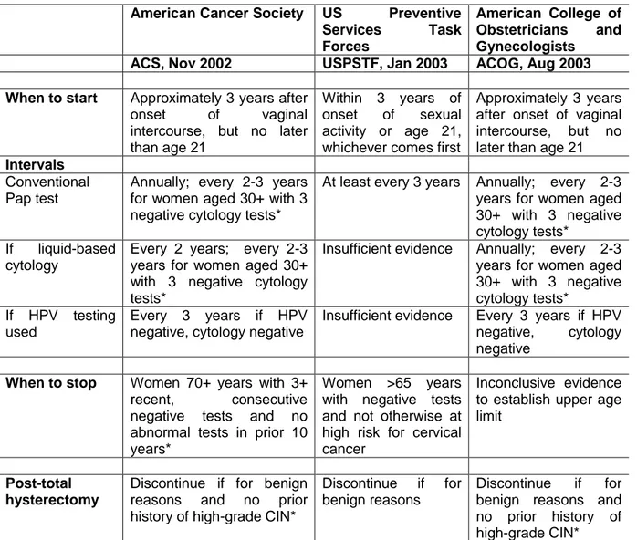
screening. Guidelines for cervical screening in the US are presented in Table 2.2 below.
WHO guidelines for screening targeted mainly for developing countries recommend at least
one screen in the 4th and 5th decade or a screen every 3 years where possible [WHO, 2006].
Conventional cytology is considered the standard method, though this could be replaced
with visual methods if possible.
Screening would result in the detection of precancerous lesions which need to be
treated. Treatment modalities include outpatient treatments such as cryotherapy, loop
electrosurgical excision procedure (LEEP), and cold knife conization.
Tertiary prevention involves the reduction of cancer mortality: treatment options are varied
15
Table 2.2: Summary of cervical cancer screening guidelines in the US
[Source CDC, From http://www.cdc.gov/std/hpv/ScreeningTables.pdf]
American Cancer Society US Preventive Services Task Forces
American College of Obstetricians and Gynecologists ACS, Nov 2002 USPSTF, Jan 2003 ACOG, Aug 2003 When to start Approximately 3 years after
onset of vaginal intercourse, but no later than age 21
Within 3 years of onset of sexual activity or age 21, whichever comes first
Approximately 3 years after onset of vaginal intercourse, but no later than age 21 Intervals
Conventional Pap test
Annually; every 2-3 years for women aged 30+ with 3 negative cytology tests*
At least every 3 years Annually; every 2-3 years for women aged 30+ with 3 negative cytology tests*
If liquid-based cytology
Every 2 years; every 2-3 years for women aged 30+ with 3 negative cytology tests*
Insufficient evidence Annually; every 2-3 years for women aged 30+ with 3 negative cytology tests*
If HPV testing used
Every 3 years if HPV negative, cytology negative
Insufficient evidence Every 3 years if HPV negative, cytology negative
When to stop Women 70+ years with 3+ recent, consecutive negative tests and no abnormal tests in prior 10 years*
Women >65 years with negative tests and not otherwise at high risk for cervical cancer
Inconclusive evidence to establish upper age limit
Post-total hysterectomy
Discontinue if for benign reasons and no prior history of high-grade CIN*
Discontinue if for benign reasons
Discontinue if for benign reasons and no prior history of high-grade CIN* *some exceptions apply (for example women who are immunocompromised, have a history of prenatal exposure to DES etc).
2.2 Cervical cancer and precancerous lesions in HIV
An estimated 33 million people were living with HIV by the end of 2007, about half of whom are women and about two-thirds of whom live in sub-Saharan Africa [UNAIDS, 2008]. The proportion of women with HIV is much higher in developing countries
16
a substantial role for homosexual and intravenous routes). In Cameroon, which has an
estimated HIV prevalence of 5.5%, women constitute up to 300,000 of the 490,000 people
15+ years living with HIV. In the US, with a prevalence of 0.6%, only 230,000 of the
1,100,000 people 15+ living with HIV are women.
The early recognition of the frequency of cervical cancer in patients with advanced
HIV disease led to its definition as an AIDS-defining condition. It is now recognized that
HIV-positive women have: a higher prevalence of HPV and that the risk of infection increases
with the extent of immunosuppression; a higher prevalence of persistent infection and
infection with multiple HR-HPV types; a greater risk of precancerous lesions; a higher risk of
developing cancer, with diagnosis up to 10 years earlier than in the general population and a
faster progression to advanced disease with poor prognosis [WHO, 2006].
HIV positive women have a higher prevalence and incidence of cervical HPV infection [Palefsky, 2006]. A recent review of over 30 studies showed that the ratio of HPV prevalence in HIV positive to negative women ranged from 1 to 9.3, with most estimates
being between 1 and 3.6 [De Vuyst et al, 2008]. In an analysis of baseline data from the HIV
Epidemiology Research Study (HERS) cohort in the US, 64% of 851 HIV positive women
had HPV infection compared to 28% of 434 HIV negative women [Cu-Uvin 1999]. In a
longitudinal study of 284 women in the US (186 of whom were HIV positive) with
semi-annual HPV testing, HPV positivity among HIV- women and HIV+ women with CD4+ > or
=200 and <200 cells/uL was 47.5%, 78.7%, and 92.9% respectively [Ahdieh et al, 2000].
While about half of these infections may be attributed to the similar sexual transmission
routes, the other half occurred in women who did not report any recent sexual exposure yet
had incident HPV-infection, suggesting that there may be a reactivation of previously
controlled HPV-infection in immunosuppressed HIV positive women [Strickler et al, 2005].
17
was 0.29 and 0.10 among HIV+ women with CD4+ > or =200 and <200 cells/uL [Ahdieh et
al, 2000]. In another study of 220 HIV positive and 231 HIV negative women in New York,
HPV was persistent in 24% of HIV positives versus only 4% of HIV negatives [Sun et al,
1997].
HIV positive women are more likely to have infection with high-risk HPV. HIV-positive women were 1.8, 2.1, and 2.7 times more likely to have high-, intermediate-, and
low-risk HPV infections, respectively, compared with HIV-negative women [Ahdieh et al,
2001]. In a recent meta-analysis of 3230 women with no cytological abnormalities any HPV
prevalence was 36.3%, multiple HPV type prevalence was 11.9% while the most common
HPV types were HPV-16, 58, 18, 52, 31 and 33 (respective prevalence of 4.5, 3.6, 3.1, 2.8
2.0 and 2.0%) [Clifford et al, 2006]. Curiously HIV positive women with HSIL were less likely
to have HPV-16 compared to other women in the general population with HSIL (OR=0.6;
95%CI: 0.4, 0.7). Also in studies from Africa and North America, HPV type 58 was more
prevalent than type 18. Furthermore HIV positive women with HSIL had a higher prevalence
of low risk –HPV types 11, 53, and 61 suggesting that these supposedly low-risk types
may have a potential to cause neoplastic changes in immunodepressed women. It is also plausible that lower but undetectable levels of high risk types could be leading to HSIL.
Both low grade and high grade precancerous lesions are more prevalent in HIV positive women. In a recent review of published studies the prevalence of LSIL in HIV positive women ranged from 9.7% to 19.0% and LSIL was 1.6 to 8.8 times as prevalent in
HIV positive women as in HIV negative women [De Vuyst et al, 2008]. HSIL was also more
prevalent in HIV positive women who had prevalence rates ranging from 2.3% to 17.6%,
representing an occurrence 1.9 to 11.7 times as prevalent in HIV positive women as in HIV
18
of HIV positive women had lesions with LSIL in 23%, HSIL in 32% and lesion suspicious of
ICC in 20% of 150 HIV positive women [Parham et al, 2006].
HIV positive women have a higher incidence of precancerous lesions. Three cohort studies conducted in the US reported four-fold incidence of SIL in HIV positive
women compared to HIV negative women [Six et al, 1998; Massad et al, 2001; Schuman et
al, 2003]. In another study conducted in Senegal 71/627 (11%) of women developed HSIL
after a median 2.2 years of follow-up[Hawes et al, 2006]. HIV-2 positive appeared less likely
to develop HSIL compared to HIV-1 infected women (HR=0.3, 95%CI: 0.1, 0.9). HIV+
women with each of CD4 counts <200 high HIV viral load appeared more likely to develop
HSIL (HR for CD4<200 versus >200=5.5, 95%CI: 2.0, 15.2; HR for each log increase in viral
load=1.4, 95%CI: 1.1, 1.7). These factors were however not significantly associated with
incident HSIL in multivariate adjustments.
HIV-positive women may have a higher progression and a lower regression of precancerous lesions. Few studies have assessed the long-term evolution of lesions in HIV positive women. In the US, Six et al [1998] and Massad et al [2001] both reported faster
progression from LSIL to HSIL in HIV-positive women. In the former study, 38.1% of LSIL
had progressed to HSIL over a year in HIV positive women while none had progressed in
HIV negative women. All the progression occurred in women with CD4 counts less than
500/uL. In the latter study, the 6-months progression was 13.6 in HIV-positive versus 6.8%
in HIV negative women. The regression rate was also slower in HIV positive women (43%
versus 66%). These studies were however not sufficiently powered to assess the role of
other factors such as CD4 count, HIV viral load on evolution.
The prevalence and incidence of invasive cervical cancer appear higher in HIV positive women. Though cervical cancer was the most frequent cancer observed in early HIV positive women, early comparative studies did not find an increased
19
population. A few cohort studies such as the Women Interagency HIV study (WIHS) did not
find a difference in cervical cancer incidence by HIV status. Initial analysis by IARC in 1996
did not find any relationship either [De Vuyst, 2008]. However the rarity of cervical cancer
suggests the best evidence of association could only be gleaned from case-control studies
or much larger population based cohort studies. The WIHS observed no case of cancer in
HIV negatives and only one case of confirmed cervical cancer in HIV positives, thus having
a very low power to detect any difference [Massad et al, 2004]. Furthermore, high
HIV-associated mortality prior to HAART may have prevented the development and subsequent
detection of cervical cancer in HIV positive women in cancer registries. In a larger
population based study, the Sentinel Hospital Surveillance System for HIV infection, the
prevalence of cancer was slightly higher in HIV positive women (10.4 versus 6.2 cases per
1000 women) [Chin et al, 1998]. A case-control study in South Africa found a relative risk
for cervical cancer of 1.6 (95%CI 1.1, 2.3) [Sitas et al, 2000], while another study in Kenya
found that among women aged 35 and less women with cervical cancer had a higher HIV
prevalence (35% versus 17% in women without cancer) [Gichangi, 2002]. Multiple other
studies have shown an increased risk of cervical cancer in HIV positive women [De Vuyst,
2008].
While HIV related immunosuppression could account for most of the aforementioned characteristics of cervical lesions in HIV, other mechanisms, such as HIV-HPV interactions, have been postulated to play a role. Multiple studies reported a higher prevalence of any HPV, HR-HPV types, persistent HPV, SIL and CD4 count less than
200/uL (or 500/ul in some studies) [De Vuyst et al, 2008]. In some studies, high HIV viral
load was independently associated with increased incidence and progression of SIL
[Massad et al, 2001] suggesting that there may be direct HIV-HPV viral interactions. The
20
E7 transcription. Furthermore, it is postulated that the inflammatory responses associated
with HIV may interfere with the effectiveness of the anti-HPV immune response.
The effect of antiretroviral therapy on HPV and HPV-associated disease is not clearly understood. Highly active anti-retroviral therapy (HAART) does not appear to reduce HPV prevalence or incidence. In a French cohort the prevalence of HPV remained at
81% 5 months after HAART initiation [Heard et al, 2001]. In some studies, highly active
anti-retroviral therapy (HAART) has been shown to reduce the progression to high-grade
lesions and increase the regression of lesions to normal [Heard et al, 2004], however this
has not been consistent. The effect of HAART appears to be best with higher CD4 levels
[Palefsky, 2003].
2.3 Need to screen for cervical cancer in HIV in resource poor settings including Cameroon
Cervical cancer could potentially be a major cause of mortality in HIV-positive women if their life expectancy is increased with the use of antiretroviral treatment and they are never screened. In developed countries effective screening and early treatment of precancerous lesions have been key in preventing cervical cancer [Franceschi and Jaffe,
2007]. In the US 81% of women on antiretrovirals successfully receive annual Pap smears
[Stein et al, 2001] and up to 94% of cervical cancers are detected at an early stage of
carcinoma-in-situ [Frisch et al, 2000]. Furthermore the long interval between precancerous
lesions and cancers allows for time to screen for cancer. In Italy 50% of HIV positive women
diagnosed with cervical cancer in 1996-2004 had had their HIV test results at least 10 years
prior to cancer diagnosis [Franceschi et al, 2006], sufficient time for the women to have
been screened and received treatment for precancerous lesions.
Access to antiretrovirals has dramatically increased in developing countries. While
the increased survival that is expected to accompany this increased access with an
21
the effect on cervical is expected to be moderate at best, as HAART has a very limited
effect, if any, on HPV persistence and the progression of lesions [Heard, 2004]. Even while
on antiretrovirals women would still need to be screened or receive other preventive care for
cervical cancer.
One of the reasons why women in resource-limited areas are not regularly screened
is the limited access to health providers or services. Presumably this could be overcome in
women on antiretrovirals who because of the need for follow-up of their HIV disease have a
greater contact with health services. This regular contact could be taken advantage of to
propose regular screening services. Once the women can be seen regularly in hospitals the
next potential barrier would be the cost of screening and the chance that most tests would
be negative. Screening could be made more cost-effective by targeting. Screening could be
targeted based on clinical demographic and or behavioral characteristics. HPV DNA tests
could also be used for screening, however the accuracy of HPV DNA testing in HIV positive
women is still unclear and it is plausible that HPV types not included in the current tests and
which are not considered high risk in the general population could well turn out to be
oncogenic in HIV-positive women. Furthermore the current costs of HPV DNA testing
remains a barrier for its widespread use in resource-limited settings. Other screening
methods such as VIA and VILI also have limited assessment in HIV positive women.
Recently developed HPV vaccines could potentially be useful, however so far their
efficacy has only been demonstrated in immunocompetent women. Screening with cervical
cytology thus offers better prospects for effectiveness in reducing cervical morbidity and
mortality in HIV positive women. Knowing the risk factors for cervical lesions could help in
22
2.4 Age and cervical cancer
The occurrence of cervical cancer and precancerous appears to be age dependent and may reflect the interaction of multiple physiological and pathological factors.
Physiological changes in the size and anatomical position of the cervical squamocolumnar junction (SCJ) may play a role in the age at which lesions occur.
Prior to puberty the SCJ is located at the external cervical os, at the intersection between
the endocervix (covered by a single-layered and thus relatively fragile columnar epithelium)
and the ectocervix (covered by a stratified squamous epithelium). At puberty, under the
influence of higher estrogen levels, the uterus and cervix grow in size, a growth that results
in the original SCJ being pushed externally such that a substantial proportion of columnar
epithelium is exposed to the vaginal atmosphere and its consequent acidity. Under the
influence of this acidity and increased estrogen levels, this area of columnar epithelium
begins undergoing a process called squamous metaplasia (which is considered normal) with
a gradual change of the columnar epithelium into a squamous epithelium. This occurs until
women are in their thirties and both the original and a new SCJ become visible on
colposcopy. The area between the new and the original SCJ is known as the transformation
zone and is the site where most precancerous lesions are detected. As women age into
menopause the influence of estrogen decreases, with a resultant decrease in the size of the
cervix that leads to the transformation zone retracting to the endocervical canal. In the
postmenopausal women both the new SCJ and the transformation zone have completely
retracted into the cervical canal and only the original SCJ is visible on colposcopy [WHO,
2006].
It can be inferred from these physiological changes that squamous lesions would be
23
puberty and incidence could increase until reaching a peak in the premenopausal ages and
then decrease after menopause with a reduction in the amount of transformation zone
exposed to the ectocervix. Although the retraction of the transformation zone in the canal
after menopause may imply a reduction in the occurrence of new lesions, it may also imply
that, particularly in settings with little or no systematic screening, lesions that occurred prior
to menopause may be retracted into the endocervical canal rendering detection by cytology
smears more difficult and thus resulting in the detection of cancers at a relatively advanced
stage in older women. It may also be inferred that, in countries with repeated regular
screening, in the absence of any lesions in the early post-menopausal period, it will be
unlikely to detect new lesions at older ages (as the transformation zone is less exposed).
The age at which lesions occur may also be related to the age-specific incidence, prevalence and persistence of HPV infection. Potentially reflecting higher exposure to HPV and increased susceptibility to HPV-infection, multiple studies have shown
that the incidence of HPV peaks amongst adolescents and women aged less than 25 years
then decreased in women in their third to fifth decades (35 to 55 years) only to have a
second peak after age 55 [Baseman and Koutsky, 2005; Herrero et al, 2000]. The latter
increase is thought to be reflective of the lesser ability to clear HPV infections, a reactivation
of latent HPV infection or a cohort effect at older ages. A recent review of age-specific HPV
prevalence, confirmed these trends to be consistent across geographical regions, with peak
ages varying to reflect sexual activity at different ages [Smith et al, 2008].
The age-specific occurrence of cervical lesions appears to reflect that of HPV infection albeit with a delay. Numerous studies report peak prevalence of lesions in women under 40[Chang et al, 1991; Sadeghi et al, 1989; Gupta et al, 2008]. The median or
mean age of occurrence however increases with severity of lesion [Chung et al, 1982;
Gupta et al, 2008]. There is no evidence that the progression or regression rates of lesions
24
[2005] found no age differences in regression or progression. However in one study of HSIL
cases in India, lesions in older women (as well as women with high parity) were more likely
to progress than those in younger women [Misra et al, 2006].
Unlike HPV-infection and precancerous lesions, the incidence of and the mortality due to cervical cancer increases with age as shown in figure 2.1 below. While the increase is gradual in countries with a well established screening system, it is very
marked in less developed countries peaking after the fourth decade of life.
The relationship between age and the occurrence of precancerous or cancerous lesions led to the consideration of age as targeting factor in screening the general population of women. As described in Table 2.2 all the major screening guidelines consider the age at which to begin screening, the age at which to modify the
interval between screening tests and the age to discontinue screening. Such is not the case
for guidelines in HIV positive women at least not explicitly. Differences in the pathogenesis
of cervical SIL and age specific mortality may imply different age considerations in HIV
positive women. The age of occurrence of lesions in HIV-positive women, as well as the
progression of lesions by age and the cost-effectiveness of age-targeted screening needs to
25
26
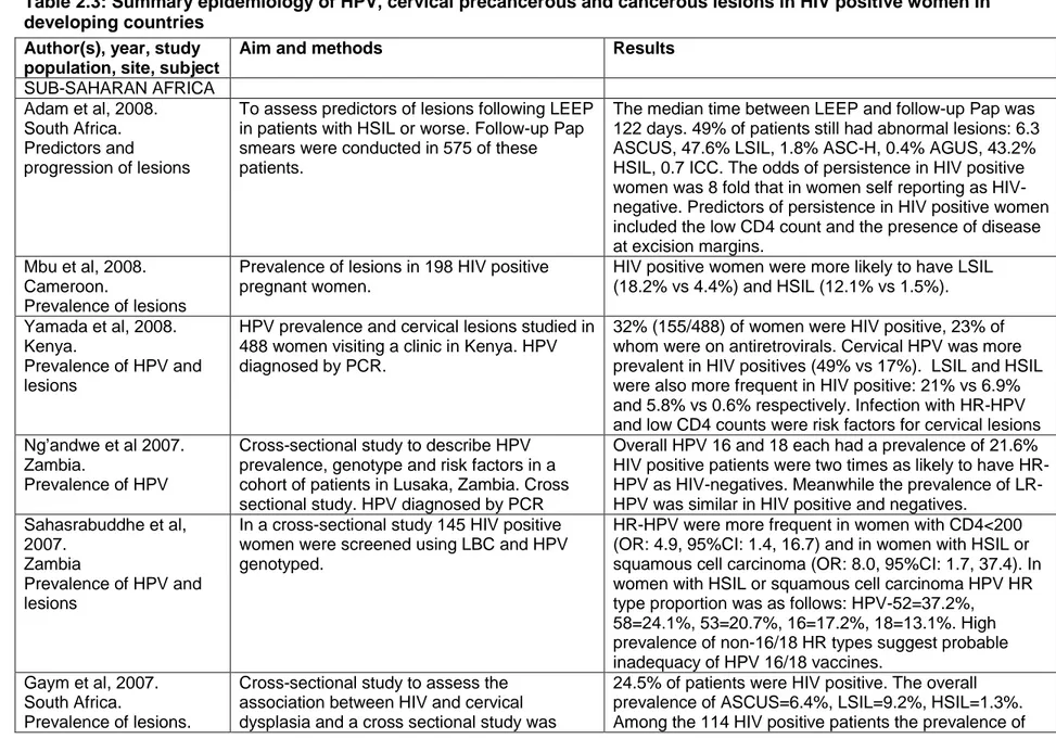
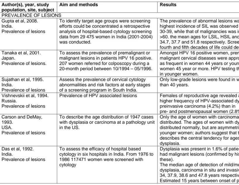
2.5 Review of previous studies
2.5.1 Studies of the prevalence and predictors of cervical lesions in HIV-positive women developing countries
Though numerous studies have documented the epidemiology of cervical squamous
intraepithelial lesions in HIV positive women in developed countries few do so for developing
countries. Table 2.3 summarizes the main studies conducted in the latter. The prevalence of
SIL was quite varied ranging from 1.2% [Mbakop et al, 1996] to 76% [Parham et al, 2006].
Because most studies were conducted to establish the association between HIV infection
and the presence of SIL, very few assessed the risk factors specific to HIV positive women.
Amongst these, immunosuppression (low CD4 count), high HIV viral load and or infection
with high-risk HPV types tended to be associated with the presence of SIL. Only one
prospective study in sub-Saharan Africa was identified in the literature and this study
indicated a SIL incidence rate of 11% over a mean of 2.2 years of follow-up [Hawes et al,
2
7
Table 2.3: Summary epidemiology of HPV, cervical precancerous and cancerous lesions in HIV positive women in developing countries
Author(s), year, study population, site, subject
Aim and methods Results
SUB-SAHARAN AFRICA Adam et al, 2008. South Africa. Predictors and
progression of lesions
To assess predictors of lesions following LEEP in patients with HSIL or worse. Follow-up Pap smears were conducted in 575 of these patients.
The median time between LEEP and follow-up Pap was 122 days. 49% of patients still had abnormal lesions: 6.3 ASCUS, 47.6% LSIL, 1.8% ASC-H, 0.4% AGUS, 43.2% HSIL, 0.7 ICC. The odds of persistence in HIV positive women was 8 fold that in women self reporting as HIV-negative. Predictors of persistence in HIV positive women included the low CD4 count and the presence of disease at excision margins.
Mbu et al, 2008. Cameroon.
Prevalence of lesions
Prevalence of lesions in 198 HIV positive pregnant women.
HIV positive women were more likely to have LSIL (18.2% vs 4.4%) and HSIL (12.1% vs 1.5%). Yamada et al, 2008.
Kenya.
Prevalence of HPV and lesions
HPV prevalence and cervical lesions studied in 488 women visiting a clinic in Kenya. HPV diagnosed by PCR.
32% (155/488) of women were HIV positive, 23% of whom were on antiretrovirals. Cervical HPV was more prevalent in HIV positives (49% vs 17%). LSIL and HSIL were also more frequent in HIV positive: 21% vs 6.9% and 5.8% vs 0.6% respectively. Infection with HR-HPV and low CD4 counts were risk factors for cervical lesions Ng’andwe et al 2007.
Zambia.
Prevalence of HPV
Cross-sectional study to describe HPV prevalence, genotype and risk factors in a cohort of patients in Lusaka, Zambia. Cross sectional study. HPV diagnosed by PCR
Overall HPV 16 and 18 each had a prevalence of 21.6% HIV positive patients were two times as likely to have HR-HPV as HIV-negatives. Meanwhile the prevalence of LR-HPV was similar in HIV positive and negatives.
Sahasrabuddhe et al, 2007.
Zambia
Prevalence of HPV and lesions
In a cross-sectional study 145 HIV positive women were screened using LBC and HPV genotyped.
HR-HPV were more frequent in women with CD4<200 (OR: 4.9, 95%CI: 1.4, 16.7) and in women with HSIL or squamous cell carcinoma (OR: 8.0, 95%CI: 1.7, 37.4). In women with HSIL or squamous cell carcinoma HPV HR type proportion was as follows: HPV-52=37.2%,
58=24.1%, 53=20.7%, 16=17.2%, 18=13.1%. High prevalence of non-16/18 HR types suggest probable inadequacy of HPV 16/18 vaccines.
Gaym et al, 2007. South Africa.
Prevalence of lesions.
Cross-sectional study to assess the association between HIV and cervical dysplasia and a cross sectional study was
2
8
conducted in 466 women at a primary health care clinic in KwaZulu-Natal.
ASCUS=10.5%, LSIL=21.0%, HSIL=4.4%. Parham et al, 2006.
Zambia.
Prevalence of lesions.
Cross-sectional study to evaluate the prevalence and predictors of SIL in 150 HIV-infected women in Zambia (age 23-49). Screening was done using LBC and HPV typing by Roche linear array PCR
CD4 ranged from 7 – 942 (median 165)/uL.
76% had SIL: of which LSIL=23.3%, HSIL=32.6% and 20% had lesions suspicious of SCC.
85.3% had HR-HPV.
Overall very high prevalence of SIL and HR-HPV. The highest in any study population.
The HR-HPV type independently predicted the presence of HSIL/SCC (adjusted OR: 12.4, 95%CI: 2.62, 58.1). Didelot-Rousseae et al,
2006.
Burkina Faso.
Prevaence of HPV and lesions
Cross-sectional study of HPV and cervical SIL in 379 high-risk women.
HIV-1 seroprevalence=36.0%.
Overall HPV prevalence=66.1% of 360 validly tested. HPV prevalence was higher in HIV (87% vs 54%; PR=1.61, 95%CI: 1.4, 1.8). Similarly HR-HPV types and multiple HPV infections were more frequent in HIV positive women.
Prevalence of SIL in 126 HIV+ women= 48.4%; LSIL=38%, HSIL= 10%
Hawes et al, 2006. Senegal.
Incidence of lesions
627 women were assessed for the persistence of HPV and incidence of HSIL over a mean follow-up time of 2.2 years.
71/627 (11%) of women developed HSIL.
HIV-2 positive appeared less likely to develop HSIL compared to HIV-1 infected (HR=0.3, 95%CI: 0.1, 0.9). HIV+ women with each of CD4 counts <200 high HIV viral load appeared more likely to develop HSIL (HR for CD4<200 vs>200=5.5, 95%CI: 2.0, 15.2; HR for each log increase in viral load=1.4, 95%CI: 1.1, 1.7).
These factors were however not significantly associated with incident HSIL in multivariate adjustments.
Moodley and Garib, 2004. South Africa.
Prevalence of lesions
To determine the prevalence of SIL in 160 HPV positive women in South Africa.
HIV prevalence=41.9%; SIL prevalence=36.9%.
Biopsy confirmation of SIL in HIV positive=49.3% vs 28% in HIV negative. But no difference by grade. Authors suggest similar management of HPV positive women whether they are HIV-positive or not.
Hawes et al, 2003. Senegal.
Prevalence of HPV and lesions
Cross-sectional study of prevalence and predictors of HSIL/ICC in 4114 outpatient women in Senegal.
HIV prevalence =10.5%, 433 women. HIV-1 only=8.1%, HIV-2=1.7%, HIV1 and 2= 0.7%.
Prevalence of HR-HPV in HIV-1 only (N=330), -2only (N=68), -1 and -2 (N=28) was respectively 53%, 41%, and 60%.
![Table 2.1: Terminologies/classification of cervical precancerous/cancerous lesions [source: WHO, 2006]](https://thumb-us.123doks.com/thumbv2/123dok_us/8261172.2188631/24.918.133.814.184.547/table-terminologies-classification-cervical-precancerous-cancerous-lesions-source.webp)
![Figure 2.1: Age-specific incidence and mortality of cervical cancer (Source: Globocan 2002, [Ferlay et al, 2004])](https://thumb-us.123doks.com/thumbv2/123dok_us/8261172.2188631/40.918.136.807.135.531/figure-specific-incidence-mortality-cervical-source-globocan-ferlay.webp)
![Figure 3.3: Summary of states in the Markov model (Adapted from an original depiction by Goldie et al, [1999])](https://thumb-us.123doks.com/thumbv2/123dok_us/8261172.2188631/79.918.134.809.430.930/figure-summary-states-markov-adapted-original-depiction-goldie.webp)