IRON DEFICIENCY ANEMIA AND THE PATHOGENESIS OF FALCIPARUM MALARIA
Morgan McFarland Goheen
A dissertation submitted to the faculty at the University of North Carolina at Chapel Hill in partial fulfillment of the requirements for the degree of Doctor of Philosophy in the Department of Microbiology and
Immunology in the School of Medicine.
Chapel Hill 2016
Approved by:
Carla Cerami
Myron S. Cohen Kristina De Paris
Raj S. Kasthuri Thomas H. Kawula
ii © 2016
iii ABSTRACT
Morgan McFarland Goheen: Iron Deficiency Anemia and the Pathogenesis of Falciparum Malaria (Under the direction of: Carla Cerami and Steven R. Meshnick)
Anemia, primarily iron deficiency anemia (IDA), affects up to 50% of pregnant women and 40% of preschool children in the developing world, significantly impacting perinatal and developmental health. However, clinical studies have found (1) iron deficiency protects from malaria, and (2) administration of iron to iron deficient individuals may increase the risk of malaria, thus complicating universal iron supplementation recommendations in malaria-endemic areas.
Our lab developed an in vitro model, obtaining red blood cells (RBCs) from IDA or healthy donors at UNC, to study mechanisms of malaria-associated IDA protection and iron treatment risk. We
demonstrated decreased P.falciparum invasion and growth in IDA RBCs and increased infection susceptibility in young RBCs and reticulocytes. Given iron is essential for the parasite, it was previously thought iron deficiency inhibited malaria through starvation. However, IDA also limits erythropoiesis and induces physiologic RBC changes. Our UNC-based studies thus generated a novel and paradigm-shifting hypothesis – namely that changes in RBC properties and the RBC population structure drive IDA
resistance to and iron supplementation risk for malaria.
Our next objective was to evaluate this hypothesis in pregnant women and children from a malaria-endemic area, via comprehensive longitudinal examination of P.falciparum pathogenesis in RBCs drawn from iron deficient Gambian children and pregnant women before, during, and after iron
iv
rates following iron supplementation paralleled increases in circulating reticulocytes and other markers of young RBCs, kinetics of which correlate with overall increased erythropoiesis. We conclude malaria growth in vitro corresponds with elevated erythropoiesis, an inevitable consequence of iron
v
ACKNOWLEDGEMENTS
vi PREFACE
The material included in Chapter 2 is a replication of our paper published in Nature
Communications (2014) and is included here with permission from Nature Publishing Group. The authors of this paper are, in order: Clark MA, Goheen MM, Fulford A, Prentice AM, Elnagheeb MA, Patel J, Fisher N, Taylor SM, Kasthuri RS, Cerami C. As second author on this paper, I was involved in much of the experimental work in terms of data collection and analysis, as well as discussion of interpretations and ideas and editing of the manuscript. As first author, Martha Clark was unquestionably the one who spearheaded the research and development of this paper.
The material included in Chapter 3 is a replication of our paper published in the British Journal of Haematology (2016) and is included here with permission from John Wiley and Sons. The authors of this paper are, in order: Goheen MM, Clark MA, Kasthuri RS, Cerami C. As first author on this paper, I was centrally involved in experimental design, data analysis and results interpretation, and writing of the manuscript.
The material included in Chapter 4 is a replication of a manuscript conditionally accepted to EBioMedicine following minor formatting revisions. The authors of this paper are, in order: Goheen MM, Wegmuller R, Bah A, Darboe B, Danso E, Affara M, Gardner D, Patel JC, Prentice AM, Cerami C. As first author on this paper, I was centrally involved in experimental design, data analysis and results
interpretation, and writing of the manuscript. I have also worked extensively with Dr. Meshnick and his epidemiology Ph.D. student Jordan Cates to include additional analyses and discussion of analytical approaches beyond the scope of our accepted manuscript at the end of this chapter.
vii
TABLE OF CONTENTS
CHAPTER ONE: INTRODUCTION TO ERYTHROCYTE ALTERATIONS WHICH IMPACT
FALCIPARUM MALARIA INFECTION……….…….…1
1.1 INTRODUCTION ... 1
1.2 HEMOGLOBINOPATHIES... 4
1.2.1 Sickle-cell Trait ... 4
1.2.2 Hemoglobin C ... 6
1.2.3 Hemoglobin E ... 7
1.2.4 Thalassemias ... 8
1.3 ENZYMATIC DEFICIENCIES ... 11
1.3.1 Glucose-6-Phosphate Dehydrogenase Deficiency ... 11
1.3.2 Pyruvate Kinase Deficiency ... 14
1.4 RBC MORPHOLOGY AND THE CYTOSKELETON ... 15
1.5 BLOOD GROUPS ... 18
1.5.1 ABO Blood Groups ... 18
1.5.2 Duffy Antigen Receptor for Chemokines (DARC) ... 19
1.6 NEW AREAS OF RESEARCH REGARDING CHANGES IN RBC PHYSIOLOGY... 21
1.6.1 Nutritional Immunity, Iron Deficiency, and Anemia ... 21
1.7 CONCLUSIONS ... 22
1.8 DISSERTATION OUTLINE ... 23
1.9 REFERNCES ... 25
CHAPTER TWO: HOST IRON STATUS AND IRON SUPPLEMENTATION MEDIATE MALARIA SUSCEPTIBILITY TO ERYTHROCYTIC STAGE PLASMODIUM FALCIPARUM………..37
2.1 OVERVIEW ... 37
2.2 INTRODUCTION ... 37
2.3 MATERIALS AND METHODS ... 38
2.4 RESULTS ... 43
2.4.1 Malaria growth is reduced in RBCs from individuals with IDA. ... 43
2.4.2 Malaria growth is increased in RBCs from iron-supplemented donors. ... 43
2.4.3 RBCs from donors with IDA are refractory to malaria infection. ... 45
2.4.4 Replacement of iron deficient RBCs increases malaria growth. ... 46
viii
2.5 DISCUSSION ... 49
2.6 TABLES AND FIGURES ... 53
2.7 SUPPLEMENTARY FIGURES ... 59
2.8 REFERENCES ... 65
CHAPTER THREE: BIOPRESERVATION OF RBCS FOR IN VITRO PLASMODIUM FALCIPARUM CULTURE………68
3.1 OVERVIEW ... 68
3.2 INTRODUCTION ... 68
3.3 MATERIALS AND METHODS ... 69
3.4 RESULTS ... 72
3.4.1 P.falciparum growth rates decline proportional to RBC storage length. ... 72
3.4.2 Differential storage media does not prolong longevity of RBCs for use in P.falciparum growth assays. ... 72
3.4.3 The decline in P.falciparum growth rates in stored RBCs is primarily due to decreased invasion. ... 73
3.4.4 Biopreserved RBCs remain suitable for P.falciparum growth and invasion assays. ... 73
3.5 DISCUSSION ... 73
3.6 FIGURES ... 75
3.6 REFERENCES ... 77
CHAPTER FOUR: ANEMIA OFFERS STRONGER PROTECTION THAN SICKLE-CELL TRAIT AGAINST ERYTHROCYTIC STAGE MALARIA IN VITRO AND THIS PROTECTION IS REVERSED BY IRON SUPPLEMENTATION……….78
4.1 OVERVIEW ... 78
4.2 INTRODUCTION ... 79
4.3 MATERIALS AND MEHTODS ... 80
4.4. RESULTS ... 82
4.4.1 P.falciparum growth is reduced in RBCs from anemic children ... 82
4.4.2 The population level impact on parasite growth is greater from anemia than from sickle-cell trait genotype. ... 83
4.4.3 P.falciparum clinical isolates exhibit decreased growth in RBCs from anemic children. ... 84
4.4.4 RBCs from anemic children are resistant to invasion by laboratory and clinical strains of P.falciparum. ... 84
4.4.5 Malaria susceptibility increases transiently with iron supplementation. ... 84
4.4.6 The population of young RBCs increases in anemic children undergoing iron supplementation. ... 85
4.5 DISCUSSION ... 86
4.6 TABLES AND FIGURES ... 89
4.7 SUPPLEMENTAL INFORMATION ... 94
ix
4.7.2 Supplemental Tables and Figures ... 96
4.8 DISCUSSION ADDENDUM ... 102
4.8.1 Linear regression and population level impact: results, interpretations, and alternative analyses. ... 102
4.8.2 Conclusions ... 114
4.9 REFERENCES ... 115
CHAPTER FIVE: ERYTHROCYTES FROM GAMBIAN PREGNANT WOMEN ARE PROTECED AGAINST MALARIA IN VITRO AND THIS PROTECTION IS REVERSED BY IRON SUPPLEMENTATION
………..118
5.1 OVERVIEW ... 118
5.2 INTRODUCTION ... 119
5.3 MATERIALS AND METHODS ... 122
5.4 RESULTS ... 127
5.4.1 P.falciparumin vitro growth rates are reduced based on level of anemia in pregnant Gambian women. ... 127
5.4.2 RBCs from anemic pregnant women are resistant to invasion by P.falciparum, but invasion deficits in anemic RBCs are not attributable to PfRh invasion ligands nor parasite attachment. ... 128
5.4.3 P.falciparum growth rates increase in RBCs from pregnant women undergoing iron supplementation. ... 130
5.4.4 Pregnant women undergoing iron supplementation exhibit increased levels of young RBCs in circulation. ... 131
5.5 DISCUSSION ... 132
5.5.1 Work in Progress ... 135
5.6 TABLES AND FIGURES ... 136
5.7 SUPPLMENTAL TABLES AND FIGURES ... 143
5.8 REFERENCES ... 148
CHAPTER SIX: DISCUSSION AND FUTURE DIRECTIONS……….152
6.1 BRIEF SUMMARY OF WORK PRSENSED IN THIS DISSERTATION ... 152
6.2 WAYS FORWARD ... 155
6.2.1 Further investigating mechanisms of reduced invasion into iron deficient RBCs. ... 156
6.2.2 Further investigating iron acquisition and utilization pathways in the falciparum malaria parasite. ... 158
6.2.3 Clinical needs: further understanding impact of anemia on malaria pathogenesis and implications for global health strategies. ... 161
6.2.4 New research into human evolution against blood stage malaria infection. ... 164
6.3 CONCLUSIONS ... 166
x
LIST OF TABLES
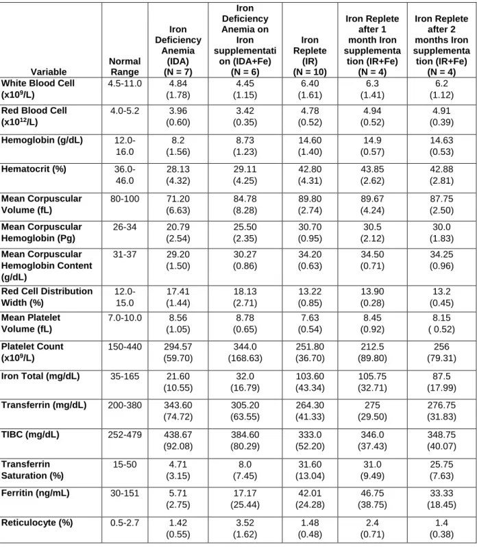
Table 2.1. Iron parameters and values of study participants ... 53
Table 4.1: Blood, inflammatory, and iron parameters of anemic donors whose RBCs were used for parasite growth assays before (Day 0), during (Day 49), and after (Day 84) iron supplementation ... 89
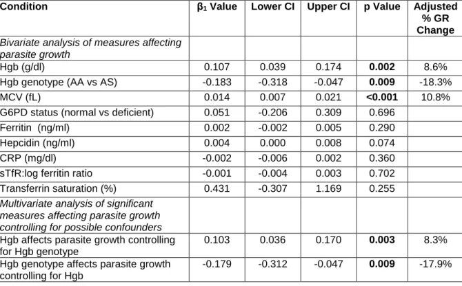
Table 4.2. Effect of host hemoglobin, iron status, and other hematological characteristics on in vitro P. falciparum growth in RBCs from anemic children (Hgb<11g/dl) at baseline ... 90
Supplemental Table S4.1: Description of subjects ... 96
Supplemental Table S4.2: Blood, inflammatory, and iron parameters of study participants with reportable parasite growth rate data versus those without ... 97
Addendum Table A4.1: Multivariate modeling with Hgb dichotomized ... 105
Addendum Table A4.2: Combinations of hemoglobin categories tested ... 106
Addendum Table A4.3: Number of people in each group for different Hgb categories ... 106
Addendum Table A4.4: Linear regression results for different Hgb categories ... 107
Addendum Table A4.5: Frequency of anemia type in Gambian children ... 110
Addendum Table A4.6: Population level impact using continuous versus dichotomized Hgb ... 111
Table 5.1: Blood, inflammatory, and iron parameters of donors whose RBCs were used for parasite growth assays before (Day 0), during (Days 14 and 49), and after (Day 84) iron treatment ... 136
Table 5.2. Effect of host hemoglobin, iron status, and other hematological characteristics on in vitro P. falciparum growth in RBCs from pregnant women (14-22 weeks gestation) at baseline ... 137
Supplemental Table S5.1: Description of subjects at baseline ... 143
xi
LIST OF FIGURES
Figure 2.1. P.falciparum growth is reduced in iron deficient RBCs and iron supplementation
eliminates growth attenuation ... 54
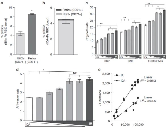
Figure 2.2. P.falciparum invasion and growth are reduced in RBCs from IDA donors ... 55
Figure 2.3. Replacement of iron deficient RBCs with iron-replete RBCs increases P.falciparum infection ... 56
Figure 2.4. The elevated P.falciparum infection supported by young RBCs is reversed as young RBCs are replaced with old RBCs ... 57
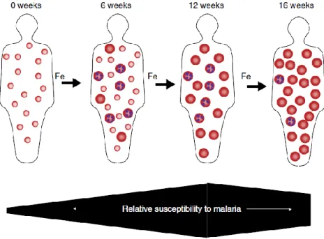
Figure 2.5. Hypothesized impact of iron deficiency and iron supplementation on host RBC population dynamics and susceptibility to erythrocytic stage malaria infection ... 58
Supplementary Figure S2.1. Serial in vitro growth assay design for P.falciparum ... 59
Supplementary Figure S2.2. Replacement of RBCs from IDA donors with RBCs from IR donors increases the invasion rate of erythrocytic stage P.falciparumin vitro (additional strains 3D7 and Dd2) ... 61
Supplementary Figure S2.3. Reticulocytes and young RBCs support greater P.falciparum growth rates and PEMRs in vitro than increasingly older RBCs ... 63
Supplementary Figure S2.4. P.falciparum plateau in invasion rate of RBC populations containing 50% to 100% young RBCs is independent of merozoites inoculum ... 64
Figure 3.1. Effects of RBC storage conditions on Plasmodium falciparum growth in vitro ... 75
Figure 3.2. Biopreserved red blood cells (RBCs) are suitable for Plasmodium falciparum growth ... 76
Figure 4.1: Description of subjects and flow chart of sample collection and assays performed ... 91
Figure 4.2: Parasite growth and invasion in RBCs from anemic children (Hgb<11g/dl) at baseline ... 92
Figure 4.3: Malaria susceptibility increases transiently during iron supplementation and anemic children receiving iron supplements have increased numbers of young RBCs... 93
Supplemental Figure S4.1: Parasite growth rates in RBCs from children categorized by different definitions of anemia at baseline... 98
Supplemental Figure S4.2: Gating strategy to highlight adaptation of RBC barcoding assay to the field setting using basic two-color flow cytometry ... 99
Supplemental Figure S4.3. Changes in parasite growth, invasion, and reticulocytosis in RBCs from anemic children before and after daily iron supplementation ... 100
xii
Figure 5.1: Description of subjects and flow chart of sample collection and assays performed ... 138 Figure 5.2: Parasite growth and invasion in RBCs from pregnant women (14-22 weeks
gestation) at baseline ... 139 Figure 5.3: P.falciparum PfRh invasion ligands and attachment rates are not involved in
mechanisms of differential invasion into RBCs from anemic versus non-anemic donors ... 141 Figure 5.4: Malaria susceptibility increases transiently during iron supplementation and pregnant
women receiving iron supplements have increased numbers of young RBCs... 142 Supplemental Figure S5.1: Parasite growth in RBCs from different donor groups at baseline ... 145 Supplemental Figure S5.2: Changes in parasite growth and reticulocytosis in RBCs from
pregnant women before, during, and after daily iron supplementation ... 146 Supplemental Figure S5.3: Surface markers of RBC age and integrity change in a pattern
consistent with an increase in erythropoiesis in pregnant women undergoing iron
xiii
LIST OF ABBREVIATIONS
AA – normal β-globin genotype
ABO – blood group consisting of A, B, and O antigens AC – heterozygous hemoglobin C β-globin genotype ANCOVA – analysis of covariance
ANOVA – analysis of variance
ARDS – acute respiratory distress syndrome CC – homozygous hemoglobin C β-globin genotype CR1 – complement receptor 1
AE – heterozygous hemoglobin E β-globin genotype ACD – acid citrate dextrose
ACM – asexual culture media
AS – heterozygous sickle-cell trait β-globin genotype ATP – adenosine triphosphate
CD – cluster of differentiation CD71+ – reticulocyte
CI – confidence interval
CPDA – citrate phosphate dextrose adenine CRP – C reactive protein
DARC – Duffy antigen receptor for chemokines DPG – diphosphoglycerate
EBA – family of Plasmodium falciparum erythrocyte binding-like antigens EPCR – endothelial protein C receptor
Fe – iron
G6PD – glucose-6-phosphate dehydrogenase GPA – glycophorin A
GR – growth rate
xiv EE – homozygous hemoglobin E β-globin genotype Hb – hemoglobin (in the context of the β-globin genotype) Hgb – hemoglobin
HLA – human leukocyte antigen IDA – iron deficiency anemia
ICAM – intracellular adhesion molecule IL – interleukin
IRB – institutional review board
IPT – intermittent preventative treatment of malaria
IPTp – intermittent preventative treatment of malaria in pregnancy IR – iron replete
MACS – magnetic activated cell sorting MCH – mean corpuscular hemoglobin
MCHC – mean corpuscular hemoglobin concentration MCV – mean corpuscular volume
MFI – mean fluorescent intensity MHC – major histocompatibility complex MNP – micronutrient powder
MPV – mean platelet volume MRC – Medical Research Council
MRCG – Medical Research Council The Gambia Unit NADPH – nicotinamide adenine dinucleotide phosphate NHANES – National Health and Nutrition Examination Survey NOS – nitric oxide synthase
PEMR – parasite erythrocyte multiplication rate Pf – Plasmodium falciparum
PfEMP1 – Plasmodium falciparum erythrocyte membrane protein 1
xv PK – pyruvate kinase
pRBC – parasitized red blood cell RBC – red blood cell
RDW – red cell distribution width
Rh – rhesus factor for human blood types RDT – rapid diagnostic test
RDW – red cell distribution width ROS – reactive oxygen species SAO – Southeast Asian ovalocytosis
SC – heterozygous sickle-cell trait and hemoglobin C β-globin genotype SCC – scientific coordinating committee
SD – standard deviation SI – susceptibility index
SS – homozygous sickle-cell anemia β-globin genotype sTfR – soluble transferrin receptor
Tf – transferrin
TIBC – total iron binding capacity TNF – tumor necrosis factor Tsat – transferrin saturation
UIBC – unbound iron binding capacity
UNC – University of North Carolina at Chapel Hill
UNIMMAP – UNICEF international multiple micronutrient preparation WBC – white blood cell
1
CHAPTER ONE: INTRODUCTION TO ERYTHROCYTE ALTERATIONS WHICH IMPACT FALCIPARUM MALARIA INFECTION
1.1 INTRODUCTION
Malaria has been a threat to humankind for centuries, impacting vast swaths of societal function from geographical settlement to war and colonial infiltration, to economic productivity. Still, today an estimated 438,000 people die annually (primarily children under 5 succumbing to Plasmodium falciparum in Sub-Saharan Africa) (1). The lost economic cost is substantial, and correlates directly with per capita GDP in the developing world (2). This highlights how much more work needs to be done if we are to win the battle against this formidable infectious disease.
The life cycle of malaria parasitesis complex, involving the mammalian host and the mosquito vector. The mosquito injects the sporozoite form of the parasite into the human host. Sporozoites quickly home to the liver and after a brief asymptomatic hepatic stage, merozoites burst out of hepatocytes to enter the bloodstream and begin the intra-erythrocytic infection cycle. During this cycle (which lasts 48hrs for P.falciparum), the parasite develops from the ring stage to the metabolically active trophozoites, which then mature into schizonts. When they are completely mature, the schizonts (up to 36 per RBC) burst out of the RBC and again become merozoites. Merozoites remain extracellular for a very brief time before invading new red blood cells (RBCs) and starting the 48 hour cycle again. The most severe clinical symptoms of malaria are associated with this burst of RBCs when the parasites leave one host RBC and move to the next.
2
is endemic throughout parts of Asia, Africa, and Central and South America, however, its highest infection burden falls on Sub-Saharan African children under 5.
There is much to be learned about malaria pathogenesis from uncovering the relationship between the human RBC and the malaria parasite. With drug resistance now arising to even the most important first line artemisinin-based antimalarials (3), and disappointing recent large-scale vaccine trials (the most promising vaccine candidate has been found to only offer roughly 30% reduction in severe malaria in infants and children with limited cross-strain protection and waning efficacy over time (4–7)), we clearly are still in need of new strategies to combat malaria. Turning our attention towards naturally derived alternations in the RBC that confer protection against malaria may provide much needed insight into possible drug development strategies as well as better understanding of malaria pathogenesis and transmission. Of course the immune system, endothelium, and liver also impact malaria infection; however in this introductory chapter, I will focus attention on changes in RBC physiology that impact malaria pathogenesis. One such example is sickle-cell trait, which is so well-known that the average high school student knows it as a biological example of human evolution and selective pressure. I will discuss recent findings on the cellular and molecular mechanisms that help explain how sickle-cell trait and other hemoglobinopathies impact malaria pathogenesis. In addition, I will discuss other common genetic mutations that effect RBC physiology (via RBC enzyme deficiencies and abnormalities in RBC
membranes) and thereby impact malaria pathogenesis. Finally, in my own work presented in Chapters 2-5, I will discuss how changes in RBCs secondary to anemia decrease malaria parasite growth and development. My work also raises the interesting possibility that the high prevalence of anemia in people of African descent may not only be secondary to nutritional iron deficiency, but may also be the result a genetic attempt to alter RBC physiology in an effort to adapt to malaria.
Before delving further into discussion of these topics, however, it is important to review the range of clinical presentations of malaria seen in children and non-pregnant adults.
3
replication in check (8). However, the mechanisms responsible for this non-sterile immunity are unclear. There is also controversy about whether or not patients with asymptomatic malaria should be treated (9).
(2) Uncomplicated malaria. Malaria is considered uncomplicated when symptoms are present, but there are no clinical or laboratory signs of organ dysfunction. Symptoms of uncomplicated malaria are not specific and can include: fever, sweating, general weakness, enlarged spleen, mild jaundice, liver enlargement and increased respiratory rate.
(3) Severe malaria. Malaria is considered severe when it is complicated by serious organ failures or abnormalities in the patient's blood or metabolism. The manifestations of severe malaria can include cerebral malaria, severe anemia due to hemolysis, hemoglobinuria (hemoglobin in the urine) due to hemolysis, acute respiratory distress syndrome (ARDS), abnormalities in blood coagulation, acute kidney failure, metabolic acidosis, hypoglycemia, and hyperparasitemia, where more than 5% of the red blood cells are infected by malaria parasites.
Note that the term parasitemia is not a specific term: It is typically used to describe the simple presence of parasites or to refer to the level of parasite-infected RBCs, but it does not include any clinical information. Hence, people with either asymptomatic or uncomplicated malaria can be referred to as parasitemic.
4 1.2 HEMOGLOBINOPATHIES
1.2.1 Sickle-cell Trait
Human hemoglobin is comprised of two α-globin and two β-globin proteins; the sickle-cell mutation occurs in the gene encoding β-globin, causing a glutamic acid to valine switch at the 6th codon that leads to a sickling hemoglobin phenotype (HbAS in heterozygote form, HbSS for homozygotes) (10). This single nucleotide polymorphism is commonly found in populations of sub-Saharan African descent but is present globally, reaching a prevalence of 18% in some areas and affecting nearly 6 million neonates in 2010 (11). It was first hypothesized by Allison in the 1950s to relate to malaria protection, after he observed an increase in HbAS prevalence in the lowland populations where malaria was
frequently transmitted in comparison to the prevalence in the highland areas of Kenya and Uganda where malaria was rare and he observed malaria was less common in people with sickle-cell trait versus those without (12, 13).
Since then, it has been unequivocally shown that the heterozygous HbAS genotype protects against risk of severe malaria by analysis and meta-analysis of several studies (discussed and analyzed in (14–16)). Specifically, studies in West Africa have found sickle-cell trait to afford significant reduction in severe malaria cases (with comparable protection from the sub-types of severe malaria) in the range of 90% (17–20). Meta-analysis of studies totaling over 10,000 patients revealed the odds ratio of severe malaria in HbAS populations to be 0.09, or again a roughly 90% reduction in risk of overall (14). This has been further confirmed by extensive genome wide association studies (21). The impact of HbAS
genotype on uncomplicated malaria is also clear; studies have found lower parasite densities and uncomplicated malaria rates in those with sickle-cell trait (22–26). The same meta-analysis showed that HbAS genotype protects against uncomplicated malaria by approximately 30% (14). Less clear is the impact of HbAS genotype on the risk of parasitemia. Several conflicting cross-sectional studies report a range of parasitemia prevalence in HbAS vs HbAA genotype children, from lower prevalence in HbAS (12, 27, 28), to comparable (29–32) or higher (33, 34), which together has been taken to mean that there is insufficient evidence to show that HbAS genotype protects against parasitemia.
5
of research. (1) Effect of HbAS genotype on parasite growth and development: My own work (Chapters 4 and 5) and that of many others has shown that invasion and growth of P.falciparumin vitro within HbAS RBCs is reduced in low oxygen tension growth conditions (1%- 5% gas mixture versus candle jar oxygen tension of ~18%) (15, 35–37). Some have proposed that the increased sickling of infected HbAS RBCs may be a mechanism for impaired growth (35, 38, 39), however, more recent research suggests the story may not be that simple. LaMonte et al. discovered differential trafficking and effects of host miRNAs from HbAS and HbSS RBCs. Their research shows HbAS and HbSS RBCs have higher levels of certain host miRNAs (which can specifically inhibit translation of parasite enzymes important for growth and
development) and treatment with antisense oligonucleotides partially rescued parasite growth (40) in HbAS RBCs. (2) Effect of HbAS genotype on adhesion of parasitized RBCs: Fairhurst and colleagues have shown that HbAS RBCs exhibit aberrant Plasmodium falciparum erythrocyte membrane protein 1 (PfEMP1) expression on the RBC surface (41). This parasite protein is primarily responsible for
cytoadhesion of infected RBCs to the endothelial surface and to other uninfected RBCs (termed
6
infected RBCs versus HbAA infected RBCs (48). In conclusion, the impact of HbAS genotype is
multifactorial with several of the aforementioned mechanisms working together to alter the pathogenesis of falciparum malaria (discussed in (15)).
However, this benefit comes with a cost. Individuals with the HbAS genotype are usually asymptomatic, but can develop clinical symptoms similar to a sickle cell crisis during extreme
circumstances such as dehydration. In contrast, individuals with the homozygous HbSS genotype suffer from the physiological effects of sickling RBC deformations: microvascular obstruction and hemolysis leading to many downstream negative health effects, frequent pain crises, and reduced lifespan (reviewed in (10)). The widespread prevalence of HbAS despite the cost of HbSS inheritance demonstrates the profound advantage given by HbAS in the face of malaria infection. 1.2.2 Hemoglobin C
The Hemoglobin C mutation (HbAC for heterozygotes; HbCC for homozygotes) is very similar to sickle-trait, in that it involves a point mutation at the same position in the gene encoding β-globin, however it causes a different amino acid substitution (glutamic acid to lysine) at the 6th codon. It is also most common in populations in West Africa, with prevalence reported as high as 15% in parts of Burkina Faso, though outside of that epicenter the prevalence diminishes accordingly (49). For severe falciparum malaria, evidence of protection by the hemoglobin C variant is clear. Homozygous HbCC individuals show 80% reduction in the risk of severe malaria and heterozyogous HbAC people show 30% reduction (18, 50–52). Meta-analysis of 4 case-control studies revealed odds ratio of severe malaria to be 0.27 in HbCC and 0.83 in HbAC individuals compared to normal genotype children (14) which is on par with these results. As further strong evidence for overall protection, a recent genome-wide association study
7
However, another recent longitudinal cohort study in Malian children found reduced risk of both
uncomplicated clinical malaria and lower parasitemia burden in HbAC individuals (55) and a recent cross-sectional study from Burkina Faso found significant protection from parasitemia in HbCC and HbSC individuals (odds ratio 0.04 versus HbAA), even more so than for HbAS (odds ratio 0.27), with no differences in parasitemia for HbAC versus HbAA individuals (46).
The molecular mechanisms of protection from malaria following HbC inheritance are again not fully elucidated, but, again, the same three areas of (1) parasite growth and development; (2) adhesion of parasitized RBCs to endothelial cells; and (3) immune system changes, have all been implicated. In vitro studies indicate reduced growth in HbCC RBCs (56–58), with some further evidence of normal invasion but reduced schizont lysis and merozoite egress (58), while growth in HbAC RBCs has been reported to be normal (56, 58). Similar to results with the HbAS genotype, both the HbAC and HbCC genotypes can result in altered PfEMP1 expression on the infected RBC surface (41, 42). Further microscopic analysis has revealed an altered cytoskeleton in RBCs from donors with HbAC genotype which was hypothesized to impair trafficking of parasite proteins to the RBC surface (44, 45). The impact of the HbC variants on the immune system are even less clear. Thus, exact mechanisms for protection provided by the HbCC and HbAC genotypes remain an open question (15, 59).
Clinically, the tradeoff of inheriting the most protective form, HbCC, is mild hemolysis and anemia, while those with the HbAC genotype are asymptomatic. More significant clinical issues tend to arise with HbC co-inheritance with HbS or β-thalassemia (60).
1.2.3 Hemoglobin E
The Hemoglobin E mutation encompasses a point mutation that results in a glutamic acid to lysine switch at position 26 of the β-globin gene. It is most commonly found in parts of Southeast Asia and India, even reaching prevalence of up to 60% in some areas (60).
8
reduced prevalence of HbAE and HbEE individuals compared to prevalence of these people in the population as a whole (63).
Little has been done to characterize the mechanisms by which HbE might contribute to reduced parasitemia or malaria protection. Again, three broad categories of effects come forward: reduced parasite growth and development, altered adhesion of parasitized RBCs to endothelium, and impact on the immune system. Indeed, in vitro studies of HbEE and HbAE RBCs have sometimes found reduced invasion (in HbEE (64, 65) and HbAE (65)) and growth (in HbEE (66) and HbAE (67)), sometimes not (in HbEE (68) and HbAE (64, 66, 68)), reviewed in (15). Clearly more work needs to be done to answer these questions.
The trade-off for harboring the HbE trait seems to be minimal, with mild anemia resulting from lower levels of expression of the β-globin gene due to the genetic mutation resulting in a splice site insertion that reduces mRNA stability and translation (reviewed in (15))
1.2.4 Thalassemias
Human hemoglobin is formed by a complex of two α-globin and two β-globin chains which are encoded by two alpha-globin genes (HBA1 and HBA2) and one B globin gene (HBB). The term ‘thalassemias’ collectively refers to a number of different genetic mutations that result in reduced or absent expression of one or more of these globin alleles. Specifically, individuals described as having ‘α-thalassemia’ can have the loss of one or more α-globin allele(s): αα/α- (heterozygous), α-/α-
(homozygous), or αα/-- (loss of both paired α-globin gene, also homozygous); there is also HbH disease (loss of 3 α-globin alleles, α-/--) and finally hydrops fetalis (loss of all 4 α-globin genes; incompatible with life). Individuals with mutations in the beta globin gene(s) can also have a range of genetic defects, including β-thalassemia minor (reduced expression of one β-globin allele), and β-thalassemia major (reduced expression of both β-globin alleles) (reviewed in (15)). Geographically, the thalassemias can be found worldwide, with mild α+-thalassmias reaching prevalence of 10-20% throughout a belt spanning Sub-Saharan Africa, the Middle East, India, and Southeast Asia. β-thalassemia is predominantly prevalent in the Mediterranean and a few parts of Sub-Saharan Africa (see (15, 59)).
9
geographic malaria prevalence was formulated by Haldane in the 1950s as a result of his observations on the presence of β-thalassemia in West Africa (69).
To begin with α-thalassemia protection, meta-analysis of studies which examined prevalence of α-thalassemia in children with severe malaria and healthy children found an odds ratio for severe malaria of 0.63 for homozygote α-thalassemia and 0.83 for heterozygotes (14, 18, 70–72). Interestingly, such protection from severe malaria is eliminated by co-inheritance of the HbAS allele, perhaps providing an explanation for why α-thalassemia mutations are not as prevalent in many parts of Africa (26, 73). When it comes to protection from uncomplicated malaria, consensus is less clear – some studies found
decreased incidence of malaria with any form of α-thalassemia (though consistently lower with homozygote versus heterozygote form) (73, 74), while others actually found no difference (75) or increased incidence (76) compared to normal genotype. Meta-analysis concluded there was no risk reduction of uncomplicated malaria for α-thalassemia carriers (14). Finally, there also seems to be inconclusive evidence of asymptomatic parasitemia protection with α-thalassemia, both in terms of presence and density of parasites – but most studies do not find an effect on parasitemia (75, 77, 78).
There are far fewer comprehensive studies relating to β-thalassemia malaria protection; in fact the majority of available evidence comes from 2 studies, one finding uncomplicated malaria cases were less likely to involve people with β-thalassemia (79) and the other finding upon population screening similar parasitemia prevalence amongst those with β-thalassemia as in those with normal genotype, albeit lower parasite densities were observed in those with β-thalassemia (80).
As with most of the aforementioned hemoglobinopathies, the molecular mechanisms by which the thalassemias protect against malaria disease are less clear than the clinical protection itself.
β-10
globin when cultivated at high oxygen tension (83). Interestingly, invasion is frequently observed to be reduced in RBCs harboring both a β-globin thalassemia mutation and HbE (64, 65, 67) and reduced growth has been observed here as well (81) (all reviewed in (15)). To explain these findings, it was predicted that the thalassemias resulted in loss of ability to counter oxidative stress (15, 83). (2) Effects on endothelial cell adhesion. Others observed reduced rosetting with RBCs from individuals with α-thalassemia and thalassemia and also reduced binding of α-thalassemia and β-thalassemia RBCs to endothelial cells (81, 86–88), although the findings of reduced cytoadhesion and PfEMP1 expression with α-thalassemia is conflicted and inconsistent (43, 85, 89). Interestingly, as mentioned above, there is a reduction in protection against malaria in individuals who carry both HbAS and α-thalassemia (18, 23, 73). Investigating this phenomenon further with in vitro studies, Opi et al. found that co-inheritance abrogated the reduced PfEMP1 expression, cytoadhesion, and rosetting typically observed with HbAS. The mechanism for this remains unknown, though there was some speculation it might have to do with α-thalassemia reducing the levels of HbS production, perhaps especially if the hypothesis that HbS affects intracellular PfEMP1 and other membrane protein trafficking in infected RBCs is true (43).
(3) Effects on the immune system. Studies have found that anti-malarial immunoglobulins bind thalassemic infected RBCs more than normal infected RBCs (84, 89) and undergo increased
phagocytosis (48), which may result in enhanced immune clearance of parasitized RBCs from individuals with thalassemias (discussed in (15)).
11 1.3 ENZYMATIC DEFICIENCIES
1.3.1 Glucose-6-Phosphate Dehydrogenase Deficiency
Glucose-6-Phosphate Dehydrogenase (G6PD) catalyzes the first step in the pentose phosphate shunt, which ultimately produces NADPH, a reducing agent. NADPH is a critical co-factor for glutathione reductase which generates the reduced form of glutathione and is critical for lowering oxidative stress in RBCs.
X-linked genetic mutations causing G6PD deficiencies are among the most common genetic mutations globally and affect an estimated 400 million people (reviewed in (90)). Given the distribution of mutations throughout Africa, the Mediterranean, Middle East, and Asia, and their independent
development in multiple distinct populations, G6PD deficiency has been hypothesized to have arisen due to malaria pressure (discussed in (91, 92)). Yet, despite this long standing hypothesis and a large number of studies, there is quite conflicting evidence about whether or not G6PD protects against falciparum malaria infection. There is a great deal of controversy surrounding the question of whether or not
heterozygous and homozygous females are protected, and even the previously commonly held belief that hemizygote males are protected is no longer clear cut. Even very recent comprehensive studies report nearly the exact opposite results: that heterozygote females are the only ones protected against severe malaria (21, 93, 94) or that hemizygote males and homozygote females are the only ones protected against severe malaria (95).
A descriptive review of the dozens of studies that have been done to date to examine the question of whether or not G6PD deficiency protects against clinical malaria is beyond the scope of this chapter; instead, I will discuss some of the reasons why these studies have often conflicted. There are the typical issues: many of the studies have relatively small sample sizes, vary in the immune status of the subjects, and some are village surveys while others are hospital studies. This is all further
12
sequencing versus analysis of enzymatic activity) in the epidemiological studies linking malaria and G6PD deficiency. All of the aforementioned differences and complications are compounded by the fact that many of the different studies use different definitions of and categories of clinical malaria, which makes it difficult to draw definitive conclusions and compare studies, not to mention practically impossible to systematically review this research area.
The human gene for G6PD contains 13 exons and encodes 515 amino acids. G6PD is a monomer, but the active enzyme is a dimer which requires both NADPH and FAD (riboflavin) for its activity. Over 140 mutations have been identified (90, 96, 97) that each result in various degrees of change in the enzymatic activity and over 400 variants have been characterized that have normal levels of G6PD activity (98). To make things more confusing, all of the 400 variants with normal G6PD levels are considered wild type and are referred to as G6PD B. One common variant, which is found in 20-30% of Black Africans and is therefore often discussed in studies linking malaria and G6PD deficiency, is referred to as G6PD A+. G6PD A+ has a single amino acid substitution at position 376 of an asparagine for aspartate (98). Another common variant often referred to in the malaria literature is G6PD A- and it is found in 10-15% of African-Americans. All G6PD A- variants have a mutation at nucleotide 376 (A→G), which also is the nucleotide substitution characteristic of G6PD A+. However, the G6PD A- variants have a second mutation, which is usually at nucleotide 202 (G→A) or at nucleotide 680 (G→T) or at nucleotide 968 (T→C) (98, 99).
The World Health Organization has classified the different G6PD variants into clinically important categories according to the magnitude of the enzyme deficiency and the severity of hemolysis associated with each variant (100). Class I variants, which are rare, have severe enzyme deficiency (less than 10% of normal) and have chronic hemolytic anemia. Class II variants also have severe enzyme deficiency, but there is usually only intermittent hemolysis associated with infection, drugs, or chemicals. Class III variants have moderate enzyme deficiency (10-60% of normal) with intermittent hemolysis usually
associated with infection, drugs, or chemicals. Note that most G6PD A- variants fall into Class III. Class IV variants have no enzyme deficiency or hemolysis. Class V variants have increased enzyme activity (98).
13
clinical studies and there are many conflicting results. As reviewed by Ruwende and Hill, there is evidence for two main ways which the increased oxidative stress in G6PD deficient RBCs may protect against malaria: (1) it causes increased Heinz body formation and methemoglobin formation which results either RBC lysis or enhanced phagocytosis of infected RBCs, (2) it decreases the rate of intra-erythrocytic parasite replication (91). Certainly it is logical that reduced parasitemia could be the protective
mechanism against severe clinical malaria.
To go into more detail: some original studies do indicate decreased parasite growth in G6PD deficient RBCs (101–104) though not decreased invasion (103, 105), but others did not find reduced growth (105), or only observed decreased growth with oxidative stress present (83, 102). One study looking at growth in individual RBCs from heterozygote women in which X-inactivation occurs found decreased growth in the G6PD deficient RBCs specifically (106). Indeed, as G6PD deficiency reduces a RBC’s ability to combat oxidative damage, and with evidence that oxidative stress reduces parasite growth (107) (especially in G6PD deficient RBCs (108)), this is a plausible link. Others found no
difference in parasite invasion or maturation in erythrocytes harboring G6PD deficiency, but that infected RBCs were more susceptible to phagocytosis due to increased expression of phagocytic markers on the G6PD deficient infected RBCs (109).
14
normal G6PD status cells resisting oxidative stress/lysis, and in part from the presence of X-inactivated G6PD deficient RBCs that reduce the portion of habitable RBCs (93). Others have conducted research finding replication rates in G6PD deficient RBCs only remain low for a few cycles, surmising that the parasite can adapt to G6PD deficient conditions, thus speculating heterozygote females would maintain the most protection because there would not be enough pressure to induce parasite adaptation (103, 110).
Overall, much like the clinical outcomes, there is substantial conflicting evidence and still a great deal left to be learned about the protective molecular mechanisms of G6PD deficiency. It is entirely likely that several factors combine to mediate protection. Very likely much of the confusion and conflicting evidence of malaria protection in the G6PD literature arises because of the heterogeneity of the G6PD mutations themselves, not to mention the mutations’ inconsistent impact on overall enzymatic activity within an individual, and furthermore the complications of X-inactivation in females.
The clinical consequences of G6PD deficiency include increased risks of neonatal jaundice and of extensive hemolysis induced by excess oxidative stress. The most common triggers for hemolysis in this patient population are medications (including some gametocytic antimalarials), particular foods (most famously fava beans), and several types of infections (reviewed in (90)).
1.3.2 Pyruvate Kinase Deficiency
15
examining P.falciparum cases in Thailand (116), again suggesting this is yet another human genetic variant that is likely related to malaria pressure, and will result in further studies.
1.4 RBC MORPHOLOGY AND THE CYTOSKELETON
Another important type of RBC change involves membrane defects which affect the RBC shape. There are several different kinds of these genetic mutations; most frequently mutations are seen in cytoskeletal components and associated membrane proteins such as spectrin or band 3 or 4.1 proteins, although they can also be seen occasionally in the glycophorins as well (discussed in (117–119)). The network of proteins making up the RBC cytoskeleton and junctional membrane tethering sites is
complicated to visualize. Essentially, there is a lattice-like structure underlying the RBC membrane made up of primarily of α- and β-spectrin heterodimers which associate end to end as tetramers. There are also clusters of transmembrane proteins and other associated proteins that bridge the gap between binding the spectrin-based sub-membrane network and the membrane lipid bilayer. There are two main complexes of proteins: the ankyrin complex (consisting of predominantly band 3 tetramers, protein 4.2, GPA, CD47, and Rh linked via ankyrin to the spectrin heterodimer junction sites), and the 4.1R complex (consisting predominantly of 4.1R, F-actin, tropomysin, and band 3 dimers, as well as GPC, Rh, DARC, and Kell linked to the sites of association between the actin cytoskeleton and the subsurface spectrin mesh-like lattice). These protein interactions between membrane and cytoskeleton largely determine RBC shape and deformability and ability to circulate efficiently in the microvasculature (124, 125).
Multiple categories of these morphological RBC disorders exist. This includes:
1) Hereditary elliptocytosis (a heterogeneous disorder in terms of genetics and manifestation, ranging from severe hemolytic anemia to absence of symptoms; most mutations arise in spectrin and cause issues with spectrin dimer and tetramer associations or junctional associations (119– 121))
2) Hereditary pyropoikilocytosis (a severe hemolytic anemia type of hereditary elliptocytosis, often from a spectrin deficiency (120, 121))
16
4) Southeast Asian ovalocytosis (SAO, a disorder characterized by RBCs with oval shape and decreased deformability, possibly linked to a deletion in band 3 protein, and occurring at quite high rates in certain geographic areas (118, 119, 123)).
Most of these disorders are inherited in an autosomal dominant fashion (118–120) and are classified not based on genetics, but phenotypes. Generally speaking, mutations causing these RBC disorders result in reduced integrity of the RBC cytoskeleton, such that the RBC loses its biconcave shape and appears morphologically different by microscopy. Speculation that some of these conditions evolutionarily arose due to malaria pressure first arose due to the observed prevalence of hereditary elliptocytosis in people of African descent and areas where malaria was traditionally endemic. For example, hereditary elliptocytosis was determined by molecular epidemiology to be 30 times more prevalent in black people than Caucasians (117, 125, 126). Prevalence of hereditary elliptocytosis reached levels as high as 1.6% in Benin (126), and the prevalence of hereditary ovalocytosis can reach 35% in parts of Southeast Asia (117, 127). Overall there are several different cytoskeletal genetic mutations, especially in Africa, making it difficult to pinpoint exact correlations with predicted malaria disease protection. However, as discussed, it is hypothesized that these types of mutations are highly prevalent (particularly alpha spectrin haplotypes) and were maintained in human populations because they protect against malaria (117, 120).
17
growth, altered development, and reduced invasion, and that the growth and invasion deficits were more apparent with each cumulative replication cycle (117). This is similar to what others observed, finding severely decreased growth and invasion in elliptocytes with spectrin mutations (121, 134), as well as in elliptocytes with 4.1 mutations (134, 136). As Facer speculated, the relationship between parasite growth deficiencies and hereditary elliptocytosis could very plausibly deal with RBC invasion inhibition, either for mechanical reasons (reduced deformability, stability, endocytosis), or due to cytoskeletal mutations having downstream effects such as altered sialic acid presentation on membrane glycophorins important for merozoite attachment and entry (121). Similarly, SAO RBCs are known to be less deformable (137), and Gallagher discusses the likelihood of reduced merozoite invasion playing a role in ovalocytosis protection, either from poor band 3 binding or inability of band 3 to properly cluster and allow for membrane and cytoskeletal dissociations necessary for the parasite to enter the RBC (120). Certainly there are many plausible links between cytoskeletal defects and reduced parasite growth or ability to cause disease, which will continue to be elucidated with further study.
18 1.5 BLOOD GROUPS
1.5.1 ABO Blood Groups
The ABO blood groups are most commonly discussed in the context of blood typing to determine compatible blood transfusion donors. Blood type A, B, and AB contain A and/or B antigens on the surface of RBCs which blood type O RBCs lack. Specifically, RBCs with A and B antigens express trisaccharides and RBCs with O antigens express disaccharides because individuals with Type O lack a specific glucotransferase enzyme that adds the third sugar on to the trisaccharide (138). All blood group types exist globally (albeit at different frequencies), so perhaps the connection to malaria protection is less intuitively obvious than many of the RBC mutations presented above. Early studies were conflicted: some researchers found significant associations with severe malaria in blood group A, B, and/or AB, and/or that type O was protective (139–141); others did not find an association between blood group type and malaria severity (142–144). A 2007 systematic review concluded there were poor controls and small sample sizes but likely a real association with ABO blood group type and malaria susceptibility (145). Since then, more comprehensive studies have taken place, namely (146, 147), more definitively indicating type O individuals are less susceptible to severe malaria and non-type O individuals are at increased risk in Sub-Saharan Africa and India (146–153). The correlations appear to be strongest between severe clinical disease outcomes and blood type, not simply parasitemia levels or uncomplicated infection. The protection is significant, with odds ratio for increased risk of severe malaria in non-O type blood groups ranging from 1.26 – 2.95 ((146, 147, 152)and reviewed in (146)).
19
up clinically by correlative case control studies examining rosetting frequency in parasites harvested directly from infected individuals with type O and non-type O blood (146). Others report increased hemolysis of infected RBCs with type O blood (153), so perhaps there are additional mechanisms that could contribute to malaria protection, but overall the evidence seems quite conclusive that blood group O benefits from relative protection against severe malaria due to reduction in rosetting potential of infected RBCs. The obvious question thus remains, why is blood group O not nearly universally present in areas of high malaria endemicity? As discussed in Rowe et al. (160), this condition, too, is likely under
balancing selection, as there is evidence of increased association of other infectious agents (i.e. cholera, E. coli) with type O blood group (161, 162).
There is also some indication of altered rosetting susceptibility in certain Knops groups (which comprises various antigen presentations on the complement receptor (CR1) molecule), as well as altered immune complex formation and removal, though the data is quite preliminary (reviewed in (160)). Blood groups affecting the presence of glycophorin B and C have also been preliminarily speculated to relate to malaria susceptibility, but little has been as of yet investigated (reviewed in (160)).
1.5.2 Duffy Antigen Receptor for Chemokines (DARC)
Although P.vivax malaria is not the focus of this chapter, perhaps one of the most compelling pieces of evidence for RBC evolution against malaria is the Duffy antigen receptor for chemokines (DARC) found on the RBC surface. DARC is nearly totally absent from individuals in West and Central Africa. Molecularly speaking, the Duffy blood group is determined by two co-dominant alleles (Fy*A and Fy*B) coding for FyA and FyBproteins. The phenotype found across most of Sub-Saharan Africa is FyA-FyB-, and this results from a mutation in the promoter of the DARC FY gene which affects GATA-1 transcription factor binding – specifically, the GATA-1 transcription factor for erythroid lineage cells (thus expression of DARC in other tissues such as endothelium is unaffected) ((163) and reviewed in (160, 164)). There are also mutations found in Papua New Guinea resulting in essentially heterozygotic deficiency of DARC (FyA null) in a small population (165).
non-20
human primate parasite P.knowlesi (166). Subsequent research by this group looking at voluntary vivax exposure of inmates (through mosquito feeding) found African Americans with the FyA-B- blood group were resistant, while the Caucasians were not (167). This soon became hypothesized to be the reason P.vivax was largely absent from the African continent (reviewed in (168)). As further clinical evidence of the association between Duffy antigen presence and vivax susceptibility, it has been determined that those populations in Asia with essentially half the amount of Duffy antigen (FyA null) are also
approximately half as at risk for vivax infection and less likely to have severe disease or high levels of parasitemia ((165, 169) and reviewed in (160)). Though P.vivax cell culture remains impossible to this day, it was eventually determined that closely related knowelsi parasites require the Duffy antigen for merozoite tight junction formation for invasion of RBCs (170). And although extensive in vitro vivax studies are restricted, inhibition of invasion has been shown with the presence of antibodies targeting the vivax parasite’s Duffy antigen binding protein ((167, 171, 172) and reviewed in (160, 168)). This provides a very clear mechanism for the relationship between Duffy antigen absence and vivax absence in much of Sub-Saharan Africa.
Despite the seemingly obvious connection for the existence of Duffy negative blood groups (established at near 100% frequency in most of Africa) to have arisen as an evolutionary benefit to resist P.vivax infection, this association has still been called into question (reviewed in (168, 173)). For one thing, some researchers point out that vivax malaria (at least in its current form) is rarely as severe or lethal, questioning how such a strong phenotype regarding the existence and prevalence of the Duffy antigen negative blood group could have arisen from evolutionary drive for a comparatively weak disease. However, the notion of vivax being a rather “benign” form of malaria is also a point of recent debate, with many arguing vivax infection takes a significant clinical toll even if it is not as deadly, plausibly enough to drive evolutionary adaptation for resistance to infection (reviewed in (164, 168, 174–176)). Other
21
non-human primates in Africa (179). If true, this of course allows for simple explanation of why DARC is absent from most Africans.
Regardless of vivax origin and DARC prevalence, as Howes et al. extensively investigate and assess by meta-analysis, the belief that P.vivax is totally absent from Africa is far from accurate, with evidence for widespread (albeit quite low level) infection in many areas (180). Vivax existence in Africa is due to a combination of factors: a small population of Duffy positive individuals (particularly in parts of East Africa and Madagascar), possible zoonotic infection, and also parasite exploitation of alternative invasion routes in Duffy negative individuals (reviewed in (180)). In fact, definitive evidence has developed proving Duffy antigen negative individuals can support vivax infection – coming from such diverse regions as Mauritania, Equatorial Guinea, Cameroon, Ethiopia, Angola, Madagascar, Kenya, and even Brazil (reviewed and mapped in (168, 180)), which is cause for renewed scrutiny of vivax burden potential in Africa.
1.6 NEW AREAS OF RESEARCH REGARDING CHANGES IN RBC PHYSIOLOGY 1.6.1 Nutritional Immunity, Iron Deficiency, and Anemia
22
evolved to possess the siderocalin/lipocalin2 protein which can sequester bacterial siderophores. And yet still, some bacteria have developed stealth siderophores to avoid siderocalin binding (reviewed in (186)). Such strong evidence of back and forth evolution over iron access is also seen with findings of specific, targeted, point mutations developing over time in host transferrin and bacterial transferrin binding protein (to either evade or improve binding each other, depending on the organism’s goal) (187). Clearly, iron access can play an important role in human and pathogen evolution.
Beyond bacteria, iron is also a well-known growth factor for malaria (reviewed in (188)). Iron of course also directly effects erythropoiesis and is essential for hemoglobin formation. With growing epidemiological evidence over the past decades that iron deficient people are more resistant to malaria infection (189–192) and iron supplementation possibly puts them at increased risk for infection (193–197), the idea of nutritional immunity playing a role in malaria susceptibility is very plausible. Given the
geographic overlap between prevalence of iron deficiency and malaria endemicity in the developing world, there exists a public health conundrum of how to best treat anemia (itself not an inconsequential health deficit that can significantly affect child development and pregnancy outcomes), if iron deficiency is protective and iron supplementation increases malaria risk. Yet, evidence from these epidemiological studies is inconclusive and questions remain about exactly how iron deficiency and iron supplementation may impact malaria pathogenesis. Iron is not just a growth factor for malaria; iron deficiency significantly impacts several physiological properties of the RBC and hence could alter malaria infection susceptibility in many ways.
To better address this question, we have worked to model in vitro the growth of malaria in RBCs from relevant donor populations. The crux of this dissertation thus involves investigating the degree of protection of iron deficiency anemia and the safety of iron supplementation via in vitro characterization of infection and growth parameters of the malaria parasite in RBCs from iron deficient individuals before and after iron supplementation. This research is directly applicable to the concept of RBC alterations
conferring resistance to malaria and contributes to our broader understanding of malaria pathogenesis. 1.7 CONCLUSIONS
23
beyond the RBC. This would include immunity and inflammatory related factors, such as HLA haplotypes and MHC complexes (198, 199), TNF-α (200), CR1 (201), NOS2 (202), CD40L (203), and IL22 (204), or factors that affect binding of infected RBCs to the endothelium like CD36 (205), ICAM-1 (116), and EPCR (206) variants – all of which have been published as potentially affecting malaria susceptibility. Nor have I discussed new areas of research involving GWAS looking for novel RBC or other factors associated with reduced malaria severity that are as of yet unidentified or underappreciated.
Still, as is clear from all of the above evidence, humans and Plasmodium species have been at war for centuries, and one of the most important battlegrounds is indisputably the human RBC. The work in this dissertation was designed to better understand the interaction of the falciparum malaria parasite with iron deficient and anemic RBCs, a relatively new line of interest in the long standing quest to characterize parasite-RBC interactions in the context of modified human physiology and genetic adaptations. Of further importance, the research described in this dissertation also addresses the important public health issue of how to safely correct anemia in malaria endemic areas. Better
understanding of malaria pathogenesis in the face of altered RBC physiology will ultimately help improve our strategies to combat the disease.
1.8 DISSERTATION OUTLINE
To orient the reader, the work presented in the remainder of this dissertation includes:
Chapter 2: Initial in vitro modeling using UNC-based blood donors to examine how iron deficiency and iron supplementation conspire to mediate host susceptibility to erythrocytic stage malaria infection;
Chapter 3: Development of improved long-term RBC storage methods for in vitro falciparum culture in order to better utilize valuable RBC samples, in anticipation of initiating field work using blood donors enrolled in an iron supplementation clinical trial;
Chapter 4: Evaluation of the degree of protection afforded by iron deficiency anemia and the safety of iron supplementation through in vitro erythrocytic stage infection modeling, using RBCs from anemic Gambian children enrolled in an iron supplementation trial;
24
25 1.9 REFERNCES 1. WHO | World Malaria Report 2015 WHO. Available at:
http://www.who.int/malaria/publications/world-malaria-report-2015/report/en/ [Accessed September 25, 2016].
2. Sachs J, Malaney P (2002) The economic and social burden of malaria. Nature 415 (6872):680– 685.
3. Ashley EA, et al. (2014) Spread of artemisinin resistance in Plasmodium falciparum malaria. N Engl J Med 371 (5):411–423.
4. Agnandji ST, et al. (2011) First results of phase 3 trial of RTS,S/AS01 malaria vaccine in African children. N Engl J Med 365 (20):1863–1875.
5. RTS,S Clinical Trials Partnership (2014) Efficacy and safety of the RTS,S/AS01 malaria vaccine during 18 months after vaccination: a phase 3 randomized, controlled trial in children and young infants at 11 African sites. PLoS Med 11 (7):e1001685.
6. RTS,S Clinical Trials Partnership, et al. (2012) A phase 3 trial of RTS,S/AS01 malaria vaccine in African infants. N Engl J Med 367 (24):2284–2295.
7. Neafsey DE, et al. (2015) Genetic Diversity and Protective Efficacy of the RTS,S/AS01 Malaria Vaccine. N Engl J Med 373 (21):2025–2037.
8. Bousema T, Okell L, Felger I, Drakeley C (2014) Asymptomatic malaria infections: detectability, transmissibility and public health relevance. Nat Rev Microbiol 12 (12):833–840.
9. Chen I, et al. (2016) “Asymptomatic” Malaria: A Chronic and Debilitating Infection That Should Be Treated. PLoS Med 13 (1):e1001942.
10. Rees DC, Williams TN, Gladwin MT (2010) Sickle-cell disease. Lancet Lond Engl 376 (9757):2018–2031.
11. Piel FB, et al. (2013) Global epidemiology of sickle haemoglobin in neonates: a contemporary geostatistical model-based map and population estimates. Lancet Lond Engl 381 (9861):142–151. 12. Allison AC (1954) Protection afforded by sickle-cell trait against subtertian malareal infection. Br
Med J 1 (4857):290–294.
13. Allison AC (1954) The distribution of the sickle-cell trait in East Africa and elsewhere, and its apparent relationship to the incidence of subtertian malaria. Trans R Soc Trop Med Hyg 48 (4):312–318.
14. Taylor SM, Parobek CM, Fairhurst RM (2012) Haemoglobinopathies and the clinical epidemiology of malaria: a systematic review and meta-analysis. Lancet Infect Dis 12 (6):457–468.
15. Taylor SM, Cerami C, Fairhurst RM (2013) Hemoglobinopathies: slicing the Gordian knot of Plasmodium falciparum malaria pathogenesis. PLoS Pathog 9 (5):e1003327.
16. Allison AC (2009) Genetic control of resistance to human malaria. Curr Opin Immunol 21 (5):499– 505.
26
18. May J, et al. (2007) Hemoglobin variants and disease manifestations in severe falciparum malaria. JAMA 297 (20):2220–2226.
19. Aidoo M, et al. (2002) Protective effects of the sickle cell gene against malaria morbidity and mortality. Lancet Lond Engl 359 (9314):1311–1312.
20. Williams TN, et al. (2005) Sickle cell trait and the risk of Plasmodium falciparum malaria and other childhood diseases. J Infect Dis 192 (1):178–186.
21. Malaria Genomic Epidemiology Network, Malaria Genomic Epidemiology Network (2014)
Reappraisal of known malaria resistance loci in a large multicenter study. Nat Genet 46 (11):1197– 1204.
22. Parikh S, Dorsey G, Rosenthal PJ (2004) Host polymorphisms and the incidence of malaria in Ugandan children. Am J Trop Med Hyg 71 (6):750–753.
23. Crompton PD, et al. (2008) Sickle cell trait is associated with a delayed onset of malaria:
implications for time-to-event analysis in clinical studies of malaria. J Infect Dis 198 (9):1265–1275. 24. Kreuels B, et al. (2010) Differing effects of HbS and HbC traits on uncomplicated falciparum
malaria, anemia, and child growth. Blood 115 (22):4551–4558.
25. do Sambo MR, et al. (2015) Quantitative trait locus analysis of parasite density reveals that HbS gene carriage protects severe malaria patients against Plasmodium falciparum hyperparasitaemia. Malar J 14:393.
26. Lopera-Mesa TM, et al. (2015) Effect of red blood cell variants on childhood malaria in Mali: a prospective cohort study. Lancet Haematol 2 (4):e140-149.
27. Fleming AF, Storey J, Molineaux L, Iroko EA, Attai ED (1979) Abnormal haemoglobins in the Sudan savanna of Nigeria. I. Prevalence of haemoglobins and relationships between sickle cell trait, malaria and survival. Ann Trop Med Parasitol 73 (2):161–172.
28. Colbourne MJ, Edington GM (1956) Sickling and malaria in the Gold Coast. Br Med J 1 (4970):784–786.
29. Bienzle U, Guggenmoos-Holzmann I, Luzzatto L (1981) Plasmodium falciparum malaria and human red cells. I. A genetic and clinical study in children. Int J Epidemiol 10 (1):9–15.
30. Jeremiah ZA, Jeremiah TA, Emelike FO (2010) Frequencies of some human genetic markers and their association with Plasmodium falciparum malaria in the Niger Delta, Nigeria. J Vector Borne Dis 47 (1):11–16.
31. Ntoumi F, et al. (1997) Plasmodium falciparum: sickle-cell trait is associated with higher
prevalence of multiple infections in Gabonese children with asymptomatic infections. Exp Parasitol 87 (1):39–46.
32. Danquah I, Ziniel P, Eggelte TA, Ehrhardt S, Mockenhaupt FP (2010) Influence of haemoglobins S and C on predominantly asymptomatic Plasmodium infections in northern Ghana. Trans R Soc Trop Med Hyg 104 (11):713–719.
27
34. Ringelhann B, Hathorn MK, Jilly P, Grant F, Parniczky G (1976) A new look at the protection of hemoglobin AS and AC genotypes against plasmodium falciparum infection: a census tract approach. Am J Hum Genet 28 (3):270–279.
35. Friedman MJ (1978) Erythrocytic mechanism of sickle cell resistance to malaria. Proc Natl Acad Sci U S A 75 (4):1994–1997.
36. Pasvol G, Weatherall DJ, Wilson RJ (1978) Cellular mechanism for the protective effect of haemoglobin S against P. falciparum malaria. Nature 274 (5672):701–703.
37. Pasvol G (1980) The interaction between sickle haemoglobin and the malarial parasite Plasmodium falciparum. Trans R Soc Trop Med Hyg 74 (6):701–705.
38. Roth EF, et al. (1978) Sickling rates of human AS red cells infected in vitro with Plasmodium falciparum malaria. Science 202 (4368):650–652.
39. Luzzatto L, Nwachuku-Jarrett ES, Reddy S (1970) Increased sickling of parasitised erythrocytes as mechanism of resistance against malaria in the sickle-cell trait. Lancet Lond Engl 1 (7642):319– 321.
40. LaMonte G, et al. (2012) Translocation of sickle cell erythrocyte microRNAs into Plasmodium falciparum inhibits parasite translation and contributes to malaria resistance. Cell Host Microbe 12 (2):187–199.
41. Fairhurst RM, et al. (2005) Abnormal display of PfEMP-1 on erythrocytes carrying haemoglobin C may protect against malaria. Nature 435 (7045):1117–1121.
42. Cholera R, et al. (2008) Impaired cytoadherence of Plasmodium falciparum-infected erythrocytes containing sickle hemoglobin. Proc Natl Acad Sci U S A 105 (3):991–996.
43. Opi DH, et al. (2014) Mechanistic Studies of the Negative Epistatic Malaria-protective Interaction Between Sickle Cell Trait and α (+)thalassemia. EBioMedicine 1 (1):29–36.
44. Kilian N, et al. (2015) Hemoglobin S and C affect protein export in Plasmodium falciparum-infected erythrocytes. Biol Open 4 (3):400–410.
45. Cyrklaff M, et al. (2011) Hemoglobins S and C interfere with actin remodeling in Plasmodium falciparum-infected erythrocytes. Science 334 (6060):1283–1286.
46. Mangano VD, et al. (2015) Novel Insights Into the Protective Role of Hemoglobin S and C Against Plasmodium falciparum Parasitemia. J Infect Dis 212 (4):626–634.
47. Gong L, et al. (2012) Evidence for both innate and acquired mechanisms of protection from Plasmodium falciparum in children with sickle cell trait. Blood 119 (16):3808–3814.
48. Ayi K, Turrini F, Piga A, Arese P (2004) Enhanced phagocytosis of ring-parasitized mutant erythrocytes: a common mechanism that may explain protection against falciparum malaria in sickle trait and beta-thalassemia trait. Blood 104 (10):3364–3371.
49. Piel FB, et al. (2013) The distribution of haemoglobin C and its prevalence in newborns in Africa. Sci Rep 3:1671.