THE EFFECTS OF CHRONIC AND ACUTE TOXICOLOGICAL EXPOSURES ON THE EXPRESSION OF THE TUMOR SUPPRESSOR p16INK4a
Jessica Ann Sorrentino
A dissertation submitted to the faculty of the University of North Carolina at Chapel Hill in partial fulfillment of the requirements for the degree of Doctor of Philosophy in the
Curriculum in Toxicology
Chapel Hill 2013
Approved by:
Norman E. Sharpless, MD Rebecca Fry, PhD
Hyman Muss, MD
ii
ABSTRACT
JESSICA ANN SORRENTINO: The effects of chronic and acute toxicological exposures on the expression of the tumor suppressor p16INK4a
(Under the direction of Dr. Norman E Sharpless)
In mammals, expression of p16INK4a is highly regulated. Excess expression can lead to cellular senescence and aging, while impaired activation is associated with cancer. The precise mechanism of p16INK4a regulation in vivo is poorly understood. In vitro systems have limited utility since proliferation in culture induces p16INK4a. Both extrinsic (chemotherapy and ionizing radiation) and intrinsic (telomere shortening and improper DNA damage repair) stimuli can induce p16INK4a, but the kinetics of and cellular responses to these genomic insults have not been examined in vivo. To address this question, we developed a murine strain with firefly luciferase ‘knocked-in’ to the endogenous p16INK4a
locus and under control of the p16INK4a promoter (p16LUC).
iii
weeks. We observed no differences in p16INK4a expression in mice on HFD compared to normal diet (ND, 4%) after 78 weeks. We can deduce the direct DNA damaging agents, CS and UVB, are leading to an induction of p16INK4a while in direct DNA-damaging agents (e.g. As) cause a slight induction of p16INK4a. However, non-DNA damaging agents, such as HFD, show no changes in expression when compared to ND controls.
iv
ACKNOWLEDGMENTS
I would like to express my appreciation and gratitude for Dr. Ned Sharpless with his guidance, mentorship, and support during my doctorial training. I would also like to thank my committee members Drs. Kim Rathmell, Hy Muss, Rebecca Fry, and Bill Zamboni for their time, expertise, and advice during my studies.
I could not have completed my doctorate degree without the generous support I received from my coworkers and scientific colleagues over the years. I would first like to thank my first principal investigator, Dr. Louis Trombetta, and my mentor Dr. Diane Hardej at St John’s University, for sparking my interest in the toxicology field. My deepest appreciations go to the past and present members of the Sharpless and Dr. Billy Kim labs for their kind words and encouragement, particularly Dr. Krishnamurthy Janakiraman, Dr. Christin Burd, Dave Darr, Dr. George Souroullas, Dr. William Kim, Dr. Will Jeck, and Dr. Jim Alb. Several individuals of the Curriculum in Toxicology were instrumental in guiding me through this process, and I would especially like to acknowledge Dr. Marila Cordeiro-Stone, Dr. Ilona Jaspers, and Dr. Samantha Snow for their advice and support.
v
TABLE OF CONTENTS
LIST OF TABLES ... vii
LIST OF FIGURES ... viii
LIST OF ABBREVIATIONS ... ix
CHAPTER 2: GERONTOGEN TESTING IN p16LUC REPORTER MICE ...21
2.1 Introduction ... 21
2.2 Materials and Methods ... 22
2.2.1 Exposures ... 23
2.2.2 In vivo Luminescence Imaging ... 25
2.3 Results ... 26
2.3.1 High Fat Diet... 27
2.3.2 Arsenic ... 28
2.3.3 Cigarette Smoke ... 29
2.3.4 UV Light ... 30
2.4 Discussion ... 31
2.5 Figures... 33
2.6 Supplemental Figures... 37
vi
3.1 Introduction ...43
3.2 Materials and Methods ... 46
3.2.1 Transient Inductions... 46
3.2.2 Drug Treatments ... 47
3.2.3 Flow Cytometry ... 47
3.2.4 In vivo Luminescence Imaging ... 48
3.3 Results ... 49
3.3.1 Wound Healing Model ... 49
3.3.2 UV Burn Model ... 50
3.3.3 Pharmacological Modulation of p16INK4a transient expression ... 51
3.4 Discussion ... 53
3.5 Figures... 55
CHAPTER 4: OVERALL CONCLUSIONS AND SIGNIFICANCE ...60
4.1 Principal Conclusions ... 60
4.2 p16INK4a -Luciferase Allele ... 60
4.3 Theory of gerontogens ... 64
4.4 p16INK4a regulation in vivo ... 67
4.5 Importance and Significance... 69
vii
LIST OF TABLES
viii
LIST OF FIGURES
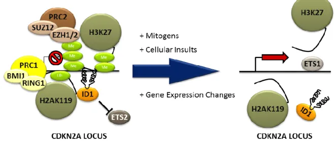
Figure 1.1: Summary demonstrating known gerontogens, mechanistic
inducers of senescence and biomarkers of a cellular senescent phenotype ...17 Figure 1.2: Schematic of p16INK4a transcriptional regulation...18 Figure 1.3: Increased expression of p16INK4a , or increased luminescence
signal, in p16LUC aging mice demonstrating the relationship between
p16INK4a and aging in vivo. ...19 Figure 2.1: High fat diet does not increase p16INK4a expression, while
arsenic moderately increases p16INK4a expression in p16LUCmice. ...33 Figure 2.2: Cigarette smoke increases p16INK4a expression in p16LUCmice. ...35 Figure 2.3: UVB light increases p16INK4a expression in p16LUCmice. ...36 Supplemental Figure 2.1: Physiologically effective doses of high fat diet
do not affect the survival rates of mice, and quantitative realtime PCR
validates p16INK4a expression as measured by TBLI. ...37 Supplemental Figure 2.2: Physiologically effective doses of arsenic do not
affect the survival rates of mice, and quantitative realtime PCR validates
p16INK4a expression as measured by TBLI. ...39 Supplemental Figure 3: Cigarette smoke does not affect the survival rates
of mice, and quantitative realtime PCR validates p16INK4a expression as
measured by TBLI ...41 Supplemental Figure 2.4: UV light leads to a senescent phenotype in skin. ...42 Figure 3.1: Wound healing leads to an induction of p16INK4a expression in
mice ...55 Figure 3.2: UV burn leads to an induction of p16INK4a expression in mice ...56 Figure 3.3: Glucocorticosteriod and NfκB inhibitor interfere with the
transient induction of p16INK4a expression in mice after UV burn ...57 Figure 3.4: Macrophage depletion interferes with the transient induction of
p16INK4a expression in mice after UV burn ...58 Figure 3.5: A working model of p16INK4a induction after wound generation
ix
LIST OF ABBREVIATIONS
8-OHdG 8-hydroxy-2'-deoxyguanosine
AA Ambient air
ARF Alternate reading frame of the INK4a/ARF locus
As Arsenic
C. elegans Caenorhabditis elegans
cA Compound A
CAD Coronory artery disease
CDKN2A Cyclin-dependent kinase inhibitor 2A Clod Clodrosome
CS Cigarette smoke
CXCL1 Chemokine (C-X-C motif) ligand 1
Dex Dexamethasone
DNA Deoxyribonucleic acid Drosophila Drosophila melanogaster FOV-24 Field of view 24
GHR/BP−/− Growth hormone receptor/binding protein Ghrhrlit/lit Growth-hormone-releasing hormone receptor GROα growth stimulating activity α
H2AX Histone γH2A.X HFD High fat diet
x
IIS Insulin/insulin-like growth factor signaling IL-10 Interleukin 10
IL1α Interleukin 1-alpha IL-6 Interleukin 6 IL-8 Interleukin 8
J/m2 Joules per meter squared LTL Leukocyte telomere length MEFS Mouse embryonic fibroblasts mRNA Messenger ribonucleic acid
ND Normal diet
NDGA Nordihydroguiaretic acid
NfκB Nuclear factor kappa-light-chain-enhancer of activated B cells p/s/cm2/sr Photons per second per centimeter squared per steradian p16LUC p16INK4a-luciferase
p16TOM p16INK4a-tdtomato
PBTL Peripheral blood T-lymphocyte PNCA Proliferating cell nuclear antigen ppm Parts per million
RB Retinoblastoma protein SA Senescence associated
xi
TM Telomere maintenance
CHAPTER 1: INTRODUCTION
Aging is a complex process with contributions from both genetic and environmental factors. There has been a great effort to determine how environmental agents are carcinogenic, but very few studies have been done to show how long term exposures can lead to aging (Goldsworthy et al., 1994; Jacobson-Kram et al., 2004). Long term studies, independent of research goal, are extremely costly, with time and mice very expensive (Long et al., 2010). Many groups have started to move towards more in vitro (e.g. ToxCast) models for their high throughput nature, however in vitro models cannot answer most physiological questions (Dix et al., 2007). A critical need in gerontologic research is an understanding of how host genetics and environmental exposures interact over the organismal lifespan to produce common phenotypes of aging such as increased risk to certain diseases, loss of regenerative capacity and frailty.
Aging and Senescence
2
accumulation of senescent cells in vivo appears to be measurable, providing a means to determine if a noxious exposure or toxicant accelerates this aspect of aging. This research will give us insight to the mechanisms of aging after toxicological exposure.
Cellular Senescence and p16INK4a Expression
3
biomarker for aging (Janzen et al., 2006; Krishnamurthy et al., 2006; Krishnamurthy et al., 2004; Liu et al., 2009; Molofsky et al., 2006).
4
molecular mechanisms of p16INK4a regulation with respect to cancer and aging, there are still many questions that need to be answered.
In vitro, p16INK4a, along with other proteins including ARF, is strongly associated with senescence. Prior work has shown that oxidative stress of many primary mammalian cell types, including WI38 (human fibroblasts) and mouse embryonic fibroblasts (MEFs), will lead to the induction of p16INK4a, and eventually lead to senescent state. Consequentially, if p16INK4a is suppressed, the cells will transform allowing for infinite passages, a neoplasmic phenotype. This same phenotype has been shown with ARF, a similar cell cycle regulator located along with p16INK4a on the CDKN2A locus (Matheu et al., 2008). Additionally, when cells are transformed with an oncogene (termed ‘oncogene induced senescence’), there is a robust induction of cells cycle inhibitors including p16INK4a
, further validating the relationship between p16INK4a and senescence. Moreover, cells with high levels of p16INK4a are shown to be cancer resistant (Matheu et al., 2004). By preventing proliferation of a cell harboring DNA damage or oncogene activation, the senescence pathway is a pivotal tumor suppressor mechanism. (Campisi and d'Adda di Fagagna, 2007; Sager, 1991).
Organismal Aging
5
that senescent cells accumulate with aging in vivo (Dimri et al., 1995). Moreover, activation of p16INK4a and senescence has been linked to replicative hypofunction in several tissues (i.e. neural stem cells (Molofsky et al., 2006), hematopoietic progenitors (Janzen et al., 2006), lymphocytes (Liu et al., 2009; Signer et al., 2008), and pancreatic β-cells (Krishnamurthy et al., 2006)). Notably, this replicative hypofunction that comes with increased p16INK4a expression with aging is markedly attenuated in the absence of p16INK4a suggesting a causal relationship. Further evidence that the senescence pathway plays a vital rule in human aging includes correlation of senescence associated markers (e.g. p16INK4a, COX-1, COX-2, SA β-gal) expression with chronological age in skin, kidney, and PBTLs (Dimri et al., 1995; Liu et al., 2009; Melk et al., 2004). Prior Altered regulation of the senescence promoting CDKN2a/b locus, which encodes p16INK4a, p15INK4b and ARF, has been linked to age-associated phenotypes including atherosclerotic disease, type II diabetes, glaucoma and several malignancies (Jeck et al., 2012).
6
et al., 2001). Pharmacologic and genetic approaches to manipulate the number of senescent cells in vivo can ameliorate age-associated phenotypes such as sarcopenia, kyphosis, and hypo-replication of T-lymphocytes and pancreatic β-cells (Berent-Maoz et al., 2012; Chen et al., 2011a; Zindy et al., 1997). One group has generated a murine model that allows drug-inducible elimination of p16Ink4a-positive senescent cells. After induction, the tissues (e.g. adipose, skeletal muscle and eye) that have p16INK4a-dependent age-related disease have delayed onset of these phenotypes with removal of p16INK4a-expressing cells (Baker et al., 2011).
The Science of Biological Aging
7
growth factor signaling (IIS) pathway and the telomere maintenance (TM) pathway are associated with human longevity (Deelen et al., 2013). Aging genetics has been well-established; however there are still answers that are necessary for understanding environment effects of aging.
8
summary, there has been some work to show the relationship between exposures and aging in numerous species, however little research has been focused on mammalian aging toxicology.
Gerontogens
In 1987, George Martin pointed out that the rate of aging is largely non-genetic (>50%), and coined the term "gerontogen" to describe an environmental agent that accelerates aging. In response to gerontogen exposure, phenotypic outcomes at the organismal level include the loss of proliferative homeostasis, the decline in the efficiency of enzyme adaptation, and immunodeficiency (Martin, 1987). Martin also discusses categories of environmental gerontogens including segmental gerontogens, which affect multiple senescent phenotypes, and unimodal gerontogens which affect only a specific tissue or organ. Environmental exposures (or therapeutic exposures such as radiation and chemotherapy) act as gerontogens when they lead to cellular insults that induce, what is now considered, cellular senescence (e.g. DNA damage or increasing reactive oxygen species). According to this theory, differential exposure to gerontogens may underlie the marked phenotypic variation in human physiological aging (Figure 1).
High Fat Diet
9
(Sone and Kagawa, 2005). Additionally, prior studies on the effects of obesity and high fat diet (HFD) on p16INK4aexpression, a cell cycle inhibitor and known marker for senescence, in vivo have given very tissue specific results. For example, HFD has been reported to increase senescence and p16INK4a expression in the murine aorta (Wang et al., 2009), whereas p16INK4a mRNA expression in peripheral blood T-lymphocytes (PBTL) does not correlate with body-mass index, a marker of obesity, in humans (Liu et al., 2009). High fat diet can be considered a gerontogen, however the mechanism of its advanced aging phenotype, is less certain. It is well known that fat causing an induction of reactive oxygen species, which can lead to a senescent phenotype (Tchkonia et al., 2010). One question that remains unanswered is which senescent pathway leads to the advanced aging phenotype in vivo.
Arsenic
Arsenic is a non-DNA damaging gerontogen. It is a common toxicant found in ground water that has been linked to age-related phenotypes including cancer (e.g. skin, bladder), type II diabetes, and atherosclerosis (Kapaj et al., 2006). Others have shown that arsenic among other heavy metals can leads to many age-related neurodegenerative diseases, including Alzheimer’s disease, Parkinson’s disease, Huntington’s disease and Ataxia telangiectasia (Migliore and Coppede, 2009). While genetic background plays a role with some of these neurodegenerative disorders, exposures to arsenic can accelerate the pathology.
10
11 UV Light
Ultraviolet light is a potential unimodal gerontogen since almost all age-related pathologies are associated with the skin. There is strong evidence to correlate the effects of
UV leading to photoaging and skin cancer (Kligman, 1989; Scharffetter-Kochanek et al.,
2000). Additionally, prior work has suggested that acute UVB exposure can rapidly induce
senescence in human skin (Pavey et al., 1999). Others have also shown that chronological
aging and photoaging share basic molecular mechanisms including inductions of cytokines, ROS, UV-associated transcription factors (e.g. AP-1). Moreover, UV-induced ROS cause negative effects on cellular components including DNA, proteins, lipids, which is similar to a chronological aging phenotype (Fisher et al., 2002).
12 Cigarette Smoke
One common segmental, or multifaceted, gerontogen is cigarette smoke. Exposure to both primary and secondary smoke can lead to a vast array of aging phenotypes, which include advance aging of the skin, bones, and teeth as well as deleterious effects of the reproduction, cardiac, and pulmonary systems, and extensive cancers to the head, neck, and lungs (Bernhard et al., 2007). In addition to its well-known impact upon various types of neoplasms, it also had deleterious effects upon reproduction, the cardiovascular and pulmonary systems, skin, and bone diseases such as emphysema, atherosclerosis and several cancers (Bernhard et al., 2007; Ito and Barnes, 2009). The relationship with chronological age was much more striking, but it remains to be seen if there is any causal relationship to mechanisms of aging.
13
humans is associated with increased p16INK4a expression and shorter LTLs in PBTL (Liu et al., 2009; Mirabello et al., 2009; Nelson et al., 2012; Song et al., 2010; Valdes et al., 2005).
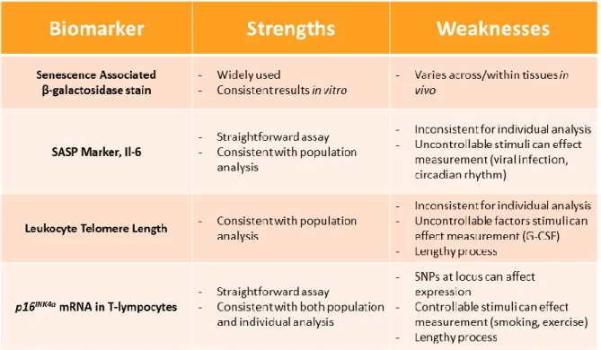
Measuring aging and senescence
Some of the markers of cellular senescence that increase with aging in humans and mice include SA β- galactosidase staining [48], SASP factors [49], telomere foci [50], and expression of the p16INK4a tumor suppressor gene [13, 46, 51, 52] (Table 1). While all have shown an increase in specific experiments, very few are substantial, global, consistent, increase in many organs over time. Historically, SA β-galactosidase staining has been used to measure senescence, since senescent cells have been shown to have higher levels of endogenous β-galactosidase expression due to a lower pH. In vitro, this marker is very consistent; however in vivo interpretation can be difficult. Since SA β-gal stain is based on a change in pH, the differences in pH can change between tissues as well as throughout a singular tissue, making the results convoluted. SA β-gal stain is also poorly suited as a longitudinal translational measure of aging because of the requirement for end-organ staining. In murine studies the animal needs to be sacrificed for proper analysis, and human studies are not feasible with rare exception because of need to biopsy.
14
confounding factors of physiological aging in humans make it difficult to control. First, despite the long held conception that telomere length is a marker for residual replicative capacity (e.g. cells with shortened telomeres will not be able to replicate), there is little evidence to substantiate that hypothesis in humans (Longo, 2009). In fact, in one of the most highly replicative organs the bone marrow in which aging is known to lead to marrow dysfunction, LTL does not predict circulating levels of leukocytes, erythrocytes or platelets, therefore does not predict the replicative capacity of their progenitors(Mollica et al., 2009). Second, while telomere length will decrease across the lifespan in population-based studies, LTL varies substantially among individuals of the same age, and the absolute change is relatively small over an organism’s lifespan it is unlikely to be a reliable aging biomarker (Longo, 2009; Muezzinler et al., 2013; Valdes et al., 2005). Additionally, the methods for determining LTL are difficult and expensive, making it less appealing as a high throughput biomarker.
15
inflammatory milieu that causes physical decline with aging, but it is poorly suited as an outcome measure of the effect of environmental exposures on aging in humans.
Expression of p16INK4a in PBTLs has been shown to a faithful biomarker for chronological age, due to a stable, dynamic increase over a lifespan, in many different organs in both humans and mice [13, 52](Table 1). Because of the ability to measure p16INK4a in people with the CD3+ T-lymphocyte assay, these exposures can be evaluated in people. In humans, known DNA damaging chemotherapy treatments lead to higher levels of p16INK4a expression in the T cells of patents after treatment. While there are some limitations for p16INK4a expression including difficultly to solate T-lymphocytes, this biomarker seems to be a promising candidate for in clinic evaluation of aging.
Measuring toxicological aging in vivo
16
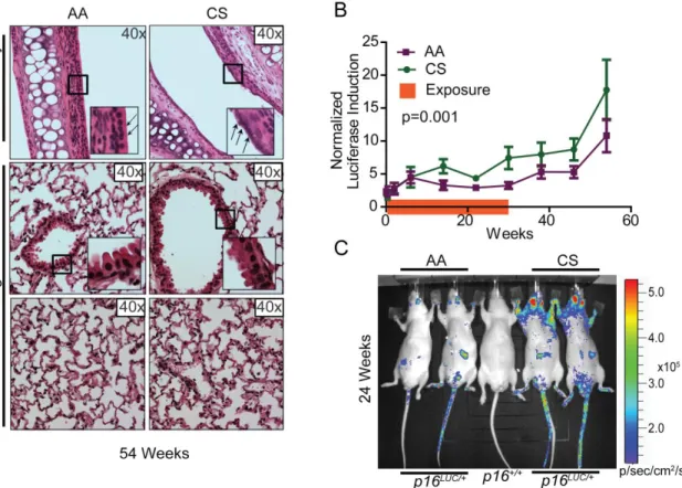
cigarette smoke, induce p16INK4a expression at a faster rate when compared to non-exposed cohorts in both human PBTLs and whole mice (Krishnamurthy et al., 2004; Liu et al., 2009). Specifically, p16INK4a increases at a faster rate over time in chronic smokers when compared to both former and non-smokers in human PBTL (Liu et al., 2009). In mice, p16INK4a expression is much higher in the upper respiratory region after four months of exposure to low chronic dose of cigarette smoke. Using the p16LUC mice, other gerontogens enhance the p16INK4a-associated aging rate include UV light and to a lesser extent arsenic, further confirming the value of this reporter allele.
17
Figure 1.1: Summary demonstrating known gerontogens, mechanistic inducers of
18
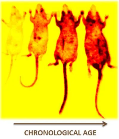
19
Figure 1.3: Increased expression of p16INK4a , or increased luminescence signal, in
20
CHAPTER 2: GERONTOGEN TESTING IN p16LUC REPORTER MICE
2.1 Introduction
Murine models have played a critical role in the identification and classification of carcinogens. For example, commercially available animal models, (i.e. BigBlueTM, MutaMouseTM and the p53+/- strain) (Goldsworthy et al., 1994; Jacobson-Kram et al., 2004; Long et al., 2010), comprise core tools in cancer toxicological assessment. These reagents have enhanced our basic understanding of carcinogenesis, and have contributed to public health by increasing the efficiency and sensitivity of carcinogen identification while reducing animal requirements and research costs. In 1987, George Martin proposed the concept of “gerontogens” (Martin, 1987); that is, environmental agents which accelerate molecular aging analogous to the way carcinogens accelerate neoplasia. While murine models for carcinogen testing are advanced, in vivo tools for gerontogen testing do not exist.
22
al., 1997). Moreover, activation of p16INK4a and senescence has been linked to replicative hypofunction in several tissues (i.e. neural stem cells (Molofsky et al., 2006), hematopoietic progenitors (Janzen et al., 2006), lymphocytes (Liu et al., 2011; Signer et al., 2008), and pancreatic beta-cells (Krishnamurthy et al., 2006)). Altered regulation of the senescence promoting CDKN2a/b locus, which encodes p16INK4a, p15INK4b and ARF, has been linked to age-associated phenotypes including atherosclerotic disease, type II diabetes, glaucoma and several malignancies (Jeck et al., 2012). Finally, pharmacologic and genetic approaches to manipulate the number of senescent cells in vivo can ameliorate age-associated phenotypes such as sarcopenia, kyphosis, and hypo-replication of T-lymphocytes and pancreatic beta-cells (Baker et al., 2011; Berent-Maoz et al., 2012; Chen et al., 2011a). Given these intimate links between aging, p16INK4a expression, and senescence induction, we sought to determine if in vivo activation of p16INK4a, measured through the use of a recently described reporter allele (Burd et al., 2013), could be used to assess the gerontogenic effects of environmental agents.
2.2 Materials and Methods
23
Histopathology Core. Quantitative TaqMan RT-PCR strategies for the detection of p16INK4a were performed as previously described (Krishnamurthy et al., 2006; Krishnamurthy et al., 2004). Statistical significance was determined using a student’s t-test or linear regression analysis for all comparisons except survival analysis, where the Gehan-Breslow-Wilcoxon test was employed.
2.2.1 Exposures
High Fat Diet: 42% Fat diet (HFD) was ordered from Harlan Laboratories (TD.88137). At 8-10 weeks of age, mouse chow was changed to HFD vs. normal diet (4% fat) after initial baseline imaging. New diet was refilled once a week. Twenty male and female SKH1-E p16+/LUC mice were analyzed per dietary cohort (42% vs. 4%) for 84 weeks.
Hepatic Steatosis Evaluation: Livers were incubated in 4% Osmium overnight, washed with diH2O, and then fixed with 10% Formalin. Tissues were then imbedded, sectioned and
stained with Hematoxilyn and Eosin using the Immunohistochemistry core facility.
Data Analysis: Linear regression analysis was completed to determine the relationship between p16LUC signal and weight in HFC cohort at 52 weeks of exposure.
24
Pancreatic Islet Measurements: Randomized 10x histological images were taken of the pancreas in each cohort. Each islet was then carefully traced and the area was measured using imageJ analysis.
Cigarette Smoke: Cigarettes were ordered from the University of Kentucky (3R4F)—through the Reference Cigarette Program at College of Agriculture in Lexington, Kentucky. Beginning at age 10-12 weeks after baseline imaging, mice were exposed to one hour of cigarette smoke/day, five days/week. For exposure, mice were put into an inExpose exposure system providing both mainstream and side stream smoke exposure. Control animals (no smoking exposure) were handled in an identical manner except were not exposed to tobacco smoke. Ten male SKH1-E p16+/LUC mice were analyzed per tobacco cohort (smoking vs. non-smoking). Animals were exposed to tobacco smoke for 24 weeks, but were imaged for an additional 32 weeks after exposure.
UV light: A UVB lamp from UVP was used for these studies, with an emission spectrum of 290-350 nm light, and with a peak emission at 312 nm. At 8-10 weeks of age, mice were exposed to 353 J/m3 of UVB light three times per week for 24 weeks. UV exposure began after initial baseline imaging. Fifteen male SKH1-E p16+/LUC mice were analyzed per UVB cohort (exposed vs. unexposed) for 16 more weeks after exposure. UV exposed mice were sacrificed for morbidity or tumor burden in accordance with established laboratory protocols.
25
Data analysis: All H2AX cells in the epithelial layer of the exposed skin were divided by the total number of epithelial cells in the microscopic field. Fisher’s exact test was used to determine statistical significance.
Senescence-Associated ß-galactosidase (SA-ß-gal) activity: Skin was assayed using Cellular Senescence Assay Kit (KAA002; Millipore, USA) according to manufacturer's instructions.
2.2.2 In vivo Luminescence Imaging
Luciferase Imaging: To serially monitor luciferase induction, mice were imaged as previously described (Burd et al., 2013) using a Xenogen IVIS LUMINA/KINETIC Imaging System. Imaging was completed under anesthesia, immediately after D-luciferin injection (10 mg/ml in sterile PBS). Sequential imaging was used to determine optimal luminescence. A sequence of 2 min, 2 min, 2 min, 2 min, 30 sec, and 1 min were used. The 4th two minute image (8 minutes after initial injection) was used for all reported imaging measurements. For arsenic, high fat diet, and cigarette smoke exposure images were taken of the ventral side of the mouse. For UV light, the dorsal (exposed) and ventral (unexposed) sides of each mouse were taken. Living Image 3.1 Software (Caliper Life Sciences) was used to analyze the images at the same image exposure time point (8 min) over the life span of the mouse. Representative images are shown with exposed and unexposed animals for each cohort, as well as appropriate imaging scales to account for imaging settings.
26
although some animals were imaged more frequently during the initial stages of each exposure period. Each mouse was circled to measure average radiance values (p/s/cm2/sr) over the region of interest. The area for each mouse was held constant for each experiment. A blank area on the image was also circled to allow for subtraction of background noise. To omit background noise from each individual mouse image, the background circle was subtracted from the mouse circle. To graph the luciferase induction over time, all murine luciferase signals in a specific cohort were averaged at time zero and normalized to a value of one. All subsequent mouse images were then normalized relative to the average time zero luminescence.
2.3 Results
27
light, arsenic, and cigarette smoke) on senescence, we performed serial analysis of p16LUC cohorts. Hairless SKH1-E mice were used for all studies to further enhance in vivo imaging.
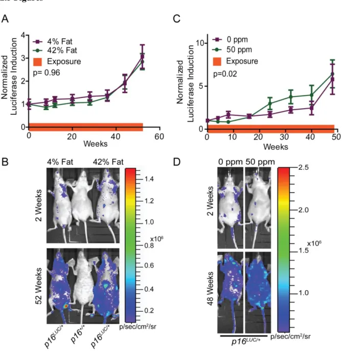
2.3.1 High Fat Diet
28
accelerate the accumulation of senescent cells in vivo, at least to the level of sensitivity of luciferase detection.
2.3.2 Arsenic
29 2.3.3 Cigarette Smoke
30 2.3.4 UV Light
Chronic UVB exposure causes photoaging and skin cancer (Kligman, 1989;
Scharffetter-Kochanek et al., 2000). Prior work has suggested that acute UVB exposure can
rapidly induce p16INK4a expression in human skin (Pavey et al., 1999). To directly assess the
effects of chronic UVB on the induction of senescence in vivo, the dorsal surface of p16LUC
mice were exposed to 350 J/m2 of UVB light three times a week for six months starting at 10-12 weeks of age. Littermate control mice were contemporaneously aged in the absence of
UVB. After six months of exposure, mice were aged without further UVB exposure until
they reached one year of age. After six months of exposure, mice exposed to chronic UV
light had more epidermal hyperkeratosis and metaplasia than the non-exposed cohort (Figure
2.3A, upper panel). The UV-exposed cohort exhibited more inflammatory infiltration into
the reticular dermis and hypodermis of lymphocytes and neutrophils than non-exposed
controls (Figure 2.3A, middle panel). A marker of DNA damage, phospho-H2AX staining,
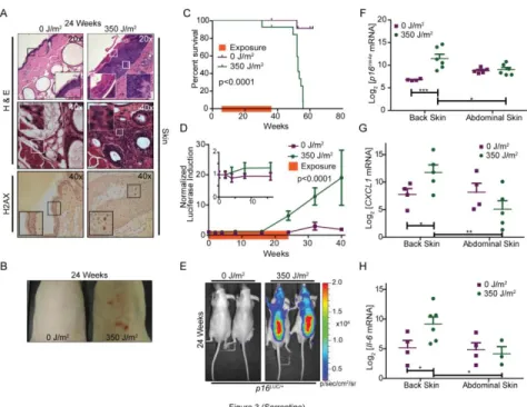
was significantly increased in the epithelial layer of skin in UV exposed mice than non-exposed mice, suggesting a persistent DNA damage response (Figure 2.3A, lower panel, and Supplemental Figure 2.4A). Additionally, UVB exposed mice developed skin neoplasms starting five months into the window of exposure (Figure 2.3B) which were associated with
increased mortality (Figure 2.3C). Accumulation of luciferase was higher in p16LUC animals treated with UV than unexposed controls prior to development of visible neoplasms and
TBLI was almost ~8 times greater in UV-treated animals after 32 weeks of exposure (Figure
2.3D). In accord with the TBLI results (Figure 2.3E), p16INK4a mRNA levels from abdominal
31
of 16INK4a expression (Figure 2.3F). Additionally, the UV exposed dorsal skin exhibited higher expression than unexposed abdominal skin from the same mouse (Figure 2.3F).
To confirm the induction of senescence in vivo using additional markers, we analyzed exposed and unexposed skin tissue for senescent associated (SA) cytokines, CXCL-1 and IL-6, as well as SA-β-galactosidase staining (SA-β-gal). Expression of CXCL-1 and IL-6 increased in mice exposed to UV light when compared to non-exposed control mice. Additionally, the increase of these mRNAs only occurred in skin that was directly exposed to UV light (back) when compared to non-exposed skin (abdomen) of the same animals (Figure 2.3, G and H). Lastly, we observed a similar pattern of SA-β-gal staining. Increased staining was observed in the skin from the back but not abdomen of the UV-treated mice (Supplemental Figure 2.4B). These data suggest that chronic DNA damage from UV light accelerated the accumulation of senescent cells in vivo.
2.4 Discussion
32
as opposed to lung parenchyma (Figure 2.2C) and that the most marked effects of UVB are on exposed rather than unexposed skin (Figure 2.3, E and F).
It is important to note two limitations of this system. First, while luciferase imaging is considerably more sensitive than optical methods, the p16LUC allele may be less useful for the detection of senescence induction in deeper organs or in rare cellular subtypes. Therefore, while we observed no effect of HFD on p16INK4a induction at the total-body imaging level, it is possible HFD activates senescence in important cellular compartments (e.g. pancreatic beta-cells (Sone and Kagawa, 2005), and vascular cells (Shi et al., 2007; Wang et al., 2009)), but not to a level detectable using this system. Secondly, activation of p16INK4a expression is only one component of molecular aging, which is a complex and multi-faceted process. Therefore, the p16LUC system does not detect cellular aging processes that occur independently of p16INK4a expression; and likewise cannot be used to identify p16INK4a-independent gerontogens.
33
2.5 Figures
Figure 2.1: High fat diet does not increase p16INK4aexpression, while arsenic moderately
increases p16INK4a expression in p16LUCmice.
34
35
Figure 2.2: Cigarette smoke increases p16INK4a expression in p16LUCmice.
36
Figure 2.3: UVB light increases p16INK4aexpression in p16LUCmice.
37
2.6 Supplemental Figures
Supplemental Figure 2.1: Physiologically effective doses of high fat diet do not affect the
survival rates of mice, and quantitative realtime PCR validates p16INK4a expression as
38
39
Supplemental Figure 2.2: Physiologically effective doses of arsenic do not affect the
survival rates of mice, and quantitative realtime PCR validates p16INK4a expression as
measured by TBLI.
Kaplan-40
41
Supplemental Figure 3: Cigarette smoke does not affect the survival rates of mice, and
quantitative realtime PCR validates p16INK4aexpression as measured by TBLI
42
Supplemental Figure 2.4: UV light leads to a senescent phenotype in skin.
43
CHAPTER 3: REGULATION OF TRANSIENT INDUCTION OF p16INK4a
3.1 Introduction
44
Research has shown that there is an induction of p16INK4a at the site of wound closure in both cells and humans. More specifically, others have shown that induction of matricellular protein, CCN1, will cause p16INK4a-dependent fibroblast senescence at site of wound repair and inhibit fibrosis during tissue repair (Jun and Lau, 2010a, b). Additionally, p16INK4a has been associated with keratinocyte migration during wound repair. Laminin5, a basement membrane protein, is co-expressed with p16INK4a along the edges of a wound in culture and in vivo (Natarajan et al., 2005; Natarajan et al., 2003). Additionally, during migration, p16INK4a is induced to lessen the potential for invasion and transformation (Natarajan et al., 2006). Chronic wound healing has been shown to cause squamous cell carcinoma with loss of known senescence markers including p16INK4a and MMP-19 (Impola et al., 2005; Telgenhoff and Shroot, 2005).
45
wound healing, UV light and p16INK4a expression, we sought to determine the magnitude and duration of transient p16INK4a activation upon insult, measured through the use of a recently described reporter allele in vivo.
46
3.2 Materials and Methods
Experiments were performed under protocols approved by the institutional care and use committee (IACUC) of the University of North Carolina. Hairless SKH1-E p16LUC/+ mice were used for all experiments (Burd et al., 2013). Genotyping was performed as previously described (Burd et al., 2013). For histologic analysis, tissues were fixed with 10% formalin overnight, and then transferred to 70% ethanol for paraffin blocking and staining. All slides were generated and stained with Hematoxylin and Eosin at the UNC Histopathology Core. Statistical significance was determined using a student’s t-test for all comparisons. All gross anatomy photos were taking with 6.0 megapixel Canon SD630.
3.2.1 Transient Inductions
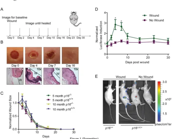
Wound Generation: At 8-10 weeks of age, mouse p16LUC/+ (SKH1-E background) or p16+/+ and p16-/- (C57/B6 background) were anesthetized with inhaled 2% isofluorane. Once anesthetized and after cleansing the 1 sq cm area with iodine, a sterile 3 mm punch was used to create one full-thickness wound on the dorsal side of each mouse, just below the shoulder blades. SKH1-E mice were imaged for luciferase following a specific schedule (Figure 1A) until the wound healed. C57/B6 mice wounds were measured weekly using a caliper until completely healed.
47
Mice were imaged for luciferase following a specific schedule (Figure 2A) until wound healed.
3.2.2 Drug Treatments
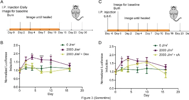
Dexamethasone: Mice were given a daily intraperitoneal injection (I.P.) of dexamethasone (Sigma D4902) at dose of 7 mg/kg diluted in Phosphate Buffered Solution (PBS) for the first 16 days after wounding (Figure 3A).
IKK-β Inhibitor: Mice were given an intraperitoneal injection (I.P.) of compound A (Dr. Al Baldwin’s drug) at dose of 10 mg/kg dose diluted in dimethylsulfoxide (DMSO) starting 6 days before wounding and ending 16 after wounding (Figure 3C).
Clodrosome: Mice were given a daily intravenous injection (I.V.) of liposomal clodronate (Encapsula NanoSciences SKU #STLMDK) at dose of 100 uL/per mouse for the three days before the wounding (Figure 4A).
3.2.3 Flow Cytometry
Bone marrow, splenic, and peripheral white blood cells were isolated as previously described (Kiel et al., 2005). Once the cells were isolated, cells were stained with B220-violet (B cell marker), Gr1-APC-Cy7 (granulocyte marker), F4/80-FITC (macrophage marker), and Mac1-PE-Cy7 (monocyte marker). Flow cytometry analysis was completing using Beckman-Coulter (Dako) CyAn ADP machine with the strategy shown in Figure 4B.
48
B220. Then, all B220- cells where gated for Gr1. Lastly, all Gr-1- cells were gated for Mac1 and F4/80. All Mac1+F4/80+ cells were used for analysis.
3.2.4 In vivo Luminescence Imaging
Luciferase Imaging: To serially monitor luciferase induction, mice were imaged as previously described (Burd et al., 2013) using a Zenogen IVIS LUMINA Imaging System. Imaging was completed under anesthesia, immediately after D-luciferin injection (10 mg/ml in sterile PBS). Sequential imaging was used to determine optimal luminescence. A sequence of 2 min, 2 min, 2 min, 2 min, 30 sec, and 1 min were used. The 4th two minute image (8 minutes after initial injection) was used for all reported imaging measurements. For all exposures, images were taken of the dorsal side of the mouse. Living Image 3.1 Software (Caliper Life Sciences) was used to analyze the images at the same image exposure time point (8 min) over the experiment. Representative images are shown with wounded and non-wounded animals for each cohort, as well as appropriate imaging scales to account for imaging settings. For data analyses, the raw average radiance values (photon/sec/cm2/steradian) from the original images were used. A wide angle lens (FOV-24) was used to capture images of 3 or more mice.
49
luciferase induction over time, all murine luciferase signals in a specific cohort were averaged at time zero and normalized to a value of one. All subsequent mouse images were then normalized relative to the average time zero luminescence.
3.3 Results
The p16-LUC allele includes firefly luciferase ‘knocked-in’ to exon 1α of the murine Cdkn2a locus, placing luciferase under the control of the endogenous p16INK4a promoter as well as cis-regulatory elements, which are known to extend for several hundred kilobases around the locus (Burd et al., 2010; Burd et al., 2013; Visel et al., 2010). Expression of p16INK4a can potentially increase to more than 300-fold on a per cell basis making p16LUC a valuable tool for in vivo imaging of transient induction of p16INK4a (Burd et al., 2013; Krishnamurthy et al., 2004). To assess the kinetics and duration of p16INK4a expression after dermal insult, we performed serial analysis of p16LUC cohorts. Hairless SKH1-E mice were used for all studies to further heighten in vivo luciferase signal.
3.3.1 Wound Healing Model
50
until the wound was healed (Figure 1A). Through observation and histological analysis, wounds healed as previously recorded (Figure 1B) (Bradshaw et al., 2002) . There was clear induction of p16INK4a at the site of the wound by 2 days after wound generation, with peak induction by 4 days (Figure 1, D and E). p16INK4a expression returned to baseline by 16 days, when the wound had healed (Figure 1D). To determine if p16INK4a played a role in wound closure rate, C57/B6 mice at varying ages (5 months and 10 months) with and without p16, were wounded and closure was measured. Older mice (10 months old), have an initially slower wound closure than the younger mice (5 months old), regardless of p16INK4a status, suggesting p16INKa-independent age-related factors, including other age-dependent cell cycle inhibitors (e.i. ARF) play a role in wound regeneration (Figure 1C).
3.3.2 UV Burn Model
51
anatomic specific expression (Figure 2D). These results suggest that UV light leads to a rapid, transient expression of p16INK4a.
3.3.3 Pharmacological Modulation of p16INK4a transient expression
Pharmacological inhibitors were employed to determine the mechanism of p16INK4a transient induction after UV exposure. Others have shown that inflammation can lead to the induction of p16INK4a (Freund et al., 2010). Moreover, glucocorticoids, are shown to suppress components of the senescence associated secretory phenotype, including IL-6, IL-8, GM-CSF, and MCP-2 expression (Laberge et al., 2012). In vivo, glucocorticoid receptors have been correlated with p16INK4a levels in tumors, suggesting a potential relationship with dexamethasone, a glucocorticoid, and p16INK4a expression (Theocharis et al., 2003). However, how dexamethasone affects p16INK4a expression after cellular insult is unknown. To address this question, SKH1-E mice were given a daily I.P. injection of dexamethasone every day, starting from the day of UV burn. The mice were then serially imaged for 30 days (Figure 3A). Luciferase signal was recorded and dexamethasone-treated and non-treated mice were compared. Interestingly, dexamethasone treatment hindered induction of p16INK4a to almost baseline levels, suggesting an inflammatory response after UV exposure leading to the transient induction of p16INK4a (Figure 3B).
52
NfκB in conjunction with p38, a known upstream inducer of p16INK4a, regulate the senescence associated secretory phenotype (Freund et al., 2011). To understand the relationship between NfκB and p16INK4a pathways, we treated UV exposed mice with compound A, an IKK-β inhibitor. IKK-β is part of the IKK complex, which is the initial step in the NfκB cascade. By inhibiting IKK complex’s ability to phosphorylate the IkB complex, the NfκB pathway is hindered. Our results show mice treated with compound A, every other day starting six days before UV exposure (Figure 3C) exhibited lower p16INK4a induction after UV burn, further strengthening the hypothesis that inflammatory response after UV exposure leads to the induction of p16INK4a(Figure 3D).
53
modest decrease in peripheral blood macrophages, with no change in bone marrow derived macrophages, validating the data generated from this experiment (Figure 4C). Our imaging results demonstrate the treatment with clodrosome lessened the p16INK4a inducing effects of UV burn, suggesting that macrophages play a role in the transient induction of p16INK4a (Figure 4D).
3.4 Discussion
In this work, we demonstrate through the use of the p16-LUC allele, we are able to transiently induce p16INK4a through wounding and UV burns. Moreover, we have effectively modulated expression of p16INK4a after exposure of UV burns by altering the inflammatory response through treatments with dexamethasone, a glucocorticoid, compound A, an IKK-β inhibitor of the NFκB pathway, and clodrosome, a macrophage inhibitor. These data suggest
the transient induction of p16INK4a is due to the inflammatory response, potentially through recruitment of macrophages to wound site after a cellular insult, such as a UV burn (Figure 5).
54
mice that have wildtype p16INK4a with bone marrow from mice that have heterozygous for the p16LUC allele. If there is induction at the site of the insult (luciferase glow at the burn site),
p16INK4a expression is from the circulating white blood cells. If induction is seen, the white blood cells with be sorted into specific populations using the protocol described, and adoptively transferred into wildtype p16INK4a mice, to determine cell specific induction. To strengthen the theory that macrophage infiltration leads to a stronger p16INK4a expression, we will inject mice with KRAS transformed cells that have extras copies of colony stimulating factor 1 (CSF-1), a macrophage recruitment signal, and measure tumor growth and luciferase signal. If our hypothesis is correct, then we should see higher levels or faster induction of p16INK4a in mice that have the extra CSF-1 when compared to the empty vector controls.
In summary, we have shown that both wounding and UV burn lead to a transient induction of p16INK4awith maximum expression at day 4. We have also shown using the UV burn model that inflammatory responses, including macrophage infiltration and induction of the NFκB pathways, affect p16INK4atransient expression. While there are many questions
55
3.5 Figures
Figure 3.1: Wound healing leads to an induction of p16INK4a expression in mice
56
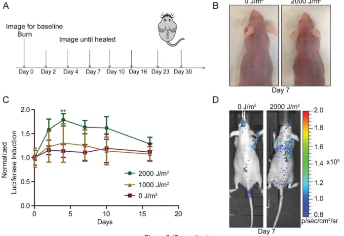
Figure 3.2: UV burn leads to an induction of p16INK4aexpression in mice
(A) Model describing the imaging protocol for burned mice. (B) Gross images of exposed and non-exposed mice at day 7. (C) Luciferase levels were measured periodically of p16LUC mice wound as measured by TBLI (0J/m2, n=4, 1000 J/m2, n=5, 2000J/m2, n=9). Results were recorded, analyzed, and displayed as in Figure 1D. (D) Representative images (day 7) of the study described in ‘C” are shown. Error bars indicate standard error of the mean. P values were determined through student’s t-test analysis comparing 2000J/m2
57
Figure 3.3: Glucocorticosteriod and NfκB inhibitor interfere with the transient
induction of p16INK4aexpression in mice after UV burn
(A) Model describing the imaging and dosing protocol for dexamethasone-treated, burned mice. (B) Luciferase levels were measured periodically of p16LUC mice wound as measured by TBLI (0J/m2, n=4, 2000J/m2, n=9, Dexamethasone+2000J/m2, n=4). Results were recorded, analyzed, and displayed as described in Figure 1D. (C) Model describing the imaging and dosing protocol for the IKK-β inhibitor compound A-treated, burned mice. (D) Luciferase levels were measured periodically of p16LUC mice wound as measured by TBLI (0J/m2, n=4, 2000J/m2, n=9, compoundA+2000J/m2, n=3). Results were recorded, analyzed, and displayed as described in Figure 1D. Error bars indicate standard error of the mean. P values were determined through student’s t-test analysis comparing 2000J/m2
58
Figure 3.4: Macrophage depletion interferes with the transient induction of p16INK4a
expression in mice after UV burn
(A) Model describing the imaging and dosing protocol for clodrosome (liposomal clodronate) treated, burned mice. (B) Flow cytometry strategy to determine the macrophage population (Mac1+F4/80+). See methods for further description. (C) Graph depicting the levels of Mac1+F4/80+ cells of clodrosome-treated vs. non-treated in the peripheral blood, spleen, bone marrow (BM) on day 4 (bolded arrow in Figure 4A). (D) Luciferase levels were measured periodically of p16LUC mice wound as measured by TBLI (0 J/m2, n=4, 2000 J/m2, n=9, clodrosome+2000 J/m2, n=3). Results were recorded, analyzed, and displayed as described in Figure 1D. Error bars indicate standard error of the mean. P values were determined through student’s t-test analysis comparing 2000J/m2
59
Figure 3.5: A working model of p16INK4a induction after wound generation or UV burn.
60
CHAPTER 4: OVERALL CONCLUSIONS AND SIGNIFICANCE
4.1 Principal Conclusions
There have been great strides in developing better assays to understand the molecular underpinnings of carcinogens, but the field of gerontogens is not as established. My work describes a way to evaluate the severity of a gerontogen in vivo using a novel mouse model which has allowed us to systemically evaluate four different exposures in mice (Chapter 2). Contrary to the known literature, my work also shows how p16INK4a does not always accumulate in an organism, and might have a niche of transient induction after a wounding event, such as a UV burn (Chapter 3). In summary, my work provides an innovative method for measuring molecular age after chronic exposure to gerontogens and begins to clarify a novel role for p16INK4a transient expression in vivo.
4.2 p16INK4a -Luciferase Allele
61
region of a tumor before tumor palpation in vivo using the p16LUC allele (Burd et al., 2013). Additionally, we show that the p16LUC allele will detect tumorigenesis in many different tumor types including breast and melanoma. Chapter 2 defines multiple gerontogens and verifies the allele’s ability to measure p16INK4a-associated aging in vivo. In this paper, we also validate the expression of p16INK4a after chronic exposure, and correlated the expression with a senescent phenotype through SA-β gal staining and mRNA expression of SA factors including Il-6 and CXCL1. Currently, we are completing experiments to understand the mechanisms of transient induction of p16INK4a using the p16LUC wound healing model through pharmacological modulation of the p16INK4a expression after a UV burn. The p16LUC reporter allele has made it easier to understand the relationship between p16INK4a-associated cancer and aging.
Not only has the p16LUC allele made advancements from a scientific standpoint, this allele is a novel in vivo model that can serially measure a specific protein in the same organ over time. Historically, aging in vivo studies are very long and expensive since there are tremendous costs for mice, as well as cage costs for having the mice aging for months and possibly years. Additionally, there are incredible costs for the time invested, and proper storage of all tissues after the conclusion of the experiment. By using a mouse model that can measure the protein of interest (p16INK4a) in real time, it decreases the costs exponentially and lessens the time commitment significantly.
62
period of time will show a slow, but steady, induction of luciferase signal correlative with endogenous p16INK4a expression. However, this signal will not be linear, with slight spikes and drops throughout the life of the mouse. Additionally, measuring a set of mice at the same time point (e.g. same age) will also yield some variability, which is characteristic of p16INK4a expression in human PBTLs (Liu et al., 2009). To fix these issues, internal controls were used per mouse to correct for any background induction (transient exposures), and higher cohort numbers (15+) were used if variability was expected to be higher than normal (e.g. chronic exposures). Secondly, since the luciferase allele required an I.P. injection of luciferin before each image set, technical variation of injection placement, amount of luciferin injected, and time to image taken can also confound results. To address these variations, multiple sets of mice were taken over many months to make sure it was not a dose dependent response of the luciferin. Each image taken included mice from various cohorts if space permitted. To stabilize the time to image variation, experiments were completed to determine steady state of luminescence after injection, which was 8 minutes. Additionally, we determined that 48 hours were needed before mice could be re-imaged with maximal luminescence potential. Mice were also weighed before each luciferin injection to confirm the proper luciferin serum concentrations. Though both minor, these shortcomings were taken into consideration during the course of the studies and addressed to maintain accurate, reliable data.
63
64
4.3 Theory of gerontogens
George Martin developed a new philosophy about exposures that lead to an aging phenotype and coined the term gerontogens (Martin, 1987). Gerontogens are defined as an exposure that leads to an advanced aging phenotype. While this opened the door to a new thought process about aging toxicology, there were a few questions that were left unanswered; What is an “aging phenotype”? How do you measure aging? Can a gerontogen also be a carcinogen?
65
light (photo aging, e.g. slower wound repair, lack of elasticity), and cigarette smoke (a wide-array of pathologies, including immune suppression and CAD) as my gerontogen panel.
After gerontogens are defined, there are many ways to measure the aging phenotype, which includes whole organism measurements such as decrease in lifespan and onset of age-related pathologies as described above. As described in the Chapter 1, there are also many different molecular markers to measure the aging phenotype. To utilize our novel p16LUC system, I measured p16INK4a mRNA expression levels as my molecular marker after exposure to the gerontogens of choice. I also tested of validity of p16INK4a expression as an aging marker, and senescence activator, by evaluating CXCL1 and IL-6 mRNA levels (SASP factors) as well as SA β-gal stain (senescence marker). The evaluation of p16INK4a
66
including atherosclerosis and cancer (Giovannucci and Goldin, 1997; Keys, 1952; Mosby et al., 2012; Nishina et al., 1990; Smith et al., 1992).
Future experiments for this project would take two different directions. Firstly, I would better characterize the “gerontogeness” of the two more dynamic p16INK4a
expressing gerontogens. I would solidify that exposure to either UV light or cigarette smoke would lead to induction of p16INK4a, which leads to a lack of proliferation. For this, cell culture analysis would be employed on organs with the highest levels of p16INK4a, including 3T9 cellular passaging assays. Secondly, I would also better define the type of “gerontogeness” the other two less p16INK4a associated gerontogens. For this, I would test other markers of aging, including telomere length and oncogene activation. I would also more closely analyze potential specific organs that are hotspots for age-related pathologies. For example, in HFD fed mice, I would look at p16INK4a expression in the aorta and pancreatic β cells. In the arsenic exposed mice, I would analyze the pancreatic β cells for p16INK4a expression as well as measure characteristics of skin aging including elasticity and wound healing. In summary, detecting changes in p16INK4a expression is not the only way to measure aging in vivo.
Finally, there is some overlap between “carcinogens” and “gerontogens”, including the relationship between both classes and p16INK4a expression. As addressed briefly in Chapter 1, cancer is considered an age-related pathology along with other highly proliferative diseases, including atherosclerosis. They occur more frequently at the later stages of life, but the opposite of the “aging” mechanism, or lack of regenerative potential. p16INK4a
67
β cells and neural progenitors (Krishnamurthy et al., 2006; Molofsky et al., 2006). Mechanistically, p16INK4a has been shown to be induced after oncogenic stimuli, leading to cell cycle arrest and ultimately senescence, giving this protein tumor suppressor status (Bartkova et al., 2006; Serrano et al., 1997). However, many tumors have p16INK4a silenced, through methylation of the promoter for example, allowing the cell to bypass the tumor suppression and hyper proliferate leading to cancer (Herman et al., 1995). DNA damage, either direct or indirect, has also been shown to induce p16INK4a as described in Chapter 2. Chronic DNA damage can also lead to suppression of the CDN2A locus, allowing the transition into a tumorigenic state (e.g. UV light-induced melanoma) (Kannan et al., 2003). The commonality with the different relationships of p16INK4a induction and cancer is that somehow the original insult, whether it is oncogenic stimuli or DNA damaging agent, causes an induction of p16INK4a to senesce the cell before transition into a malignant state. The mechanism as to how the cells moves from extremely high levels of p16INK4a to a silenced CDKN2A locus, bypassed senescent state, and subsequently tumorigenesis is still unclear. Additionally, our group has shown the p16INK4a is produced in the stromal cells around a tumor, suggesting some signal for normal cells to stop proliferating. As my recent work proposes (Chapter 3), macrophage infiltration with high levels of p16INK4a expression may explain the influx of p16INK4a localized around the tumor or wound.
4.4 p16INK4a regulation in vivo
68
known that p16INK4a expression leads to a permanent growth arrest, it is still unclear how the cell switches from a paused state in the cell cycle to a senescent state. A tremendous effort has been made to determine the regulation of p16INK4a expression. Prior work has shown that many transcription factors, including the polycomb group proteins, contribute to p16INK4a modulation (Bracken et al., 2007). The immediate upstream transcription factors have been discovered, however, relationship between environmental exposure and modulation of transcription factors remains unclear. Moreover, very little work has been completed to understand the transient induction in vivo.
Our newest experiments have shown that p16INK4a is transiently induced four days after a wound is generated (Chapter 3). The transient induction can be caused by one or a combination of the following hypotheses: 1) transcription factors that regulate p16INK4a expression to turn on momentarily, 2) there is an influx of a specific cell type that has high level of p16INK4a to the wound site, suggesting a concentrated induction, and 3) the cells are recruited to the site of insult, then leading to an induction of p16INK4a. There is strong evidence to show that p16INK4a accumulates over time in both mice and humans with no translational repression, suggesting that our first hypothesis is possibly but unprecedented (Krishnamurthy et al., 2004; Liu et al., 2009). Both our second and third hypotheses seem plausible especially with our data indicating a relationship between p16INK4a transient expression and an inflammatory response.
69
generation. Additionally, peak induction of p16INK4a induction correlates with macrophage infiltration during wound healing (Leibovich and Ross, 1975), suggesting p16INK4a regulation is dependent on macrophage recruitment during an inflammatory response. The experiments described in Chapter 3 will help solidify our hypothesis that p16INK4a is found in the macrophages at the site of cellular insult, however, they will not determine if the macrophages have high levels of p16INK4a before recruitment to injury or once macrophages reach the wound site then p16INK4a expression occurs. Potentially, single cell tracking can be employed to determine the p16INK4a expression mechanism during macrophage recruitment. Additionally, further experimentation is necessary to determine if p16INK4a is required for proper wound healing or rather simply a biomarker for the inflammatory response.
4.5 Importance and Significance
70
71
4.6 Figures
Figure 4. 1 Design of of p16TOMallele
72 REFERENCES
Andrew, A.S., Burgess, J.L., Meza, M.M., Demidenko, E., Waugh, M.G., Hamilton, J.W., and Karagas, M.R. (2006). Arsenic exposure is associated with decreased DNA repair in vitro and in individuals exposed to drinking water arsenic. Environmental health perspectives 114, 1193-1198.
Andrew, A.S., Karagas, M.R., and Hamilton, J.W. (2003). Decreased DNA repair gene expression among individuals exposed to arsenic in United States drinking water. International journal of cancer Journal international du cancer 104, 263-268.
Baker, D.J., Wijshake, T., Tchkonia, T., LeBrasseur, N.K., Childs, B.G., van de Sluis, B., Kirkland, J.L., and van Deursen, J.M. (2011). Clearance of p16Ink4a-positive senescent cells delays ageing-associated disorders. Nature 479, 232-236.
Banerjee, M., Sarma, N., Biswas, R., Roy, J., Mukherjee, A., and Giri, A.K. (2008). DNA repair deficiency leads to susceptibility to develop arsenic-induced premalignant skin lesions. International journal of cancer Journal international du cancer 123, 283-287.
Bartkova, J., Rezaei, N., Liontos, M., Karakaidos, P., Kletsas, D., Issaeva, N., Vassiliou, L.V., Kolettas, E., Niforou, K., Zoumpourlis, V.C., et al. (2006). Oncogene-induced
senescence is part of the tumorigenesis barrier imposed by DNA damage checkpoints. Nature 444, 633-637.
Belluzzi, J.D., Lee, A.G., Oliff, H.S., and Leslie, F.M. (2004). Age-dependent effects of nicotine on locomotor activity and conditioned place preference in rats. Psychopharmacology 174, 389-395.
Berent-Maoz, B., Montecino-Rodriguez, E., Signer, R.A., and Dorshkind, K. (2012). Fibroblast growth factor-7 partially reverses murine thymocyte progenitor aging by repression of Ink4a. Blood 119, 5715-5721.
Bernhard, D., Moser, C., Backovic, A., and Wick, G. (2007). Cigarette smoke--an aging accelerator? Experimental gerontology 42, 160-165.
Bracken, A.P., Kleine-Kohlbrecher, D., Dietrich, N., Pasini, D., Gargiulo, G., Beekman, C., Theilgaard-Monch, K., Minucci, S., Porse, B.T., Marine, J.C., et al. (2007). The Polycomb group proteins bind throughout the INK4A-ARF locus and are disassociated in senescent cells. Genes & development 21, 525-530.
Bradshaw, A.D., Reed, M.J., and Sage, E.H. (2002). SPARC-null mice exhibit accelerated cutaneous wound closure. The journal of histochemistry and cytochemistry : official journal of the Histochemistry Society 50, 1-10.
Bruunsgaard, H., Pedersen, M., and Pedersen, B.K. (2001). Aging and proinflammatory cytokines. Current opinion in hematology 8, 131-136.