An Accelerated System for Melanoma
Diagnosis Based on Subset Feature
Selection
Ezzeddine Zagrouba
1and Walid Barhoumi
2y1Laboratory LIP2, Facult`e des Sciences de Tunis, Tunisia
2GRIFT, Laboratory CRISTAL, Ecole Nationale des Sciences de l’Informatique, Tunisia
In this paper we present an accelerated system for di-agnosing skin lesions based on digitized dermatoscopic color images. This system is composed mainly of three levels : lesion detection, lesion description (features
selection) and decision. The lesion detection level
consists in the preprocessing of the lesion image in order to remove the undesired objects from the original image. Then, the extraction of the lesion is done by separating it from the healthy surrounding skin. The lesion description level is based on the extraction of a set of features modeling clinical signs of malignancy. The decision level is based on the produced vector of features scores, which is used as input to a multi-layer perceptron classifier in order to assign the lesion to the class of benign lesions or to the one of malignant melanomas. We focus particularly in this paper on the critical step of the features selection allowing to select a reasonable reduced number of useful features while removing redundant in-formation and approximating the properties of melanoma recognition. This permits to reduce the dimension of the lesion’s vector, and consequently the computing time, without a significant loss of information. In fact, a large set of features was investigated by the application of relevant features selection techniques. Then, the number of features for classification was optimised and only five well-selected features were used to cover the discrimi-natory information about lesions malignancy. With this approach, for reasonably balanced training/test sets, we
record a good classification rate of 77:7% in a very
promising CPU time.
Keywords: computer-aided diagnosis, melanoma, per-ceptron, feature subset selection, sequential floating search methods.
1. Introduction
The incidence of skin cancer is rapidly increas-ing through-out the world and it becomes one of the deadliest form of cancers specially in white-population countries. Malignant melanoma is the third most frequent type of skin cancer and one of the most malignant tumors 3]. It is the
least common but the most deadly skin cancer, accounting for only about 4% of all cases but 79% of skin cancer deaths 1]. Two main types
of factors risk has been identified for the malig-nant melanoma: individual factors (numerous
naevus, atypical naevus, reached antecedents)
and a behavior factor which is the abused exhi-bition to the sun and to the artificial ultraviolets. Their incidence is on the order of 10;12 per
100 000 persons in Europe, 18;20 per 100 000
persons in the United States of America and 30;40 per 100 000 persons in Australia 29].
Fortunately, if detected early, even malignant melanoma may be treated successfully. Many studies have clearly shown that the prognosis of malignant melanoma is directly depending on an early diagnosis followed by an appropriate surgical excision. However, the recognition of malignant melanoma is a difficult task, even for the trained dermatologists, since other skin le-sions can have similar physical characteristics. A study at Karolinske hospital of Stockholm
(Sweden), has shown that newly educated
der-matologists with less than 1 year of experience
Campus Universitaire, 1060 Tunis, Tunisia.
y
detect only 31% of the melanoma cases they are presented with, while dermatologists with more than 10 years of experience are able to detect just 63% 15] of the melanoma cases they are
presented with.
Thus, seen the gravity of the melanoma pres-ence and the doubt that reigns about its visual diagnosis, many dermatologists perform a med-ical procedure (called biopsy), which consists
in appropriating a part of patient lesion, in order to ascertain whether the skin lesion is benign or malignant. The problem is that only 10% of these procedures reveal a cancerous pathology. It means that 90 % people undergo a useless surgical intervention 34]. However, this
opera-tion which aims to avoid mistakes of diagnosis and it is no valid in most cases, involves some expenses and morbidity. It has some human effects, psychologic and physical(anesthesia),
on the patients, particulary for those with mul-tiple lesions, since it could cause unnecessary surgery. Besides, it has some material effects since it is necessary to pay for operations and hospitalization expenses.
For all these reasons, in recent years there has been a rising interest in the development of quantitative diagnosis support methods, with computer-aided diagnosis tools. In fact, the computer tool has one certain advantage on the human eye: a bigger perception of colors. In-deed, the human melanoma diagnosis is always done according to several criteria such as the color. However, human eye distinguishes two to three dozens of nuances of gray and about one thousand colors. The utilization of the dig-itized image in true colors permits the distinc-tion of more than sixteen million colors and this number can progress very quickly with the fast evolution of the computer material. Thus, a computer uses other information that human eye cannot provide. Besides, with the auto-mated data processing, it is possible to perform more deepened geometric and colormetric anal-yses of the lesion, subject of the diagnosis. It remains to note that the computer tool, that pro-vides precious help to the clinicians, does not pretend to replace the physician. In fact, every doubt must be taken seriously into account, con-sidering the gravity of the melanoma pathology. Thus, over the last few years, much work has been done, producing useful computer-aided di-agnosis systems for malignant melanoma
recog-nition. The main objective of such systems is to assist dermatologists in different analysis steps, such as detection of the lesion boundary, quan-tification of diagnostic features, classification into different lesions types, visualization, etc 29]. In the following section, we will present
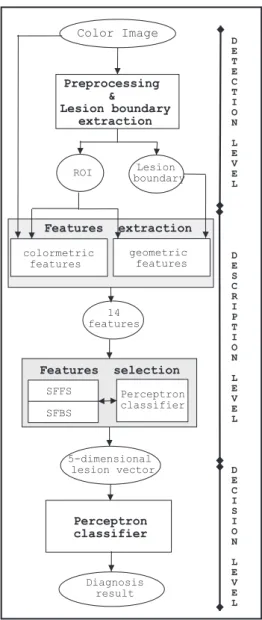
an overview of the literature of computer-aided melanoma diagnosis systems based on image processing techniques. We also present an ac-celerated system for melanoma diagnosis. In section 3, we present the architecture of our system(Figure 1)while retailing the lesion
de-tection, the lesion description and the decision levels. The lesion detection level consists of preprocessing the lesion image followed by the extraction of the lesion by separating it from the healthy surrounding skin. The lesion descrip-tion level is based on the feature extracdescrip-tion pro-cess and on a subset feature selection technique. This permits to define a pertinent descriptor vec-tor relatively to the lesion. This lesion’s vecvec-tor will be then the raw input to a multi-layer per-ceptron classifier(decision level)in order to
as-sign the lesion to the malignant melanoma class or to the benign nevi class. In section 4, we will discuss the relevance of our proposed system by analysing results of the experiments in terms of true-positive and true-negative rates, hence per-mitting to evaluate the global classification ac-curacy of our diagnosis system. Finally, conclu-sion and suggestions for future work are given in section 5.
2. Overview of the Literature
This section presents a review of the existing literature on computer-aided diagnosis systems for melanoma recognition. A classic system of melanoma diagnosis is principally composed of three stages. First, after the acquisition and the preprocessing of the dermatoscopic image, the segmentation process aims to extract the lesion
L from the Healthy Surrounding Skin (HSS).
In a previous work 34], we presented a
pre-limary approach for the automated recognition of malignant melanoma. In order to localize suspicious lesion regions, we introduced an au-tomated approach to segmentation by region growing, after a preliminary step based on fuzzy sets. Then, relatively to every detected skin le-sion, we measured a series of attributes model-ing the clinical malignancy symptoms. Finally, the selected attributes were supplied to an ar-tificial neural network in order to differentiate melanomas from benign lesions. This approach allowed us to obtain 79:1% of correct
classifi-cation of malignant and benign lesions on a test set consisting of 200 real skin lesions images. In 15], Hintz-Madsen developed an efficient
system for diagnosing skin lesions images. He used an optimal thresholding technique to de-termine two thresholds separating the light skin background from the dark skin lesion. In the case that the segmentation produces several skin lesion candidates, due to other small non-lesion objects, the largest object is selected as the skin lesion. To realize the lesion classification, a neural network in a probabilistic framework was designed and it allowed to detect 73:2% of
be-nign skin lesions and 75% of malignant skin lesions on a test set of 200 images.
In 30], the authors have introduced a
knowledge-based system for supporting the early diagno-sis of melanoma. It detects the lesion using a thresholding technique applied on the blue and on the saturation images. Then, the system, called MEDS(MElanoma Diagnosis System),
extracts a set of colorimetric and geometric fea-tures. It then yields a diagnosis based on a voting schema integrating the results produced by three different classifiers: k-nearest neigh-bors(KNN), discriminant analysis and decision
tree. The system is trained and validated on a set of 152 skin images. The average sensitiv-ity and specificsensitiv-ity of the designed system were 82% and 71% respectively.
In 29], the authors presented a preliminary
computer-aided diagnosis system for pigmented skin lesions, with solutions for lesion boundary detection and for the quantification of the asym-metry degree. To detect the lesion boundary, they used a segmentation technique based on the clustering of a two dimensional color space. To perform the clustering, a modified Fuzzy C-Means (FCM) technique (called
Orientation-Sensitive FCM: OS-FCM)was introduced.
Le-sions detection results were validated by expert dermatologists, who also provided hand-drawn boundaries of the lesions. The authors showed that dermatologists were not able to reproduce their results. In particular, the boundaries of any expert taken alone showed higher diver-gence from those of remaining experts as well as from the developed automatic techniques. In this paper feature extraction is restricted to the quantification of degree of symmetry by a six-dimensional vector, which was used to classify, with a linear classifier, pigmented skin lesions as being benign or malignant. This classifier achieved a sensitivity of 60% and a specificity of 70% on a test set of 100 skin lesion images. Vannoorenberghe et al 33]proposed a color
im-age segmentation method based on the Dempster-Shafer theory. The tristimuli Red, Green and Blue are considered as three independent infor-mation sources, imprecise and uncertain, per-mitting separation of the pigmented lesion from the healthy surrounding skin by computing a threshold obtained by means of the Maximum Entropy Principle(MEP). Then, the three
rel-ative decisions are combined by the Dempster-Shafer theory technique. In the second step, features concerning the lesion are extracted us-ing color information and all these primitives are considered as uncertain information sources on the lesion malignancy. Then, these infor-mations are combined using Dempster-Shafer theory to bring out a particular behavior for the malignant melanoma. This classification pro-cedure was applied on a set of 120 skin lesion images with a general success rate of 63%. In 22], the authors proposed a similar
classi-fication method based on the Dempster-Shafer theory and information criteria. In fact, after an original basic belief assignment with probabil-ity densities obtained by learning, an attenuation factor based on the dissimilarity between prob-ability densities is introduced. The proposed method allows to obtain 81:1% of good
classi-fications. However, the major drawback of this method resultes in increased CPU time.
Ercal et al 7] presented a neural network
supplied to an artificial neural network for clas-sification. This approach permitted to obtain above 80% correct classification of malignant and benign tumors on real skin tumor images. Ganster et al 12]developed a system to enhance
an early recognition of malignant melanoma. As an initial step, the binary mask of the skin lesion is determined by several basic segmen-tation algorithms, which are combined together with a fusion strategy. A set of 122 features con-taining shape and radiometric features, as well as local and global parameters, was calculated to describe the malignancy of a lesion. Using statistical feature subset selection methods, 21 significant features were selected from this set. Finally, a k-nearest neighbors classifier is used for the classification with a sensitivity of 81%. Some other non-complete systems were also in-troduced. For example, in 8], the authors used
color-based segmentation approach applied on the Karhunen-Lo`eve transform of the rgb color vectors to separate pigmented lesions from the skin and proceed with histogram equalisation and greyscale morphology to enhance and filter the pigmented lesions. They demonstrated that this approach allows to enhance pigmented skin lesions visually, and make them accessible to further analysis and classification tasks.
In 28], the authors introduced a region-based
approach to segment lesion images into areas of different colors. Initially, the process di-vided the dermatoscopic image into rectangular regions, small enough to be considered as hav-ing only a shav-ingle color. This is followed by conservative merging, where adjacent regions whose colors are very similar are coalesced. The segmentation is then completed by an it-erative optimal merging process, the two most similar regions being merged at each step until the stopping condition is reached.
In 1995, Lee et al. 20]presented an algorithm
to identify skin lesions from the digitized color images. After the application of a multi-stage median filter on the original image, in order to suppress the noise, the algorithm consists of the computation of threshold values for the le-sions and the background normal skin. Then, the application of a rule-based system permits identification of the lesions.
In 14], the authors defined an approach by
adap-tive thresholding for the automated
segmenta-tion of color skin lesion images. This approach combines six different thresholding techniques which are applied on the principal component of the Karhunen-Lo`eve transform, on the rgb space and on the spherical coordinates.
Finally, we can conclude that many existing sys-tems produced high rates of malignant melanoma recognition. But, their major inconvenience is too high computing time. The use of more than one algorithm for segmentation and/or
classifi-cation to be combined, and the use of a large set of features, is the main cause of the non-realistic computing time. In many works 34] 29], it was
confirmed that the task of detection of a spe-cific pattern in dermatoscopic images is both, the most interesting and very difficult. In other words, computing the features that model the clinical signs of malignancy is the step with the largest CPU time consumption in a computer-aided melanoma diagnosis system. In fact, a fundamental problem of automated melanoma recognition is the complexity of the classifica-tion scheme, because a large number of fea-tures can be found in the feature design process. In order to keep the complexity low, a reason-able number of useful features, that remove re-dundant information and approximate proper-ties of the melanoma diagnosis task, should be selected. A general theory for the design of such features does not exist 11]. However, reduction
of the feature set dimension is very important and must be addressed in the right way. On the one hand, smaller feature set reduces the cost of the measurements. On the other hand, reduction in the number of features may lead to a loss of the discriminatory power of the classification and consequently may reduce the accuracy of the resulting system.
Roß et al. 27] performed selection of features
by application of the “sequential forward selec-tion algorithm” that reduced the set of features, starting with 87 features, to five features. In 15], the authors achieved feature selection with
a neural network by application of node prun-ing. This approach allowed to reduce the set size from 21 lesion features to only six. However, Ganster et al. 12] started with 122 features
and used three statistic approaches to reduce the number of the selected features. These ap-proaches are: Sequential Floating Forward Se-lection(SFFS), Sequential Floating Backward
Finally, a subset size of 21 is chosen for further classification experiments.
3. The Proposed System
In this section we will establish the scheme of our proposed computer-aided diagnosis system for skin lesions recognition. This system is pre-sented in Figure. 1 and is composed mainly of a lesion detection, a lesion description and a decision levels. The input to our system are color images (rgb) which are digitized
photo-graphic slides of dermatoscopic images 34].
The lesion detection level consists of the re-alization of two steps. The first step concerns the preprocessing of the lesion image in order to eliminate the noise and the undesired objects in the original image. The second step con-sists of the extraction of the lesion by separat-ing it from the healthy surroundseparat-ing skin. This step produces a binary image(lesionvs.healthy
surrounding skin)which will be treated by the
border extraction process, in order to define a polygon representing an approximation of the lesion boundary. The lesion description level is based on the realisation of two steps. The first step is the features extraction process produc-ing numerical features which designate clinical symptoms of malignancy. Then, in order to re-move redundant information and approximate the properties of melanoma recognition, selec-tion of features allows to select a reasonably reduced number of useful features, that permits to reduce the dimension of the lesion’s vector, and consequently the computing time, without significant loss of information. Finally, the de-cision level consists of the presentation of this lesion’s vector as an input to a multi-layer per-ceptron classifier in order to assign the lesion either to the class of malignant melanoma or to the class of benign lesions.
Finally, let’s note that the proposed system rep-resents an extension of the system presented in 34]. Indeed, here our interest will be focused on
the step of features selection which represents the main amelioration in our system. This step will allow to reduce considerably the comput-ing time of our computer-aided diagnosis sys-tem without significant loss of the classification performance. Color Image Preprocessing & Lesion boundary extraction ROI Lesion boundary SFFS SFBS Perceptron classifier colormetric features geometric features 14 features Diagnosis result Perceptron classifier 5-dimensional lesion vector D E T E C T I O N L E V E L D E S C R I P T I O N L E V E L D E C I S I O N L E V E L Features selection Features extraction
Fig. 1.Architecture of the proposed system.
3.1. Lesion Detection Level
The first step of the lesion detection level is the preprocessing of the dermatoscopic original im-age aiming to ameliorate the imim-age quality for ulterior tasks. The second step is based on the preprocessed image and it aims to define the le-sion by a region of interest(ROI)and to extract
the correspondent boundary.
3.1.1. The Original Image Preprocessing
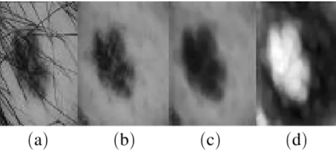
color images are impulsive-like artifacts. These artifacts can be considered as impulsive noise and may thus be reduced using a median filter 13] while preserving edges. The median filter
is very effective in removing noise spikes that only cover a few pixels compared to the kernel neighborhood size. Thus, it can remove blond hairs and small air bubbles from skin images
(Figure 2.c). Besides, as we were working with
color images, we applied the median filter to all three color components(r, g, b)separately.
The next preprocessing step aims to facilitate the segmentation process by enhancing the edges in the image. For this purpose, we applied the Karhunen-Lo`eve(KL)transform 23]on the
re-sulting filtered images in order to enhance the edges which permits eventually to ease the sep-aration between the lesion and the surrounding healthy skin. The Karhunen-Lo`eve transform is a linear transform that correlates the input vari-ables by employing an orthonormal basis. This basis is found by an eigenvalue decomposition of the sample covariance matrix of the input variables. The covariance matrix Cov is com-puted with a vectorV enclosing all three color vectors of the image. In fact, we use projections of the color vectors onto the eigenvectors of the covariance matrix that models the variation of the color components. Due to the decreasing ordering of the eigenvalues and the correspond-ing eigenvectors, the first principal component will contain the maximum variance 34]. Thus,
since most variations occur at the edges between the lesion and surrounding skin, the first prin-cipal component is a natural choice for further segmentation(Figure 2.d).
Besides, some skin images contain dark hairs with similar color hue to the lesion, which may occlude the lesion and mislead the segmentation
(a) (b) (c) (d)
Fig. 2.Image preprocessing.(a)original image;(b)
hairs removal;(c)median filtering;(d)first principal
component of the Karhunen-Lo `eve transform.
process. In fact, while blond hairs can be left without problem with the median filter, this fil-ter is insufficient for dark hairs suppression and can even intensify some undesired dark hairs
(Figure 3.). To remove these dark hairs from
the dermatoscopic image, we implemented the program solution called “DULLRAZOR” 21]
pro-posed by Lee et al. This solution consists of identifying the dark hair locations by applying a generalized morphological closing operation to the three color bands separately, and replac-ing the determined hair pixels with the nearby no-hair pixels 34](Figure 2.b).
(a) (b)
Fig. 3.Insufficiency of median filter for dark hair removal. (a)original image;(b)median filter result.
3.1.2. The Lesion Boundary Extraction
The lesion boundary extraction is a difficult task in dermatoscopic images since the tran-sition between the lesions and the surrounding healthy skin is often smooth and ill-defined. In-deed, most skin lesions possess fuzzy bound-aries with slow and extended transition from the dense core region to the surrounding tissues. In the literature, various region-based segmenta-tion methods are applied for the skin lesion de-tection and the most commonly used technique has been color and grayscale thresholding 18].
However, the majority of these methods are un-able to define the criterion to precisely separate pigmented lesion from the surrounding healthy skin.
To accomplish detection of the skin lesion, we used our automated approach of segmentation, introduced in 34]. It consisted of performing a
fuzzy plane, using a membership transforma-tion functransforma-tion. The goal is to generate an image of higher contrast than the input image(the first
principal component of the median filtered der-matoscopic image)by giving a larger weight to
the gray levels that are closer to the lesion repre-sentative gray level than to those that are farther from the lesion representative gray level. In fact, after a succession of intensity histogram equalizations, we obtained a bimodal curve char-acterized by two peaksp1and p2and their cor-responding gray levelsg1andg2assumed to be representative of the gray levels of the lesion and the surrounding healthy skin. Then, as the le-sion is always darker than the surrounding skin, its gray levelgLshall be the minimum ofg1and g2. Given the lesion gray levelgL, a pixel whose
gray level is similar to gL could be assigned
a high intensity in the resulting fuzzy image. To perform the enhancement of this fuzzy im-age an appropriate membership transformation function, that evaluates the similarity between the gray levelgl(pix)of the pixelpixbeing
con-sidered and the representative gray level (gL)
of the lesion, may be achieved. This function must be symmetric, it decreases monotonically from 1 to 0 and assigns a membership degree equal to 1 to those pixels that possess the same gray level as the lesion(gL). We have
consid-ered two functions,=1(eq. 1)and=2(eq. 2),
verifying these properties.
=1(pix)=(1+β jgl(pix);gLj) ;1
: (1)
=2(pix)=1;β jgl(pix);gLj: (2)
We deduce that better results are obtained with a normalized combination,= (eq. 3), of these
two functions=1 and =2 (Figure 4.). Finally,
the resulting fuzzy matrix could be used as a scale factor to obtain gray levels and display the resulting fuzzy imageIf uzzy(eq. 4).
=(pix)=
=1(pix)+=2(pix)
2 : (3)
If uzzy(pix)==(pix):max
p2=
gl(p) (4)
Value β defines opening of the membership function. For largeβ the opening is narrow and the function behavior is strict. However, for small β the opening is wide, and the function
(a) (b)
(c) (d)
Fig. 4.Comparison between the membership functions results. (a)original image;(b)lesion detection with the
function=1;(c)lesion detection with the function=2;
(d)lesion detection with the function=.
presents a more permissible behavior (Figure
5., Figure 6.).
To perform the region growing algorithm, a rep-resentative seed pixel must be defined inside the skin lesion. This seed pixelgis defined by the pixel havinggLas intensity while being the
center of a homogeneous neighborhood V(g)
(eq.5). Finally, starting from the determined
seed pixel, a classic region growing algorithm 13]is applied on the resulting fuzzy matrix and
it permits to define precisely the lesion as a
re-(a) (b)
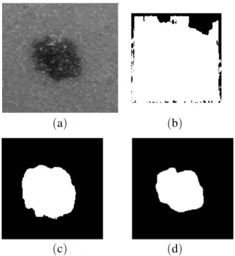
Fig. 5.Depending on lesion extraction relatively toβ.
(a)β =0:006;(b)β =0:011(binary plane relative to
(a)
(b)
Fig. 6.Depending on the function=behavior relatively
toβ(gL=128).(a)β =0:006;(b)β =0:012.
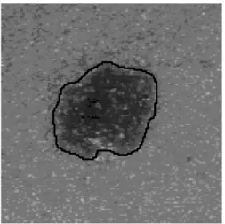
Fig. 7.Detection of the lesion boundary of the lesion in Figure 4.(a).
gion of interest(ROI). The output of the region
growing procedure is the so-called binary plane
(Figure 5.), i.e. 1-bit image that separates lesion
(1)from the healthy surrounding skin(0). The
resulting binary image(lesionvs. healthy
sur-rounding skin) is then treated by an algorithm
of follow-up applied on the border pixels of the lesion which are characterized by a local maxi-mum of gradient. This allows the definition of a closed boundary relating to the lesion(Figure
7.).
max
q2V(g)
jgl(g);gl(q)j=
=min
p2ξ
max
q2V(p)
jgl(p);gl(q)j
: (5)
where,ξ =fp2==gl(p)=gLg.
3.2. Lesion Description Level
The first step of the lesion description level is based on the binary plane defining the skin le-sion by a region of interest and on the corre-sponding boundary of the lesion which is pro-duced by the lesion detection level. This step consists of the extraction of a set of numerical features modeling clinical signs on the lesion malignancy. Then, in order to remove redun-dant information and to approximate the proper-ties of melanoma recognition, a features subset selection step allows to select a reduced num-ber of features. This step allows to reduce the dimension of the lesion’s vector without losing information about the lesion malignancy.
3.2.1. Lesion Features Extraction
The objective of the lesion features extraction step is to quantify clinical signs used by a der-matologist for the melanoma diagnosis by a set ofmnumerical features. Thus, given its defini-tion by a region of interest(ROI)and by a closed
boundary, every lesion will be described by am -dimensional feature vector. The extraction of these features is based on the so-calledABCD
rule of dermatoscopy. This rule, originally in-troduced by Friedman et al. 9], is a very useful
In the literature, many attributes were used to describe characteristics of the lesions ma-lignancy relatively to the ABCD rule. How-ever, as it was always proved 29], a unique
attribute is not sufficient to diagnose precisely melanoma cancer. Combination of different cri-teria is the key to an early detection of
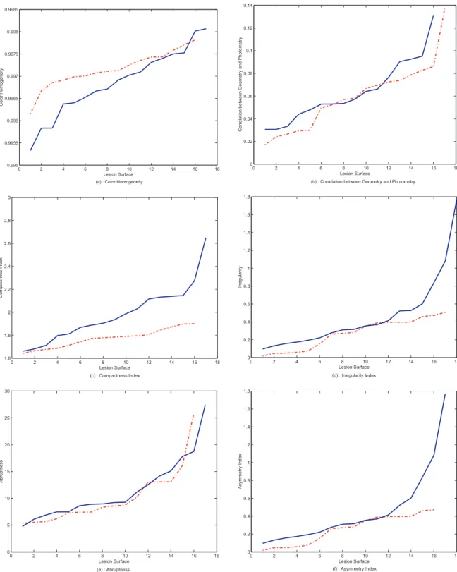
malig-nant melanoma. In this sense, we carried out a study of many significant attributes, accord-ing to the surface of the lesion. The means of these parameters per interval of lesions surfaces were calculated on randomly selected images of malignant and benign lesions(Figure 8). We
deduced an important correlation between most
of the attributes and the melanoma diagnosis. The two types of lesions differ slightly in their characteristics relatively to these attributes. For example, consider the color homogeneity at-tribute(Figure 8.a.). The value of this attribute
is normally more elevated for the benign lesions that for the malignant ones. However, it is of-ten possible to encounter malignant lesions of which the value of the color homogeneity at-tribute is more elevated than the ones recorded with some benign lesions and the same behav-ior can be deduced with other attributes(Figure
8.). Thus, we can conclude that it is
impossi-ble to separate linearly the two types of lesions using every attribute separately. From there, it is necessary to perform a combination of a set of suitable attributes in order to obtain precise classification of the lesion.
As the next step, we will perform feature selec-tion by choosing to use a relatively large set of possible attributes. After literature overview of the more used attributes to quantify theABCD
rule and after many discussions with trained der-matologists, we selected 14 attributes modeling the lesion malignancy symptoms used by ex-perts in clinical practice. These attributes are 34] 7] the asymmetry index, the lesion
diam-eter, the lengthening index, the compactness index, the mean square error between the im-age and its reflected version, the fractal dimen-sion, the edge abruptness, the mean and the variance of the gradient magnitude along the le-sion boundary, the color homogeneity, the rel-ative chromaticity of the red, green and blue components and finally the index of correlation between geometry and photometry.
3.2.2. Features Subset Selection
The goal of this step is, given the lesions set described bym(m =14)attributes, to find the
minimum numbern (n << 14)of relevant
at-tributes which describe the lesions malignancy as well as the original set of attributes do 19]. In
fact, there is often a need to minimize the num-ber of attributes actually used for classification, since each used attribute increases the running time of the computer-aided diagnosis system. At the same time, there is a need to achieve high recognition rates. This has led to the de-velopment of a solution for finding an “optimal” subset of features from a larger set of possible
features. The produced subset of features would achieve high melanoma recognition rates, while at the same time it decreases cost and running time of the computer-aided diagnosis system. In the literature, two main categories of features selection are used 32]. The first approach
se-lects features independently of their effect on the classification performance. However, mere evaluation of the discriminatory power of the initialm features individually is not sufficient, but all subsets must be considered to find the best combination 5]. Thus, the second
ap-proach, called subset features selection, selects directly a subset of the available m features, without significant decrease of the recognition performance. Several algorithms are used for the realization of the second approach and as the complexity of the algorithm grows expo-nentially withm 26], it is not possible to derive
the optimal performance by exhaustive search and all these algorithms are sub-optimal. Be-sides, we note that the feature selection process is always followed by a measure of the dis-criminatory power of subsets given a separabil-ity criterion, which measures the discriminatory power of the subset under consideration. Given the dimension of the desired subset of features, probably the most effective feature se-lection techniques are the sequential floating search methods (SFSM) 16] 25]. There are
two main categories of floating search methods: forward and backward. In the case of sequen-tial floating forward search (SFFS), the
algo-rithm starts with a null feature set and, for each step, the best feature satisfying some criterion function is included with the current feature set. Besides, at every step, the worst feature con-cerning the criterion is eliminated from the set. Therefore, the SFFS proceeds dynamically, in-creasing and dein-creasing the number of features, until the desired set dimension is reached. The backward search(sequential floating backward
search: SFBS) works analogously, but
start-ing with the full feature set and performstart-ing the search until the desired dimension is reached. These algorithms have been improved, leading to the adaptive floating search methods(AFSM)
31]. The main difference is in the number of
desired features that can be determined dynam-ically. In 4], the authors proved that the
In this work, we perform an automatic subset features selection technique where subsets of the initial set of 14 attributes are investigated in order to derive a combination of a reduced set of pertinent attributes modeling theABCDrule. To do this, we applied two sequential search tech-niques: Sequential Floating Forward Selection
(SFFS)and Sequential Floating Backward
Se-lection(SFBS). These algorithms use stepwise
inclusions and exclusions of features into/from
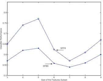
the subset of consideration. As separability cri-terion, we used the performance of a multi-layer perceptron classifier using the selected features. We performed the SFFS and SFBS techniques with various size of the designed features subset. Our experiments showed that the best perfor-mance is recorded with a subset of five features determined by the SFFS technique and the cor-respondence between the selected features and the clinical criteria is clearly shown(Figure 9).
Fig. 9.Results of subset feature selection with SFFS and SFBS algorithms.
3.3. Decision Level
The final level of our melanoma computer-aided diagnosis system is the lesion screen-ing decision, lookscreen-ing for the association be-tween the studied skin lesion and the malignant melanomas class, or the benign lesions class. This level is based on the supplying of the le-sion’s vector, produced by the previous level, to a multi-layer perceptron classifier. In the liter-ature, many classical methods such as the near-est neighbors classification 12], fuzzy c-means
(FCM) 6]as well as neural networks 11] 30]
7], have been applied for the purpose of skin
lesions classification. However, it was always showed that the recognition rates are slightly higher for the neural networks classification, compared to the classical methods 11]. Thus, to
perform the classification procedure, as neural network classifier we used a multi-layer percep-tron trained with the standard back-propagation algorithm 10]. The input to this perceptron
is the lesion’s vector produced by the previous level. However, as the features composing the lesion’s vector have different unit and different value ranges, an objective scaling, between 0 and 1, of the feature values is achieved by cal-culating the correspondentz-scores 17].
There-fore, given a featurej and the set of t samples
(tis the size of the training or the test sets)for
the featurej, theithsample measure of featurej
(Xij)will be replaced by the valueYijgiven by:
Yij=
Xij;mj
σj
: (6)
where,mjis the mean value of thetsamples for
the featurej andσj is the correspondant mean
absolute deviation.
The perceptron architecture is formed of only one hidden layer 34], whose number of units
is determined manually, by evaluating the clas-sification performance(Table. 1.). The output
layer consists of one linear output unit produc-ing a binary result (1 for a malignant lesion
and 0 for a benign lesion) which corresponds
to the target class. The defined perceptron is trained with supervision, given a training set of dermatoscopic images, using the standard back-propagation algorithm 10].
4. Tests and Results
This architecture allows to obtain above 95% of good classifications on the training set and above 82% of good classifications on the test set.
hidden units training set(%) test set(%)
1 78 73.7
2 75.7 72.5
3 84.9 74.6
4 95.3 82.4
5 91.8 78.1
Table 1.Classifier performance with reference to employed hidden units.
The main inconvenience of this classifier is its higher calculating time caused mainly by the large number of features used to describe a le-sion. Thus, the legitimate solution for reducing the complexity of the classification process was to reduce the number of features without losing information about the malignancy of the lesion. Hence, we used the feature subset determined by the SFFS technique permitting decrease of the size of feature set from 14 to 5 while pre-serving similar significant information on the lesion malignancy. In fact, a recognition rate of 77:7% is achieved with five features and it does
not increase over 82:4% by including the 14
features. However, decreasing the size of the feature set allows to reduce considerably the computing time for both, features computing
(tFC)step and classification step, including the
training (tT1) and the test (tT2) modes (Table.
2.).
features set dimension tFC(s) tT1(s) tT2(s)
14 98 3028 57
5 43 1059 13
Table 2.Comparison of the classification computing time with reference to the features set dimension.
In Table. 3., we present the results achieved with the selected 5 attributes determined by the SFFS technique. Besides, we compare the ob-tained classification accuracy on a test set of
new dermatoscopic images, while using 14 fea-tures(subsection 3.2.1.), 5 features(subsection
3.2.2)and using the 9 features used in our
pre-vious work (34]). Accuracy of the
classifi-cation on the test set is evaluated in terms of “true-positive” (TP) rate (called also
sensitiv-ity)and “true-negative”(TN)rate (called also
specificity).
features set dimension TP(%) TN(%)
14 71.3 93.5
9 75.1 83.1
5 74.8 80.6
Table 3.Comparison of the classification accuracy with reference to the features set dimension.
At the end, we can conclude that the step of fea-tures selection with the SFFS technique allows us to reduce the number of features to five. This step allows to reduce considerably the dimen-sion of the ledimen-sion’s vector without significant loss of information about the lesion malignancy. In fact, the system with only five features per-mits to obtain a correct classification rate of 77:7% (with true-positive rate of 74:8% and
true-negative rate of 80:6%) in a very
accept-able CPU time (less than one minute for both
steps of feature calculation and of the lesion screening). The recorded correct classification
rates are very comparable with the detection rates recorded with 9 features (79:1% of
cor-rect classification rate with true-positive rate of 75:1% and true-negative rate of 83:1%)and also
with 14 features(82:4% of correct classification
rate with true-positive rate of 71:3% and
true-negative rate of 93:5%). However, the relative
reduction of the number of features describing the lesion malignancy reduces the CPU time of a complete diagnosis of a lesion for about 50%
(198 seconds with 14 features vs. 99 seconds
with 5 features)and reduces the training time of
the multi-layer perceptron for about 65%(3028
5. Conclusion and Future Work
Different sections of this paper proposed solu-tions to specific problems related to computer-aided diagnosis of pigmented skin lesions. Thus, an accelerated system for melanoma diagnosis was presented. In this study, we focus particu-larly on the important step of feature selection by modeling the clinical signs of lesions ma-lignancy to enable the classification into either benign or malignant lesions class. Indeed, a large set of features was investigated by the ap-plication of feature selection techniques. The number of features relevant for classification was optimised and five well-selected features were sufficient to cover the major of discrim-inatory information about lesions malignancy. Results on real data proved the effectiveness of the proposed system by recording a good clas-sification rate of 77:7% in a very encouraging
computing time.
However, while the lesion detection problem is practically resolved, there is still much to do with regard to the extraction of diagnosis fea-tures. For example, we think to use a larger set of features as the input set for the subset fea-ture selection techniques. This will probably permit to deduce more generalized descriptors for the lesion malignancy. Besides, we plan to use the genetic algorithms(GA)techniques for
the subset feature selection with larger range of possible dimensions for the subset of features. Genetic algorithms can be used to explore the space of all possible subsets of features, so as to obtain, in the minimum of computing time, a set of features which optimise predicitive accuracy for the melanoma diagnosis.
6. Acknowledgment
The authors would like to thank sincerely the anonymous reviewers of this paper who made many important suggestions and helpful contri-butions that enabled us to improve the quality of the paper.
References
1] American Cancer Society, www.cancer.org, (2002).
2] A. BRESLOW, Thickness, cross-sectional area and
depth of invasion in the prognosis of cutaneous melanoma,Annals of Surgey, 172(5), pp. 902–908, (1970).
3] G. BURG, Das Melanom, Serie Gesundheit:
Piper/VCH,(1993).
4] T.E. CAMPOS, R.S. FERISAND ANDR.M. CESAR,
Im-proved face x non-face discrimination using Fourier descriptors through feature selection, 13th
Brazil-ian Symposium on Computer Graphics and Image Processing, Gramado, Brazil,(2000).
5] T.M. COVER, The best two independent
measure-ments are not the best, IEEE Transactions on Systems, Man and Cybernetics, SMC, 4(1), pp.
116–117,(1974).
6] R. CUCCHIARA AND C. GRANA, Using the
topo-logical tree for skin lesion structure description. Knowledge-Based Intelligent Information Engi-neering Systems and Allied Technologies (KES
2002), Frontiers in artificial intelligence and
appli-cations,IOS Press, 82(1), pp. 166–170,(2002).
7] F. ERCAL, A. CHAWLA, W.V. STOECKER, H. LEE AND
R.H. MOSS, Neural Network diagnosis of malignant
melanoma from color images, IEEE Transactions on Biomedical Engineering, 41(9), pp. 837–845,
(1994).
8] S. FISHER, F. SCHMID ANDJ. GUILLOD, Analysis
of skin lesions with pigmented networks, Interna-tional conference on Image Processing (ICIP’96),
Lausanne, Switzerland,(1996).
9] R.J. FRIEDMAN, D.D RIGEL AND A.W. KOPF, The
continued importance of early detection of malig-nant melanoma,CA, 41, pp. 201–226,(1999).
10] K.I. FUNASHASHI, On the approximate realization
of continous mappings by neural networks, Inter-national Journal of Neural Networks, pp. 183–192,
(1989).
11] H. GANSTER, R. ROHRER, L. PALETTA, T. EBNER, A.
PINZ ANDM. BINER, Comparison of neural networks
and statistical methods in melanoma classification, Proc. 22nd OAGM/AAPR Workshop, Illmitz,
Aus-tria,(1998).
12] H. GANSTER, A. PINZ, R. ROHRER, E. WILDLING,
M. BINDER ANDH. KITTLER, Automated Melanoma
Recognition,IEEE Transactions on Medical Imag-ing, 20(3), pp. 233–239,(2001).
13] R.C. GONZALEZ ANDR. E. WOODS,Digital Image
14] G.A. HANCE, S.E. UMBAUGH, W.V. STOECKER AND
R.H. MOSS, Unsupervised color image
segmenta-tion: With application to skin tumor borders,IEEE Engineering in Medicine and Biology Magazine, Vol 15, pp. 104–111,(1996).
15] M. HINTZ-MADSEN,A probabilistic frame-work for
classification of dermatoscopic images, ph.D. the-sis, IMM,(1998).
16] A.K. JAIN AND D. ZARGKER, Feature selection:
Evaluation, Application and Small Sample Per-formance, IEEE Trans on Pattern Analysis and Machine Intelligence, 19(2), pp. 153–158,(1997).
17] L. KAUFMAN ANDP.J. ROUSSEEUW,Finding groups
in data: An introduction to cluster analysis, New York, USA,(1990).
18] I. KHANFIR, A. KALLEL, K. TAOUIL, M. S. BOUH
-LEL ANDL. KAMOUN, Segmentation d’images par seuillage d’histogramme: Application ´O l’analyse des m `Ulanomes,JTEA 2002, Tunisia,(2002).
19] P.L. LANZI, Fast feature selection with genetic
algo-rithms: a filter approach,IEEE International Con-ference on Evolutionary Computation (ICEC97),
pp. 537–540,(1997).
20] T. LEE, V. NG, D. MCLEAN, A. COLDMAN, R. GAL
-LAGHER ANDJ. SALE, A multi-stage segmentation
method for images of skin lesions, IEEE Pacific Rim Conference on Communications, Computers and Signal Processing, pp. 602–605,(1995).
21] T. LEE, V. NG, R. GALLAGHER, A. COLDMAN AND
D. MCLEAN, Dullrazor: A software approach to
hair removal from images, Computer in Biology and Medicine, 27, pp. 533–543,(1997).
22] E. LEFEVRE, O. COLOT ANDP. VANNOORENBERGHE,
A classification method based on the Dempster-Shafer’s theory and information criteria, FU-SION’99, California, USA,(1999).
23] M. LOEVE` , Fonctions al ´Uatoires de second
or-dre, In P. Levy, editor, Processus stochastiques et mouvement brownien, Hermann Eds,(1998).
24] M.C. MIHM, A.J. SOBER ANDT.B. FITZPATRICK,
Pri-mary melanoma of the skin: recognition and man-agement, Journal of the American Academy of Dermatology, 2(3), pp. 179–197,(1980).
25] P. PUDIL, J. NOVOVIOVCA ANDJ. KITTLER,
Float-ing search methods in feature selection, Pattern recognigion letters, 15, pp. 1119–1125,(1994).
26] R. ROHRE, H. GANSTER, A. PINZ ANDM. BINDER,
Feature selection in melanoma recognition, ICPR’98, 2, pp. 1668–16670,(1998).
27] T. ROSS, H. HANDELS, J. KREUSCH, H. BUSCHE,
H.H. WOLF ANDS.J. POPPL, Automatic
classifica-tion of skin tumors with high resoluclassifica-tion surface profiles,Computer Analysis and Patterns, pp. 460– 465,(1995).
28] A.J. ROUND, A.W. DULLER AND P.J. FISH, Colour
segmentation for lesion classification, 19th
Interna-tional Conference – IEEE/EMBS, Chicago, USA, pp. 582–585,(1997).
29] P. SCHMID-SAUGEON, J. GUILLOD ANDJ.P. THIRAN,
Towards a computer-aided diagnosis system for pig-mented skin lesions,Computerized Medical Imag-ing and Graphics, Vol 27, pp. 65–78,(2003).
30] A. SBONER, E. BLANZIERI, C. ECCHER, P. BAUER,
M. CRISTOFOLINI, G. ZUMIANI AND S. FORTI, A
knowledge based system for early melanoma sup-port, Intelligent Data Analysis in Medicine and Pharmacology, UK,(2001).
31] P. SOMOL, P. PUDIL, J. NOVOVICOVA ANDP. PACLIK,
Adaptive floating search methods in feature selec-tion,Pattern recognigion letters, 20, pp. 1157–1163,
(1999).
32] H. VAFAIE ANDK.D. JONG, Genetic algorithms as
a tool for feature selection in machine learning, Proceedings of the4thInternational Conference on
Tools with Artificial Intelligence, Alington, USA,
(1992).
33] P. VANNOORENBERGHE, O. COLOT AND D. DE
BRUCQ, Dempster-Shafer’s theory as an aid to color
information processing: Application to melanoma detection in dermatology, 10th International
Con-ference on Image Analysis and Processing, Italy,
(1999).
34] E. ZAGROUBA ANDW. BARHOUMI, A prelimary
ap-proach for the automated recognition of malignant melanoma,Image Analysis and Stereology Journal, 23(2), pp. 121–135,(2004).
Received:August, 2004 Accepted:November, 2004
Contact address: Ezzeddine Zagrouba D`epartement des Sciences de l’Informatique Facult`e des Sciences de Tunis 1060, Tunis, Tunisia e-mail:[email protected]
EZZEDDINEZAGROUBAreceived his PhD from the Polytechnic National Institute of Toulouse(France)in 1994. Since 1995, he is a Computer
Sciences Professor in FSM(Facult`e des Sciences de Monastir)and in
FST(Facult`e des Sciences de Tunis). His research within the laboratory
LIP2-FST is focused on image analysis and computer vision.
WALIDBARHOUMIreceived his Master’s degree from the Polytechnic National Institute of Toulouse(France)in 1998. Currently, his is
prepar-ing a PhD thesis in computer sciences at the ENSI(Ecole Nationale des
Sciences Informatiques de Tunis). His research within the laboratory