http://dx.doi.org/10.11594/jtls.08.03.02
Research Article
Genotyping of
Toxoplasma gondii
in Cerebral and Ocular Toxoplasmosis
Dearikha Karina Mayashinta 1, Ryan Halleyantoro 2, Ika Puspa Sari 3, Agnes Kurniawan 3 *
1 Department of Parasitology, Faculty of Medicine, Universitas Brawijaya, Malang 65145, Indonesia 2 Department of Parasitology, Faculty of Medicine, Universitas Diponegoro, Semarang 50275, Indonesia 3 Department of Parasitology, Faculty of Medicine, Universitas Indonesia, Depok 16424, Indonesia
Article history:
Submission January 2018 Revised March 2018 Accepted May 2018
ABSTRACT
Toxoplasma gondii is an obligate intracellular protozoon, spread world-wide and ca-pable of infecting birds and mammals. Genetic information on T. gondii type that causes human toxoplasmosis is limited. In this study, genetic analysis of SAG2 locus was used to determine the genotype of T. gondii from cases with cerebral and ocular toxoplasmosis in Indonesia. Genotype determination was done directly on the clinical samples. A number of 28 cerebrospinal fluid and 8 vitreous humor positively infected with T. gondii, underwent PCR-RFLP to classify each isolate into one of three geno-types of T. gondii. Type I was the most common found suggesting that cerebral and ocular toxoplasmosis in Indonesia is mostly caused by type I strain of T. gondii.
Keywords: Toxoplasma gondii, genotype, PCR-RFLP, cerebrospinal fluid, ocular fluid
*Corresponding author:
E-mail:
agnes.kurniawan@ui.ac.id
Introduction
Toxoplasma gondii is an obligate intracellular protozoan of phylum Apicomplexa infecting birds and mammals. It is estimated that almost one-third of global population is infected with T. gondii [1, 2]. Clinical manifestation of toxoplasmosis varies and influenced by several factors, such as duration of exposure, geographic, parasite genetics, im-mune status of the host, and time of infection [3]. In immunocompetent individuals, Toxoplasma in-fection only shows minor symptoms or even asymptomatic. On the other hand, if the infection occurs in immunodeficient individuals, clinical manifestation might be fatal or even lethal [2, 4].
Information on dynamics of toxoplasmosis cases, either continuously or periodically, is still limited and very few study reports on T. gondii
genotype infecting human, hence T. gondii type is related to its pathogenic and biological character which is considered to play role in the pathogene-sis and management of toxoplasmopathogene-sis [5].
Toxoplasma encephalitis (TE) is one of the most common neurological infection in AIDS
pa-tients. It causes significant morbidity and mortal-ity [6]. Besides TE, T. gondii is one of the most common cause of retinochoroiditis both in immu-nocompetent and immunocompromised individu-als [7]. Variation in biological characteristics of each T. gondii type is considered to cause different clinical manifestations in human.
Virulence of T. gondii in animals varies, de-pending on the type [5]. More than 95% type of T. gondii isolated from North America and Europe belongs to one of three lineages, which referred to type I-III (Howe and Sibley, 1995). Genetically, the difference amongst each type is only 1 – 2%, but virulence differs significantly [2].
Material and Methods Study sample
Samples consisted of 88 cerebrospinal fluid from AIDS patients with cerebral disorder, as part of neuroaids study. While the 64 vitreous humor, obtained from patients clinically diagnosed as atypical uveitis and has been screened for Toxo-plasma infection. All samples were stored in -30oC in Parasitology Laboratory, Faculty of
Med-icine, Universitas Indonesia. The study was ap-proved by the ethical committee of Faculty of Medicine, Universitas Indonesia number 721/UN 2.F1/ETIK/2016.
DNA isolation
DNA extraction was done by boiling the sam-ples for 10 minutes at 100oC [11]. For cerebral
fluid, it was preceded by centrifugation of 1 mL of samples for 10 minutes at 10,000 rpm and leaving 150 µL sediment for boiling. For vitreous humor, samples were directly boiled due to very limited volume of samples obtained (≤ 100 µL).
Detection of T. gondii by PCR
To confirm T. gondii infection in cerebrospi-nal fluid and vitreous humor, nested PCR were performed targeting B1 gene, following the proce-dure by Alfonso et al. (2008) [11].
Target DNA amplification for genotyping Strain type of T. gondii was determined by PCR-Restriction Fragment Length Polymorphism (RFLP) of SAG2 gene with PCR cycle condition in accordance to the protocol used by Fuentes et al. (2001), Howe et al. (1997) [4, 10]. The PCR was optimized and run in 20 µL reaction using
TopTaq Polymerase Master Mix Kit (Qiagen,
GmbH, cat no. 200403) and 2 µL DNA template on MJ Research PTC 200 thermocycler. The pri-mers used are presented in the Table 1. Negative control (pure water) and positive control (T. gondii
RH strain, generous gift from Indonesian Research Centre for Veterinary Science) were always in-cluded in every PCR.
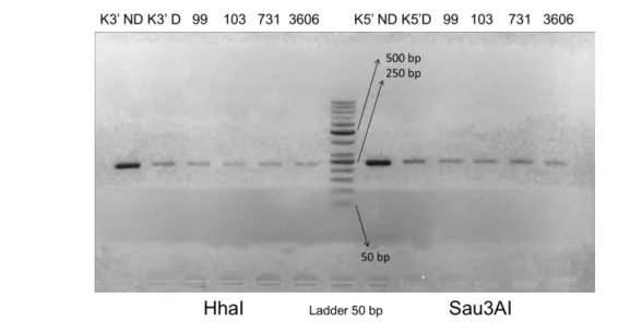
Optimum nested PCR condition for 5’-SAG2 Figure 1. Genotype determination based on RFLP analysis of SAG2 gene; A) Schematic drawing of SAG2
gene; B) Sau3AI enzyme cuts allele III sequence. B) HhaI enzyme cuts allele II sequence [10].
Table 1. Primer sequences used [4, 12]
Target genes Forward Reverse Amplicons
5’-SAG2 nest 1(SAG2.F4):
GCTACCTCGAACAGGAACAC nest 2 (SAG2.F)
GAAATGTTTCAGGTTGCTGC
nest 1(SAG2.R4):
GCATCAACAGTCTTCGTTGC nest 2 (SAG2.R2):
GCAAGAGCGAACTTGAACAC
241 bp
3’-SAG2 nest 1(SAG2.F3):
TCTGTTCTCCGAAGTGACTCC nest 2 (SAG2.F2):
ATTCTCATGCCTCCGCTTC
nest 1 (SAG2.R3):
TCAAAGCGTGCA TTATCGC nest 2 (SAG2.R):
AACGTTTCACGAAGG CACAC
and 3’-SAG2 genes is presented in Table 2. The last cycle was extended for 10 minutes at 72°C. The 241 bp and 221 bp of 5’-SAG2 and 3’-SAG2 respectively PCR products were visualized on 2% agarose gel electrophoresis.
The PCR products of 3’-SAG2 was digested with Hhal restriction enzymes by 4 hours incuba-tion at 37°C, followed by inactivaincuba-tion at 65oC for 20 minutes while 5’-SAG2 PCR product was di-gested with Sau3AI, incubated at 37°C for 3 hours and inactivated at 80°C for 20 minutes. The reac-tion was performed in 15 µL volume. The result was analyzed by electrophoresis on 2% agarose gel with TBE buffer solution and visualized by ex-posure to UV light. GeneRuler 50-bp DNA Ladder (Thermo Scientific) was used as molecular size marker. Interpretation of the result to identify the
T. gondii type I/II/III.
Results and Discussion
The PCR examination of B1 gene revealed 28 out of 88 (31.82%) cerebrospinal fluid samples and 8 out of 64 (12.5%) ocular fluid were positive for T. gondii infection. PCR of the 28 spinal fluid previously B1 gene positive, showed fewer posi-tive result which were 26/28 and 4/28 samples
res-pectively on 5'-SAG2 and 3'-SAG2. Similar result was obtained for the 8 ocular fluid samples, which were 100% positive on the 5'-SAG2 PCR however only 2/8 samples were positive on 3'-SAG2 gene.
RFLP of gene SAG2 PCR products
The size of amplicon for 5’-end was 241 bp and for 3’-end was 221 bp. PCR products were then digested by endonuclease restriction enzyme, HhaI to digest 3’-end and Sau3AI to digest 5’-end. Genotype analysis results by RFLP shown in Ta-ble 3.
Until recently, there is no report of human T. gondii genotypes in Indonesia and whether certain genotype is related to certain clinical manifesta-tion is still a debate. Several methods can be used to investigate T. gondii types, from isoenzyme electrophoresis, restriction fragment length poly-morphism (RFLP), multiplex PCR, DNA micro-satellite analysis, and DNA sequencing [5, 13, 14, 15]. Of all above, PCR-RFLP is the most common method used in more than 1,000 isolates in the world and had produced significant data on T. gondii genetic variation [16].
This work is part of a study on neuroaids where Toxoplasma laboratory screening has been Table 2. PCR condition for 5’-SAG2 and 3’-SAG2
No Condition 5’-SAG2 3’-SAG2
1. Denaturation Nested I & II: 94oC, 5 minutes Nested I & II: 94oC, 5 minutes 2. Annealing nested I: 60oC, 45 seconds nested II:
58oC, 45 seconds nested I: 58
oC, 45 seconds; nested II: 55oC, 45 seconds
3. Elongation Nested I & II: 72 oC, 1 minutes Nested I & II: 72 oC, 1 minutes
4. Cycle number 40 40
Table 3. Genotyping of T. gondii from spinal and oc-ular fluid
Samples Type I (%)
Type I or II (%) Type III (%) Total (%)
CSF 4
(15.38%) (84.62%) 22 (0%) 0 (100%) 26 Ocular fluid 2 (25%) 6 (75%) 0 (0%) 8 (100%)
performed by PCR on B1 gene, resulted in 28/88 (31.82%) cerebrospinal fluid samples and 8/64 (12.5%) of aqueous humor were positive for T. gondii. The incidence of active cerebral mosis in our study is higher than ocular toxoplas-mosis, as well as to similar study on HIV infected patients in Malaysia, which was 11.3% [17].
Studies about prevalence of ocular toxoplas-mosis in Asia showed various results. In India, the incidence varied from 1.7 – 12%, while in Japan,
T. gondii is only responsible for 1.1% of uveitis cases; 8.7% of uveitis cases in Thailand were caused by T. gondii [18]. Mahalakshmi et al. (2010) also reported that B1 gene is generally more sensitive to detect T. gondii DNA compared to SAG2, though SAG2 is able to rapidly disti-nguish T. gondii genotype as it has polymorphic sites specific for each type [19].
Studies on genetic analysis of T. gondii, re-ported that amplification of SAG2 required only small amount of DNA sample, which make it pos-sible to be applied directly on clinical samples [4]; thus, important information about various T. gondii strain from patients suspected from clinical toxoplasmosis can be analyzed [10]. The Surface Antigen Gene 2 (SAG2) codes T. gondii P22 pro-tein, the main surface protein showing good anti-genicity and immunoanti-genicity characteristic. It is suitable for rapid genotyping because it has spe-cific polymorphism for each type of T. gondii [19].
Our study showed that amplification of 5’ -SAG2 gene of T. gondii was very good, resulted in 92.8% of 28 cerebrospinal fluid and 100% of 8 aqueous / vitreous humor were positive. The good result on 5'-SAG2 gene was not replicable for PCR of 3'-SAG2, which gave positive result in only 14.3% of 28 spinal fluids and 25% of 8 samples aqueous/vitreous humor previously positive on B1 screening. The failure to amplify locus SAG2 might be caused by the small amount of parasite in the samples [10]. The presence of polymorphism
in primer attachment site of the studied isolates is also possible [20].
Amplicons of SAG2 gene, both 5' and 3'ends were then subjected to RFLP to reveal the geno-type based on digestion by Sau3AI and HhaI restriction enzymes. Sau3AI recognize restriction site on T. gondii type III, whereas HhaI enzyme can recognize restriction site on T. gondii type II. Samples which are not digested by both enzymes suggested the type I [10].
Genotyping by PCR-RFLP on cerebrospinal fluid samples could only be accomplished in 4 samples which were successfully amplified the 5'- and 3’ SAG2 genes, resulted as type I T. gondii
(figure 3). The rest 22 spinal fluids which were PCR positive merely on 5'-SAG2, the RFLP could distinguish between type III and non-type III (I/II), resulted all to be non type III. No type III was found in the spinal fluid samples.
As genotyping of T. gondii from aqueous/vit-reous humor could only be performed on 2 samples which completely amplified both SAG2 genes, resulted in type I. The rest 6 samples which only amplified 5'-SAG2 were found to be either type I or II strain. None of the aqueous/vitreous humor samples was of type III strain.
Various studies reported that type I was the most pathogenic strain in immunocompromised patients. A study in Brazil showed 46% patients were found to be infected by type I strain [6]. Similar results were also reported by Khan et al. (2005), among 11 samples obtained from HIV in-fected patients and diagnosed as Toxoplasma en-cephalitis, were infected by type I T. gondii [15].
Identification of T. gondii type in aqueous/vit-reous humor in this study showed similar result as with the spinal fluids i.e. Type I or type II strains of T. gondii. This result is supported by prior study result in Brazil which found only type I strain in all 11 ocular samples positive for T. gondii [21]. There were also three other reports documenting isolation of T. gondii from immunocompetent adults with ocular toxoplasmosis. Two of the iso-lation, which were performed in mice were viru-lent towards mice, so it is likely type I, or at least not type II Nor type III, suggesting that type I strain is the most related to ocular toxoplasmosis in immunocompetent adults [7].
with the notion that type II is the most related to toxoplasmosis [22]. It is likely to be caused by limitation in sample analysis which used only one gene target to determine the parasite genotype, and it is strongly suggested that the strain which causes lesion in brain or eye is a mixed infection or be-cause the genetic difference amongst strain is in-deed very little [21]. Genetically, the difference amongst each type is only 1 – 2%, but the viru-lence differs significantly [2]. SAG2 gene is useful in distinguishing type I, II, and III, but in this study, we did not find atypical strain as it is com-monly found in Southern America [6]. Besides, characterization of genotype with gene SAG2 can be performed directly in clinical samples, thus it prevents bias in prevalence of certain type contrib-uted by duplications of certain type in culture pro-cess [20].
Other aspect to be noted in direct genotyping is the failure in amplification of some samples us-ing standard protocol as described by Howe et al (1997) [10]. It could be due to the presence of pol-ymorphism in primer attachment sites among the Indonesian isolates, which are different geograph-ically compared to other countries isolates [20].
Conclusion
Type I and likely type II are the most common strain found in T. gondii infection of brain and eye. This study is the first research on genotyping of T. gondii in Indonesia. A genotyping method with higher resolution is needed to understand the ef-fect of various genetic background amongst T.
gondii strain with clinical manifestation and trans-mission pattern in Indonesia.
Acknowledgment
Authors would say thanks to:
1. Master Program in Biomedical Sciences, Fac-ulty of Medicine, Universitas Indonesia. 2. Parasitology Department, Faculty of
Medi-cine, Universitas Indonesia.
3. Post-graduate scholarship program from Mini-stry of Research, Technology and Higher Ed-ucation.
References
1. Robert-Gangneux F, Darde ML (2012) Epidemiology of and di-agnostic strategies for toxoplasmosis. Clinical Microbiology Reviews 25 (2): 264 – 296. doi: 10.1128/CMR.05013-11. 2. Weilhammer DR, Rasley A (2011) Genetic approaches for
un-derstanding virulence in Toxoplasma gondii. Briefings in Func-tional Genomics 10 (6): 365 – 373. doi: 10.1093/bfgp/elr028. 3. Fekkar A, Ajzenberg D, Bodaghi B et al. (2011) Direct
geno-typing of toxoplasma gondii in ocular fluid samples from 20 pa-tients with ocular toxoplasmosis: Predominance of type II in France. Journal of Clinical Microbiology 49 (4): 1513 – 1517. doi: 10.1128/JCM.02196-10.
4. Fuentes I, Rubio JM, Ramirez C, Alvar J (2001) Genotypic characterization of Toxoplasma gondii strains associated with human toxoplasmosis in Spain: Direct analysis from clinical samples. Journal of Clinical Microbiology 39 (4): 1566 – 1570. doi: 10.1128/JCM.39.4.1566-1570.2001.
5. Subekti DT, Arrasyid NK (2006) Imunopatogenesis Toxo-plasma gondii berdasarkan perbedaan galur. Wartazoa 16: 128 – 145.
6. Ferreira IMR, Vidal JE, Costa-Silva TA et al. (2008) Toxo-plasma gondii: Genotyping of strains from Brazilian AIDS pa-tients with cerebral toxoplasmosis by multilocus PCR-RFLP markers. Experimental Parasitology 118 (2): 221 – 227. doi: 10.1016/j.exppara.2007.08.006.
7. Grigg ME, Ganatra J, Boothroyd JC, Margolis TP (2001) Unu-sual abundance of atypical strains associated with human ocular toxoplasmosis. The Journal of Infectious Diseases 184 (5): 633 – 639. doi: 10.1086/322800.
8. Howe DK, Sibley LD (1995) Toxoplasma gondii comprises of parasite three clonal lineages: Correlation with human disease genotype. The Journal of Infectious Diseases 172 (6): 1561 – 1566. doi: 10.1093/infdis/172.6.1561.
9. Fazaeli A, Carter PE, Darde ML, Pennington TH (2000) Molec-ular typing of Toxoplasma gondii strains by GRA6 gene se-quence analysis. International Journal for Parasitology 30 (5): 637 – 642. doi: 10.1016/S0020-7519(00)00036-9.
10. Howe DK, Honore S, Derouin F, Sibley LD (1997) Determina-tion of genotypes of Toxoplasma gondii strains isolated from patients with toxoplasmosis. Journal of Clinical Microbiology 35 (6): 1411 – 1414.
11. Alfonso Y, Fraga J, Cox R et al. (2008) Comparison of four DNA extraction methods from cerebrospinal fluid for the detec-tion of Toxoplasma gondii by polymerase chain reaction in AIDS patients. Medical Science Monitor 14 (3): 1–7. 12. Zakimi S, Kyan H, Oshiro M et al. (2015) Genetic
characteriza-tion of GRA6 genes from Toxoplasma gondii from pigs in. Jour-nal of Veterinary Medical Science 68 (10): 1105 – 1107. doi: 10.1292/jvms.68.1105.
13. Su C, Zhang X, Dubey JP (2006) Genotyping of Toxoplasma gondii by multilocus PCR-RFLP markers: A high resolution and simple method for identification of parasites. International Jour-nal of Parasitology 36 (7): 841 – 848. doi: 10.1016/j.ijpara. 2006.03.003.
14. Ajzenberg D, Cogne N, Paris L et al. (2002) Genotype of 86
Toxoplasma gondii isolates associated with human congenital toxoplasmosis, and correlation with clinical findings. The Jour-
nal of Infectious Diseases 186 (5): 684 – 689. doi: 10.1086/342663.
15. Khan A, Su C, German M et al. (2005) Genotyping of Toxo-plasma gondii strains from immunocompromised patients re-veals high prevalence of type I strains. Journal of Clinical Mi-crobiology 43 (12): 5881 – 5887. doi: 10.1128/JCM.43.12.588 1-5887.2005.
16. Shwab EK, Zhu XQ, Majumdar D et al. (2014) Geographical patterns of Toxoplasma gondii genetic diversity revealed by multilocus PCR-RFLP genotyping. Parasitology 141: 453 – 461. doi: 10.1017/S0031182013001844.
17. Nissapatorn V, Lee C, Quek KF et al. (2004) Toxoplasmosis in HIV/AIDS patients: A current situation. Japanese Journal of In-fectious Diseases 57:160 – 165.
18. Furtado JM, Winthrop KL, Butler NJ, Smith JR (2013) Ocular toxoplasmosis I: Parasitology, epidemiology and public health. Clinical and Experimental Ophthalmology 41 (1): 82 – 94. doi: 10.1111/j.1442-9071.2012.02821.x.
19. Mahalakshmi B, Therese KL, Kirthika R et al. (2010) Evalua-tion of nested PCRs targeting the BI and SAG2 genes for detec-tion of Toxoplasma gondii genome in aqueous humor from HIV positive toxoplasma retinochoroiditis patients in a tertiary eye hospital. Current Research in Medicine 1 (2): 157 – 163. doi: 10.3844/amjsp.2010.157.163.
20. Gallego C, Saavedra-Matiz C, Gomez-Martin JE (2006) Direct genotyping of animal and human isolates of Toxoplasma gondii
from Colombia (South America). Acta Tropica 97 (2): 161 – 167. doi: 10.1016/j.actatropica.2005.10.001.
21. Vallochi AL, Muccioli C, Martins MC et al. (2005) The geno-type of Toxoplasma gondii strains causing ocular toxoplasmosis in humans in Brazil. American Journal of Ophthalmology 139 (2): 350 – 351. doi: 10.1016/j.ajo.2004.07.040.
22. Boothroyd JC, Grigg ME (2002) Population biology of Toxo-plasma gondii and its relevance to human infection: Do differ-ent strains cause differdiffer-ent disease?. Currdiffer-ent Opinion in Micro-biology 5 (4): 438 – 442. doi: 10.1016/S1369-5274(02)00349-1.