DEVELOPMENT AND CHARACTERIZATION OF MOUSE MODELS
OF HUMAN BREAST CANCER
Malini Mukherjee
A dissertation submitted to the faculty of the University of North Carolina at Chapel Hill in partial fulfillment of the requirements for the degree of Doctor of Philosophy in the
Department of Biochemistry & Biophysics, School of Medicine.
Chapel Hill 2008
“Ah! How harshly the youth of the student passes, While all around her, with passions ever fresh, Other youths search eagerly for easy pleasures! And yet in solitude
She lives, obscure and blessed, For in her cell she finds the ardor That makes her heart immense.”
“But the blessed time is effaced. She must leave the land of science To go out and struggle for her bread On the grey roads of life.
Often and often then, her weary spirit Returns beneath the roofs
To the corner ever dear to her heart Where silent labor dwelled
ABSTRACT
MALINI MUKHERJEE: Development and Characterization of Mouse Models of Human Breast Cancer
(Under the direction of Terry A. Van Dyke)
While previous studies using genetically engineered mice (GEM) have indicated potential effects of several aberrations observed in human breast cancer, the combined role of loss of RBf, P53 and BRCA1 has not been assessed before. As these events frequently occur
together in human breast cancer, we use GEMs to show that these pathways have a synergistic tumor suppressor role in the mammary gland. We show that loss of Rbf alone is not enough to promote mammary tumors, but combined loss of Rbf and p53 can lead to
mammary adenocarcinoma with a reduction in apoptosis and low latency. In addition to p53 we studied the role of Brca1 loss either in conjunction with loss of Rbf or with both Rbf and
p53. We found that these three important tumor suppressors have a synergistic effect in
mammary tumor progression and combined loss of all three further reduces mammary tumor latency and leads to progression to distant metastasis. Genomic analysis suggests that the combined loss of Rbf, p53 and Brca1 results in increased genetic instability, overexpression of metastasis promoting genes and an altered micro RNA profile that may also contribute to increased malignancy.
So far analysis of genetic lesions in human cancers has focused on the accumulation of multiple events within the epithelial cell. However it is still unresolved as to why human
TECHNICAL ACKNOWLEDGEMENTS
I would like to thank Karl Simin for his scientific advice and mentoring in the conception of this project. I learned to isolate the first mouse mammary gland from Karl and have since then shared all the successes and failures in this project with him. I shall remain eternally grateful to him for being a wonderful mentor and a good friend.
I would like to thank Hua Wu for the construction of the MFT121 transgene that
formed the core of my work.
I would like to thank the NIH grant awarded to Dr. Terry Van Dyke that helped support my work during my time in the lab. I received tremendous technical help from the members of the Van Dyke lab over the last four years of my work and would like to thank Anne Wolthusen, Ginger Muse, Drew Fogarty and the rest of the lab members for their help. My great thanks goes out to all senior members in the Van Dyke lab whose intellectual help and support taught me many things in science. Specifically I would like to acknowledge Dale Cowley, Reginald Hill and Yurong Song for many scientific discussions and great intellectual support.
would like to thank Dr. Chad Livasy for his help in analyzing the tumor histopathology data of the mice. I would like to thank Jay Debnath in the Brugge Lab, Harvard University, MA, for teaching me how to do three dimensional cultures of mammary epithelial cells. I would like to thank Dominic T. Moore at the Lineberger Cancer Center for doing statistical analysis on my data.
PERSONAL ACKNOWLEDGEMENTS
In the long and arduous road of achieving a PhD I was fortunate to receive the emotional help and support of many people whose efforts equal mine in getting this degree. I am truly blessed for their presence in my life.
I would like to thank my parents for making my dream their dream. Their undying spirit and will power got me here. They picked me up every time I fell and never let me give up. I dedicate this dissertation to them.
I would like to thank my husband. His life joined mine in the toughest of times and he was my greatest advocate, my best friend and the only person to constantly lift up my spirits and show me the light of hope. No words can describe my gratitude to him for his patience and his support.
I would like to thank my son. His toothless smiles and his first gurgling words and his complete and unequivocal demand of my constant time and attention were both my greatest joy as well as challenge while completing my dissertation. He showed me what happiness is really all about!
I would like to thank my sister-in-law for being there every time I needed her. She saw me through my ups and my downs and gave me her unwavering support always. I am very lucky to have a sister like her.
I would like to thank my parents-in-law for their constant encouragement and their prayers for my success. Their love and support helped me get through difficult times.
I would like to thank Dr. Dhiren Thakker, my mentor and my guide. His presence in my life at Chapel Hill made all the difference. He never lost faith in me, never let me give up and was always, always there for me. I learned as much from him in science as in personal values and will cherish them always.
I would like to thank Dr. Leslie Lerea. I was lucky to come in touch with her and to get to know her. She is an amazing person. Her complete faith in my potential and her constant, unwavering support of my efforts has touched my life for ever.
I would like to thank Dr. Beverly Koller, who played the role of my academic advisor in the last few months of my graduate career. Her complete selflessness and generosity in guiding me and spending time with me made this road much easier for me.
I would like to thank Dr. Melinda Manning for her support and her encouragement. She is going to remain in my mind as one of the most wonderful people I met. I would like to thank Dr. Stephanie Schmitt for her support of my work and her encouragement.
I would like to thank Dr. Christiana Glen Tugman for everything she has done for me. Words cannot describe her contribution to my life and I will always remember her as a dear friend and a most wonderful person.
I would like to thank Dr. Karl Simin. He was my mentor and my friend and I learned so much from him about science and about life.
TABLE OF CONTENTS
List of Tables ... xv
List of Figures ... xvi
List of Abbreviations ... xix
Chapter I. INTRODUCTION ...1
1.1 What is Cancer? ...1
1.2 Cancer Cause and Statistics ...2
1.3 Breast Cancer Types and Statistics...5
1.4 Landmarks of Cancer Research ...7
1.5 Biology of the Mammary Gland...9
1.6 Breast Cancer Research - A History ...16
1.6.1 Cell culture studies of breast cancer...17
1.6.2 Mouse models of breast cancer ...19
1.6.3 Mammary specific promoters play an important role in mouse models of breast cancer ...22
1.7 Microarray Classification of Breast Cancer ...26
1.8 Genetic Instability and Cancer...28
1.9 Making a Good Pre-clinical Mouse Model ...31
1.10.1 The RB1 pathway is frequently mutated in human
breast cancers ...32
1.10.2 Rb family proteins show functional overlap in multiple cell types...33
1.10.3 pRb mutations in breast cancer...34
1.10.4 T121 specifically binds to and inactivates pRbf...35
1.11 The P53 Tumor Suppressor Gene is Frequently Mutated Concomitant to RB1 Pathway in Human Breast Cancer ...37
1.12 Loss of BRCA1 Combined with Loss of P53 and RBf Pre-disposes to Highly Malignant Breast Cancers ...41
1.13 Transgenic Mice Strains Used in Current Project ...44
1.14 Dissertation Chapter Sequence ...46
References...47
II. Rb FAMILY INACTIVATION IN MAMMARY TUMORIGENESIS ...67
2.1 Abstract...67
2.2 Introduction...68
2.3 Results...71
2.4 Discussion...79
2.5 Material and Methods ...82
References...86
III. p53 HAPPLOINSUFFICIENCY CAUSES MAMMARY TUMOR ACCELERATION...90
3.1 Abstract...90
3.2 Introduction...91
3.3 Results...97
3.5 Material and Methods ...114
References...118
IV. Brca1 AND p53 ACT SYNERGISTICALLY TO PROMOTE MAMMARY TUMORIGENESIS IN A Rbf INACTIVATED MOUSE MAMMARY TUMOR MODEL...123
4.1 Abstract...123
4.2 Introduction...124
4.3 Results...126
4.4 Discussion...144
4.5 Material and Methods ...154
References...158
V. GENOMIC ANALYSIS OF Brca1 INACTIVATED TUMORS SHOW A NOVEL mRNA, miRNA AND CGH SIGNATURE ...164
5.1 Abstract...164
Part I: Microarray Analysis...165
5.2 Introduction...165
5.3 Results...168
5.4 Discussion...174
5.5 Methods ...178
Part II: CGH Analysis ...180
5.6 Introduction...180
5.7 Results...182
5.8 Discussion...187
5.9 Method for Array CGH ...189
5.10 Introduction...190
5.11 Results...190
5.12 Discussion...192
5.13 Methods ...194
References...197
VI. ESTABLISHMENT OF A 3-D EPITHELIAL CULTURE SYSTEM USING PRIMARY MAMMARY EPITHELIAL CELLS ...207
6.1 Abstract...207
6.2 Introduction...208
6.3 Results...215
6.4 Discussion...223
6.5 Materials and Methods ...227
References...232
VII. CONCLUSIONS AND FUTURE DIRECTIONS...236
7.1 Introduction...236
7.2 Rbf Inactivation Does Not Promote Mammary Tumorigenesis in This Model...237
7.3 “Layering on” of Loss of p53 Results in the Formation of Mammary Adenocarcinomas with No Distant Metastasis ...239
7.4 Acquisition of Mutations in Brca1 in Addition to Loss of p53 and Rb Function Decreases Tumor Latency and Increases the Frequency of Tumor Metastasis...241
7.5 Loss of Rbf and p53 Does Not Promote Genetic Instability in the Mammary Gland but Synergistic Loss of Brca1 Leads to Increased Genetic Instability ...242
7.6 What Comes First? Genetic Instability or Cancer? ...245
7.8 Micro RNA Signature - What More To It? ...247 7.9 Three Dimensional Culture of Primary ME Cells ...249 7.10Location is Important - Targeting the Mammary
LIST OF TABLES
Table
3.1 The predominant histological features of each genotype of mice ...108 4.1 Tumor phenotypes in the different genotypes ...140 4.2 Human analysis of mouse mammary tumors...142 4.3 Summary of timing of appearance post day one lactation and
histopathology of mammary tumors in different genotypes ...151 4.4 Summary of previously generated mouse models with germline or
LIST OF FIGURES
Figure
1.1 Multiple pathways of tumor suppressors and oncogenes are
involved in the initiation and progression of cancer...2
1.2 A timeline for cancer ...9
1.3 A schematic representation of the mammary gland...10
1.4 Structure of the mammary gland terminal end bud ...12
1.5 Signaling pathways involved in the development of mammary gland at several stages is frequently altered in breast cancer...13
1.6 MFT121 transgene design...21
1.7 Common cancer causing mutations in the pRb pathway ...33
1.8 The truncated SV40 T antigen binds to pRBf and inactivates it functionally ...37
1.9 The signals activating p53 and their downstream effects ...38
1.10 Brca1 protein and its binding partners ...42
2.1 Schematic diagram of the MFT121 transgene and predicted protein ...72
2.2 eGFP expression in MFT121 virgin mammary ...73
2.3 T121 expression is detected in day one lactation mammary glands of WAP-Cre/ MFT121 mice ...74
2.4 T121 expression causes increased proliferation on day 1 lactation...75
2.5 A,B. Proliferation but not Apoptosis increases in WAP-Cre/ MFT121+/- mammary glands on Day 1 lactation ...76
2.6 Delayed involution is observed in T121 expressing mammary glands ...78
3.2 WAP-Cre; MFT121/, p53cf/f mice develop heterogeneous broad
spectrum mammary adenocarcinomas that stain positive for T121...99
3.3 PCR shows expression of T121 in mammary tumors and Cre mediated
deletion of p53 ...100 3.4 Proliferation and apoptosis levels remain unaltered in
WAP-Cre; TgMFT121+/- /p53cf/f mice ...101 3.5 Hyperplasia and early MIN lesions appear in mammary glands
not expressing p53 ...104 3.6 Keratin classification of Day 14 mammary glands in
WAP-Cre; TgMFT121+/- / p53cf/f mice show luminal filling
by Keratin 8/18 positive cells ...105 3.7 Histopathology and marker characterization of different
mammary tumor types from WAP-Cre; TgMFT121+/-/p53cf/+
and WAP-Cre; TgMFT121+/-/ p53cf/f mice ...107 3.8 Mammary Tumor Progression in TgMFT121+/- mice is
accelerated and diversified by p53 loss ...117 4.1 Mammary tumor latency is shortened by p53 reduction and further
accelerated by combined loss of p53 and Brca1...128 4.2 PCR shows detection of T121 and loss of p53 and Brca1 in the
mammary tumors ...129 4.3 Histological characterization and progression of mammary
tumorigenesis in TgMFT121 mammary glands of distinct p53
and/ or Brca1 genotypes ...132 4.4 Keratin staining of progressively hyperplastic mammary glands
from triple null and double mammary glands show increased
luminal cell expansion ...133 4.5 Loss of Brca1 activity increases proliferation and apoptosis
in day one lactation mammary glands ...135 4.6 Loss of Rbf and Brca1 results in persistent mitotic arrest in
day one and day fourteen (post day one) lactation mammary glands...137 4.7 Marker Characterization of Brca1 mutant mammary tumors
4.8 Morphologic features of Brca1 tumors...141
4.9 Distribution of Keratin 5 and Keratin 8/18 in normal mammary glands ...141
4.10 pRbf, p53 and Brca1 act synergistically in initiation, progression and metastasis of mouse mammary tumors ...153
5.1 Gene expression patterns of mouse mammary tumors analyzed by hierarchical-clustering using an "intrinsic" gene set...170
5.2 CXCR4-SDF1 expression is altered in triple mutant tumors ...173
5.3 Genome plots of two representative samples...183
5.4a CGH analysis showing loss of a large region on Chromosome 4...184
5.4b Genes lost on Chromosome 4 in the triple mutant tumors...185
5.5a Triple negative mouse mammary tumors show loss of a region of Chromosome 10 ...186
5.5b The region of Chromosome 10 that is lost in triple null tumors includes several important pro-apoptotic genes...187
5.6 Global expression pattern of micro RNA is altered in Brca1 inactivated tumors when compared to the expression of Brca1 wild type tumors...196
6.1 Wild type primary ME cells form spheroids with hollow lumen in 3D culture ...215
6.2 T121 expressing primary ME cells show delayed and incomplete Lumen formationin 3D culture ...216
6.3 Primary ME cells isolated from late pregnant TgWAPT121 mice were grown in matrigel using total embedment method...219
6.4 Complete loss of p53 in primary ME cells caused increased metastatic potential in vitro...222
LIST OF ABBREVIATIONS
AAV Cre Adeno Associated Viral Vector containing Cre
BMP Bone morphogenetic protein
BRCA1 Breast Cancer Associated Gene 1
BRCT Breast Cancer Associated Gene 1 C Terminal CCND1 Cyclin D1
CDK2 Cyclin Dependent Kinase 2
CDK4 Cyclin dependent kinase 4
CK5 Cytokeratin 5
CK8 Cytokeratin 8
CK14 Cytokeratin 14
CML Chronic Myelogenous Leukemia COP-1 Constitutively Photomorphogenic 1 CRYAB Crystallin Alpha B
CXCR4 C-X-C Chemokine Receptor 4
DCIS Ductal Carcinoma In Situ
EMT Epithelial Mesenchymal Transition
ER Estrogen Receptor
eGFP Enhanced Green Fluorescent Protein EGFR Epidermal Growth Factor Receptor
ESA Epithelial Specific Antigen
GIST Gastrointestinal Tumor H&E Hematoxylin and eosin
HDAC Histone deacetylase
HDR Homology Directed Repair
HER2 Human Epidermal Growth Factor Receptor 2 HR Homologous Recombination
IBC Inflammatory Breast Cancer
IDC Invasive Ductal Carcinoma
IHC Immunohistochemistry
ILC Invasive Lobular Carcinoma
LCIS Lobular Carcinoma In Situ
LOH Loss of heterozygosity
MEC Mammary Epithelial Cell
miRNA Micro RNA
MMHCC Mouse Models of Human Cancers Consortium
MMP Matrix Metalloproteinase
MMTV Mouse Mammary Tumor Virus
NHEJ Non Homologous End Joining
PCR Polymerase Chain Reaction
PI-MEC Pregnancy Induced Mammary Epithelial Cells
PR Progesterone Receptor
PTEN Phosphatase and Tensin Homology on Chromosome Ten
Rbl-1 Retinoblastoma Like 1
Rbl-2 Retinoblastoma Like 2
Rbf Retinoblastoma family members
RT-PCR Reverse transcription- polymerase chain reaction SDF1 Stromal derived factor 1
SMA Smooth Muscle Actin
SV40 Simian virus 40
T121 First 121 amino acids of SV40 large T antigen
TEB Terminal End Bud
TGF Transforming growth factor
TNF Tumor Necrosis Factor
TUNEL Terminal deoxynucleotidyltransferase mediated dUTP-end labeling
VEGF Vascular endothelial growth factor
WAP Whey Acidic Protein
CHAPTER ONE INTRODUCTION
1.1 What is Cancer?
In a simplistic way cancer can be defined as a disease where cells undergo uncontrolled proliferation. This leads to a breakdown of the normal differentiation pattern of the cells and the burden of tumor cells affects all organs within the body. This results in organ failure finally leading to death. Almost any cell type within the body can be affected by cancer and as such more than 100 different types of human cancer have been identified. Some of the common “hallmarks” of cancer cells that have been identified through prolonged research on this disease are: (i) increased proliferation through positive growth signaling pathways (ii) decreased sensitivity to growth-inhibitory signaling pathways (iii) decreased sensitivity to cell death (apoptotic) pathways (iv) infinite ability of the cells to self-replicate (v) increased supply of blood vessels (angiogenesis) (vi) ability to travel to distant sites through blood and lymph nodes and form colonies (metastasis) (Hanahan, Weinberg 2000).
Second only to lung cancer, breast cancer is the biggest killer in the modern world (American Cancer Society, Breast cancer facts and figures, 2007). The goal of this work is to understand some molecular and genetic aspects of human breast cancer that are currently not well understood with the hope that this work will count towards improved cancer treatment of patients in the real world.
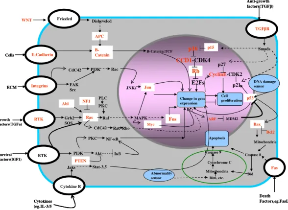
Figure 1.1. Multiple pathways of tumor suppressors and oncogenes are involved in the initiation and progression of cancer. The genes and pathways that are commonly involved in cellular signaling and are often involved in multiple cancer types are shown here. The commonly altered tumor suppressor genes and oncogenes are highlighted in red. (Figure adapted from Hanahan and Weinberg, 2000). This figure is incomplete in portraying the intricacy of cancer pathway circuitry. However it gives an idea of how complex this network is.
1.2 Cancer Cause and Statistics.
Cancer is a disease of abnormal proliferation within any cell type of the body. The list of cancers that affect human beings exceed a hundred different types of cancer, classified
p16
CCD1-CDK4
Rb
E2Fs
Change in gene expression Cell proliferation DNA damage sensor Apoptosis Abnormality sensor Frizzled E-Cadherin Integrins RTK Cytokine R p27 CyclinE-CDK2 p53 p21 Bax Mitochondria Caspase 8 Bid ARF MDM2 Bim, etc. Cytochrome C Caspase 9 WNT Disheveled APC Β
-Catenin Β-Catenin:TCF
Grb2 SOS
Ras Raf MAPK Fos
RTK PI3K Akt
PKC NF-κB FAK
Src
PLC PKC
CdC42 Rac Rho CdC42 PI3K Rac
JNKs Jun
Jaks Stat-3,5 IκB Cells
ECM
Growth Factors(TGFα)
Survival
Factors(IGF1) PTEN
Myc
TGFβR
Smads
p15
Anti-growth factors(TGFβ)
Fas Death Factors,eg.FasL Bcl2 Mitochondria Abl NF1 Cytokines (eg.IL-3/5 p16 CCD1-CDK4 Rb E2Fs
Change in gene expression Cell proliferation DNA damage sensor Apoptosis Abnormality sensor Frizzled E-Cadherin Integrins RTK Cytokine R p27 CyclinE-CDK2 p53 p21 Bax Mitochondria Caspase 8 Bid ARF MDM2 Bim, etc. Cytochrome C Caspase 9 WNT Disheveled APC Β
-Catenin Β-Catenin:TCF
Grb2 SOS
Ras Raf MAPK Fos
RTK PI3K Akt
PKC NF-κB FAK
Src
PLC PKC
CdC42 Rac Rho CdC42 PI3K Rac
JNKs Jun
Jaks Stat-3,5 IκB Cells
ECM
Growth Factors(TGFα)
Survival
Factors(IGF1) PTEN
Myc
TGFβR
Smads
p15
Anti-growth factors(TGFβ)
based on the cell type affected (American Cancer Society,
http://www.cancer.org/docroot/home/index.asp?promo=gaw, Weinberg, 2007). The causes of cancer are manifold and include life style factors like smoking or intake of tobacco products and alcohol (Ames, Gamble 1983, Hamajima et al. 2002), personal factors like obesity and diet (Ames 1983, Gridley et al. 1990), environmental factors (Augenlicht et al. 2002) and genetic factors like familial retinoblastoma (Leiderman, Kiss & Mukai 2007, MacPherson, Dyer 2007), etc. While the former factors can be somewhat controlled by altering one’s life style, it is impossible to control genetic factors that pre-dispose to cancer (Augenlicht et al. 2002, Bickers, Lowy 1989, Greenlee et al. 2000, Parkin, Pisani & Ferlay 1999, Peto 2001, Pisani et al. 1999, Preston-Martin et al. 1990). The focus of this work is to understand the molecular etiology of a sub type of familial breast cancer pre-disposed by inherited genetic mutations.
Cancer is staged clinically to gauge the extent of the disease at the time of diagnosis and to predict outcome (www.cancer.gov, (Greene, Sobin 2008). Various staging nomenclatures exist (like the TNM classification) but largely staging is usually done based on the following three criteria, (i) size of the primary tumor (T) at the time of diagnosis, (ii) amount of local advancement of the disease to surrounding tissues and lymph nodes (N) (iii) extent of distant metastasis (M), that is spreading of the cancer from primary site to distant secondary sites. Based on these criteria the cancer could be stage I (early and more curable) or stages III or IV that indicate metastatic cancer with a poor prognosis.
that is upregulated in these cancers. The focus of this research is to identify specific molecular mechanisms that are altered in a sub type of familial breast cancer with BRCA1 mutations that are currently not well understood.
1.3 Breast Cancer Types and Statistics
With more than 178,480 predicted new cases of invasive breast cancer in women in the United States estimated by the American Cancer Society in the year 2007 (accounting for about 7% of all cancer deaths), breast cancer is the second greatest threat for women in the Western world.
[http://www.cancer.org/docroot/STT/content/STT_1x_Cancer_Facts__Figures_2007.asp, (Desantis et al. 2008)]. Specific races around the world have showed greater incidence rates of breast cancer, for example the Ashkenazi Jewish population have a great susceptibility to familial breast cancers (Pereira et al. 2007, Spannuth, Thaker & Sood 2007), indicating the role of genetic modifiers in the etiology of this disease.
type and there is a great need for animal models that can accurately model distinct features of this disease. The current work focuses on developing and characterizing multiple mouse models to help understand some of the critical pathways that are deregulated in this type of breast cancer, which include the P53, BRCA1 and the RB pathways. In depth molecular characterization of this malignant cancer can potentially identify diagnostic markers that can be used to identify and treat them early on.
It is worth mentioning that mouse models provide an avenue to investigate the molecular mechanisms underlying the pathways involved in tumor evolution. But due to the heterogeneity of breast cancers, no single mouse model is able to depict all aspects of human cancer. Rather, mouse models seem to recapitulate certain key features of the human disease histopathologically and also in terms of the tumor “behavior” e.g. metastatic or non-metastatic. Engineered mouse models which incorporate genetic changes observed among human breast cancers more closely mimic their human counterparts with regard to histology and tumor progression than mouse tumors generated by older methods such as random viral insertion or carcinogen induction (Cardiff, Kenney 2007). Transcriptional profiling of mouse tumors shows that some mouse models more closely resemble ER+/PR+ adenocarcinomas while others share features of triple negative tumors, and confirm that none completely mimics the human pathology (Herschkowitz et al. 2007).
1.4 Landmarks of Cancer Research
egg by more than one sperm gave rise to cell death or the creation of an abnormal cell with aberrant chromosomal numbers. This led him to surmise that the individual chromosomes retain their “individuality” even after cell division occurs, in the daughter cells. So he proposed that aberrant chromosomal distribution might be the cause for abnormal cell behavior that ultimately results in cancer. His ideas gave rise to the earliest concept of “genetic instability” in cancer. In his book “The Origin of Malignant Tumors” published in 1914 he proposed many ideas regarding cancer, tumor progression and cell cycle that have since then been confirmed and accepted (discussed in http://en.wikipedia.org/wiki/Theodor_Boveri).
In 1971 Knudson proposed his “two hit hypothesis” from his study of the familial childhood cancer retinoblastoma (Knudson 1971). He observed that children born to parents with retinoblastoma do not always develop the disease themselves but may later have their children who develop retinoblastoma. From this observation he suggested that loss of single allele of a gene may pre-dispose to a specific cancer type but the cancer only occurs upon loss of the second wild-type allele (thus “second hit”). This hypothesis was also later confirmed to be true.
Figure 1.2. A timeline for cancer research is shown here. A timeline for some of the landmark historical discoveries that fuelled cancer research are shown here. Adapted from Balmain, 2001
It was my goal in this project to study the role of three major tumor suppressor genes,
RB, TP53 and BRCA1 in the molecular etiology of breast cancer. I used GEMs to study the
role of loss of each of these genes either alone or in combination, in the initiation and progression of breast cancer. This work has resulted in the generation of several mouse models of clinical importance that can be used in the future for drug targeting studies in breast cancer.
1.5 Biology of the Mammary Gland
The mammary gland is a branched secretory epithelium with a bilayered structure (Figure 1.3). The inner layer comprises the luminal mammary epithelium cells that are responsible for milk secretion. The outer layer is composed of the myoepithelial cells that provide scaffolding to the luminal epithelial cells as well as contract to help in the ejection of milk by the luminal epithelial cells. The myoepithelial cells have dual properties of muscle
Boveri identified
chromosome as the unit of heredity and proposed that chromosomal aberrations could result in cancer.
Knudson proposed the “two hit hypothesis”
Identification of P53 in a complex with SV40 viral protein
Cloning and identification of RB, the first tumor suppressor gene
Identification of P53 as a tumor suppressor gene and not an oncogene as it was earlier thought.
Identification of BRCA1 , the first familial breast cancer gene. 1902-1914 1971 1979 1986 1989 1994
The first draft of the human genome sequence is published
2001 Boveri
identified chromosome as the unit of heredity and proposed that chromosomal aberrations could result in cancer.
Knudson proposed the “two hit hypothesis”
Identification of P53 in a complex with SV40 viral protein
Cloning and identification of RB, the first tumor suppressor gene
Identification of P53 as a tumor suppressor gene and not an oncogene as it was earlier thought.
Identification of BRCA1 , the first familial breast cancer gene. 1902-1914 1971 1979 1986 1989 1994
The first draft of the human genome sequence is published
cells (and express smooth muscle actin) as well as epithelial cells (and express epithelial cytokeratins 5 and 14). Myoepithelial cells are thought to regulate cross talk between the luminal epithelial cells and the surrounding stroma by paracrine signaling pathways. Surrounding the mammary gland are the mammary stroma and fat pad.
Figure.1.3. A schematic representation of the components of the mammary gland is shown here. The mammary gland is composed of two cell types. The luminal epithelial cells are responsible for milk secretion. The outer myoepithelial cells are contractile in nature and help in milk secretion. The ductal structure is embedded in fibroblasts and mammary fat pad.
p53 mediated apoptosis clears the milk secreting luminal epithelial cells (Strange et al. 1992). In the second phase there occurs a collapse of the extensive ductal structures formed during late stage pregnancy and lactation, including break down of the mammary extra cellular matrix and basement membranes. The second phase is p53 independent (Medina 2005).
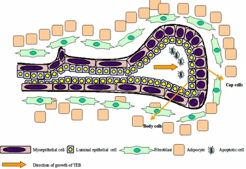
Figure 1.4. Structure of the mammary gland terminal end bud is shown here. The TEB of the mammary gland is composed of the leading edge of rapidly proliferating cap cells and the inner mass of more differentiated body cells. (Figure adapted from Lanigan et al. 2007).
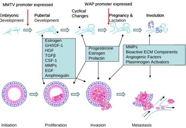
spurt of proliferation, anti-apoptotic signals, stromal invasion and angiogenesis. So it is not surprising that many of the signaling pathways involved in normal mammary gland development are hijacked by tumor cells to help in mammary tumor progression (Lanigan et al. 2007). Some of the pathways that are involved in mammary gland development and are also frequently altered in breast cancer are indicated in Figure 1.5.
Figure 1.5. Signaling pathways involved in the development of mammary gland at several stages is frequently altered in breast cancer. Hormones like Estrogen, Progesterone and Growth hormones play an important role in mammary gland proliferation and ductal morphogenesis during puberty. However overexpression of these hormones can lead to breast cancer and ER and PR are frequently overexpressed in breast cancers. Similarly, while matrix metalloproteases (MMPs) play a critical role in mammary gland remodeling during involution, overexpression of MMPs is frequently found in metastatic breast cancer. MMPs act like molecular scissors and help mammary epithelial cells invade through the stroma to distant sites. The various promoters used frequently in targeting transgenes and knockouts to the mammary gland are turned on at different stages of mammary gland development. The two most common promoters used are shown in this figure. Figure adapted from (Lanigan et al. 2007)
Cyclical Changes Embryonic Development Pubertal Development Pregnancy & Lactation Involution
Initiation Proliferation Invasion Metastasis
Estrogen GH/IGF-1 HGF TGFβ CSF-1 MMPs EGF Amphiregulin Progesterone Estrogen Prolactin MMPs
Bioactive ECM Components Angiogenic Factors
Plasminogen Activators
MMTV promoter expressed WAP promoter expressed
Cyclical Changes Embryonic Development Pubertal Development Pregnancy & Lactation Involution
Initiation Proliferation Invasion Metastasis
Estrogen GH/IGF-1 HGF TGFβ CSF-1 MMPs EGF Amphiregulin Progesterone Estrogen Prolactin MMPs
Bioactive ECM Components Angiogenic Factors Plasminogen Activators Embryonic Development Pubertal Development Pregnancy & Lactation Involution
Initiation Proliferation Invasion Metastasis
Estrogen GH/IGF-1 HGF TGFβ CSF-1 MMPs EGF Amphiregulin Progesterone Estrogen Prolactin MMPs
Bioactive ECM Components Angiogenic Factors
Plasminogen Activators
Tumor Virus (MMTV) promoter that is expressed earlier in development and does not require multiple cycles of pregnancy and lactation to be turned on (Wagner et al. 2001).
1.6 Breast Cancer Research – A History
Two common classifications of breast cancer are ductal and lobular carcinomas, which are based on morphological features shared by these tumor cells and their presumed cells of origin, either the milk ducts of the mammary gland or the mammary gland lobules, respectively. Although most human breast cancers are thought to be comprised of luminal epithelial cells and predicted to arise in the ductal compartments of the mammary gland, the cell of origin is impossible to determine at the final stage of the disease due to the great heterogeneity of the tumor. Depending on whether they are local or have spread to surrounding lymph nodes or other organs breast cancer is classified as “in situ” (local) or invasive. Progress in understanding this disease has been done by carrying out primarily “reverse engineering”, that is by trying to recreate those changes seen in patients presenting clinical forms of breast cancer, in either cell culture systems or animal models.
1.6.1 Cell culture studies of breast cancer
in a three dimensional mammary epithelial culture system that this signaling pathway acts to increase cellular proliferation and can promote cancer progression (Dillon et al. 2007, Dillon, White & Muller 2007). However, the caveat still is, that the three dimensional cultures use mammary cell lines like MCF-10A that have been grown and maintained for several years and have already accumulated unknown genetic changes. It has been a constant effort in the field to make the cell culture systems as “life like” as possible and current efforts are underway to use primary mammary epithelial cells for short-term three-dimensional cultures that would preserve the “in vivo” characteristics of the cells as much as possible. We have made significant progress in trying to establish primary mammary epithelial cells in three-dimensional culture. In this work we have shown that short term cultures with cells derived directly from the mouse mammary gland can indeed be grown in 3-D and these cells also express specific transgenes in a hormone dependant manner in culture. Such a system can be used as a high throughput assay for studying cooperating lesions without setting up multiple mouse crosses. This work will be reported in a later chapter in this thesis and the significant conclusions from it will be identified.
1.6.2 Mouse models of breast cancer
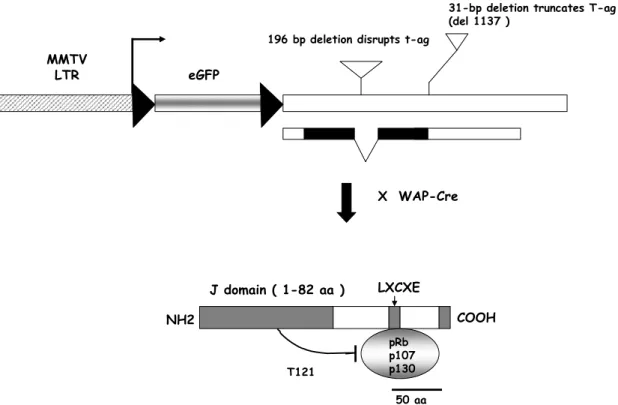
place in this spacer region leading to the precise removal of DNA in between the sites leaving a single loxP site behind (Weinberg, 2007, Van Dyke, Jacks 2002). By using this technology only those cells within the mammary gland that have undergone the recombination event will express the transgene of interest. It is possible by using the Cre-Lox-P technology to study many complex and compound genetic interactions that take place in a stochastic fashion over time and better reflect the human breast cancer scenario. We used this system to our advantage by generating the TgMMTV-Floxed-T121 (TgMFT121) (Figure 1.6) mice that expressed eGFP specifically in the mammary glands. Upon mating with mammary specific Cre strains (WAP or MMTV), excision of the eGFP cassette resulted in placing T121 expression directly under the control of the MMTV promoter. So while all cells
within the mammary gland that expressed the MMTV promoter showed eGFP expression prior to crossing with Cre strains. Upon Cre introduction, only a subset of cells within the mammary gland that expressed the WAP promoter also expressed T121, depending upon the
efficiency of Cre mediated recombination. This served two purposes. First, it allowed healthy and wild type mouse lines to be maintained in the colony that did not express T121 and could
be visually screened for strong transgene expression by the robustness of their eGFP expression. Second, it allowed somatic deletion of eGFP and expression of T121 in a specific
sub set of cells within the mammary gland that underwent the recombination event, that had not been possible in a similar model using the direct WAP-T121 transgene (Simin et al. 2004)
Figure1.6. MFT121 transgene design. The MMTV promoter drives expression of a floxed
eGFP cassette prior to crosses with mammary specific Cre strains. Upon introduction of a Cre strain (mammary or non mammary specific as indicated by WAP, beta Actin or AAV-Cre), eGFP is excised leading to the expression of T121 under the control of the MMTV
promoter in all cells that underwent recombination. The target cells that express T121would
depend on the expression of Cre. (Hua Wu in the Van Dyke Lab made the transgene).
Finally, GEMMs still have problems that need to be overcome to make them the most “ideal” system for cancer study. Some of these are the differences between mouse and human biology that make most mouse mammary tumors hormone independent while human tumors are often not. In addition, interspecies differences have made mouse mammary tumor metastasis patterns very different from human mammary tumors. While most human mammary tumors metastasize to the bones, brain and liver, most mouse tumors rarely metastasize and if they do, it is usually to the lungs. The reason for this is not clear and could be a fundamental difference in mouse and human mammary gland. Or it could be that a specific combination as well as sequence of events is necessary in a specific cell type within the mammary gland to see progression to metastasis. Modifier gene effects in the specific genetic background of mice used for these studies may also play a role (Balmain, Nagase
eGFP eGFP eGFP eGFP MMTV
MMTV MMTV
MMTV stopstopstopstop
LOXP LOXP LOXP
LOXP LOXPLOXPLOXPLOXP
LOXP
LOXPLOXP
LOXP
beta actin-CRE Recombinase
WAP-Cre or AAV-CRE
) n ( TTTTTTTT121121121121121121121121
MMTV
MMTVMMTV
1998). However it is clear that developing mouse models for breast cancer metastasis are the next barrier to be overcome to allow for good pre-clinical testing of anti-metastatic drugs. In this thesis we have described the establishment of a metastatic mouse model for breast cancer by layering on mutations within the mouse mammary gland, that occur with great frequency in human breast cancer.
1.6.3 Mammary specific promoters play an important role in mouse models of breast cancer
To target specific genetic events to a specific cell type in the mouse tissue specific promoters are required to drive expression of the gene of interest. Researchers have used several promoters to target gene expression specifically to the mouse mammary gland. The use of the Cre-Lox –P technology allows the narrowing down of gene expression or loss to a specific cell type by using a cell specific Cre recombinase. Each of these promoters has some advantages to their use, based on the scientific questions being asked. Some of the commonly used mouse mammary gland promoters and their specific advantages and disadvantages are listed below.
Whey Acidic Protein (WAP) promoter
cancer have been developed using the WAP promoter. The biggest advantage of this promoter is its expression is tightly regulated by mammary hormones and found to be present mainly in the mammary luminal epithelial cells. This prevents leaky expression of transgenes that may often pre-dispose to non-mammary phenotypes. WAP expression in the mammary gland is absent in virgin animals and peaks at around day one of lactation. Expression persists through day 10-post lactation and even after the mammary gland has undergone involution. WAP expression has also been reported in the brain but at negligible levels (Wagner et al. 1997).
The disadvantage of using the WAP promoter is the mice must undergo at least one (preferably multiple) cycles of pregnancy and lactation to turn on the transgene. So WAP promoters cannot be used to target virgin mammary epithelial cells. Also, hormonal regulation of this promoter can turn on other developmental cues that may play a role in the phenotype observed (Jonkers, Derksen 2007). This can be a confounding effect. Nevertheless, WAP has remained the promoter of choice for many mouse mammary gland researchers (Andres et al. 1987, Gallahan et al. 1996, Nielsen et al. 1991, Schoenenberger et al. 1988, Schulze-Garg et al. 2000, Simin et al. 2004) and is used in this study for restricting Cre expression to the mammary luminal epithelial cells.
Mouse Mammary Tumor Virus (MMTV) promoter
MMTV promoter are it shows strong expression in both the virgin and lactating mammary gland. Studies have detected expression of the MMTV promoter as early as 6 days post partum and strong expression in the entire mammary ductal tree is detected by 5 weeks in some mice (Wagner et al. 2001). The level of expression of MMTV peaks during pregnancy and lactation.
The disadvantage of the MMTV promoter is that it shows frequent leaky expression in other secretory tissue types, especially in the salivary glands, skin, hair follicles and seminal vesicles. The temporal and spatial expression pattern of this promoter seems to follow early developmental cues that are similar for several secretory organs like the mammary gland, hair follicles and salivary glands (Mikkola, Millar 2006, Wagner et al. 1997). This predisposes the mice to non-mammary gland tumors like salivary gland tumors. However, the MMTV promoter has been frequently used in mouse models of breast cancer due to its pre-dominant expression in both the virgin and lactating mammary glands (Guy et al. 1992, Tsukamoto et al. 1988).
In the current work, it was our goal to target T121 expression both in the virgin and
lactating mammary glands. For this we used the MMTV promoter to drive the floxed transgene expression. However, leaky expression of MMTV-Cre resulted in early lethality of the Rbf/p53 inactivated mice. This ruled out the use of the MMTV-Cre for follow up studies
and only WAP-Cre was used that expresses only in the lactating mammary epithelial cells and thus restricts T121 expression to these cells as well.
Keratin 14 Promoter
mammary myoepithelial cells. The myoepithelial (basal) like profile of human BRCA1 mutated familial breast cancers made it necessary to target this cell type in the mouse, in an effort to model this cancer sub type. To do this the Cytokeratin 14 promoter that targets the basal cells of many organs, including mammary gland, skin, lung epithelium, etc. was used. This promoter was successfully used to target the mammary myoepithelial cells and was been able to recapitulate some features of the human basal like breast cancer upon loss of
BRCA1 (Liu et al. 2007). The advantage of this promoter is it does not require pregnancy of
the mice to turn on expression of the gene of interest. The disadvantage is, this promoter is not mammary gland specific and the mice get a high percentage of non-mammary (particularly skin) tumors. Other keratin promoters that target specific cell types within the mammary gland like the Keratin 8/18 promoter to target the luminal epithelial cells and Keratin 5 promoter to target the mammary myoepithelial cells are currently being developed by many labs and may be used in the near future.
1.7 Microarray Classification of Breast Cancer
The advances in DNA microarray technology in breast cancer stratification have made it possible to predict the outcomes of the different classes of breast cancer. Instead of the older method of histological stratification of breast cancers that was often deceptive and did not reflect the true nature of the cancer (Bertucci et al. 2008), this method uses the molecular signature of each cancer subtype to predict its outcome (Bertucci et al. 2008). Using this technology Perou et al identified four broad categories of human breast cancer (Perou et al. 2000). They were either ER positive or ER negative. Among the ER positive tumors were the Luminal A type that resembled the normal mammary gland closely and had a good prognosis. Also in the ER positive tumors were the Luminal B tumors that had higher proliferation levels and were more aggressive. However both these sub types expressed the cell lineage markers keratin 8 and 18 that are normally expressed in the mammary luminal epithelial cells. These tumors also respond well to hormonal treatment by Tamoxifen. In the ER negative tumors, there are the Her2 (erb-B-2) positive tumors that respond well to Trastuzumab. The basal tumors are the second group within the ER negative tumors that do not express ER, PR or Her 2 and have very poor prognosis. These basal tumors were also found to express the mammary myoepithelial cell markers, keratins 5 and 14. The basal type tumors also show frequent mutations in BRCA1 and will be the focus of much of our studies in this thesis.
pathways. As Rb pathway inactivation leads to higher expression of E2F target proliferation genes this is a highly relevant result of mouse models reflecting human cancer biology. It also indicates that loss of RB pathway, P53 and BRCA1 may have a potential synergistic role in the promotion of familial human cancers. Also significant is similar profiles of cell lineage markers in mouse and human tumors when both sets of tumors are compared by array analysis. Both mouse and human basal-like tumors showed high expression of keratins 5, 14, 15, c-KIT and CRYAB. Some of these results were confirmed by immunohistochemistry, for example keratin 5 staining identified keratin 5 as marker for both human and mouse basal-like tumors. A subset of GEMMs display heterogeneous phenotypes and do not represent any single subtype of human tumor. However these mouse models may be useful for appreciating the scope of possible outcomes, and helpful for identifying new pathways involved in the biology of mouse mammary progression that may also play a key role in human mammary tumorigenesis. Some types of human breast cancer still remain very difficult to model in mice due to inherent mouse-human differences, like the ER + human cancers. The sporadic “basal-like” and BRCA1 mutated familial breast cancers have also been difficult to replicate in mice due to their unknown cell of origin and the possible complex genetic events necessary to generate these tumors. Several groups have tried to model this class of breast cancer, and some have been able to replicate certain key features (Liu et al. 2007).
specific combination of genetic lesions in the mouse models generated to further understand the etiology for breast cancer progression.
1.8 Genetic Instability and Cancer
Genetic instability in the germline of living organisms has been a common phenomenon since the earliest evolution of eukaryotic cells. According to Darwinian laws of evolution, cells that accrued mutations that conferred them better survival potential were carried on in the germline to create new organisms. However a great increase in genetic instability could lead to loss of viability of cells and therefore the organisms. Especially instability in somatic cells (cells that form specific tissues with the organism) results in the accumulation of mutations that can ultimately fuel cancer (discussed by Weinberg, 2007).
Mouse cells have much longer telomeres than human cells. Because of this mouse cells rarely undergo the “crisis” caused by complete telomere erosion in human cells. Also the much shorter lifespan of mice results in their rarely undergoing complete telomere erosion in a lifetime. So it has been suggested that the mouse genome is more stable than the human genome and does not easily undergo genetic instability (Artandi et al. 2000, Artandi et al. 2002).
Genetic instability is a common feature in human solid tumors (Pihan, Doxsey 2003). The cause for genetic instability is primarily damage of the fundamental genetic material within cells, DNA. DNA damage in cells can occur through multiple mechanisms. Chemically reactive molecules within the cells like reactive oxygen species can affect DNA bases. External agents like chemicals in consumed food products and environmental pollutants can cause slow but significant DNA damage. Radiation in any form can cause major DNA damage if given in large doses (Ayouaz et al. 2008). Besides damage to DNA structure chromosomal instability (CIN) within cells can also be created by alterations (increase or decrease) in their chromosome number, leading to aneuploidy (which means a deviation from the normal or euploid karyotype of a cell). This is caused by the missegregation of chromosomes due to an ineffective mitotic checkpoint control in the M phase of cell cycle. A large contribution to genomic instability and aneuploidy in human cancers is made by double strand break repair defects where damaged DNA is replicated without being repaired.
genome”) and BRCA1 that is an important DNA damage repair protein. P53 acts as a cell cycle checkpoint control protein at both the G1/S and G2/M phases. Cells that lack P53 display an elevated level of genetic instability (Chin et al. 1999, Sharpless et al. 2002). Similarly, BRCA1 functions by forcing cells that have undergone DNA double strand breaks to be repaired by the error-free Homologous Recombination Process (HR). In the absence of
BRCA1 cells are repaired by the error-prone Non Homologous End Joining Process (NHEJ)
that leads to the accumulation of mutations leading to widespread genetic instability and ultimately cancer (Deng 2006). An ongoing debate in the cancer community is whether genetic instability is the cause or the consequence of cancer. It is possible that random mutations resulting in the accumulation of genetic instability sets the stage for cancer progression. Support for this comes from studying early polyps in colorectal cancers that have abundant genetic instability both as aneuploidy as well as microsatellite instability (Bardi et al. 1997, Lengauer, Kinzler & Vogelstein 1997). This suggests that genetic instability may be a process that cancer cells that have undergone mutations acquire to get a growth advantage. However future work is required to find out if this is the case for all cancer types.
to mammary tumors generated by p53 loss alone. This gave us reason to believe that loss of
Brca1 in addition to the Rb1 pathway inactivation and loss of p53 in mice would lead to
genetic instability and result in mammary gland tumors that better emulate human tumors in numerous aspects. We show here that the loss of Rb1-family, p53 and Brca1 in the mammary gland results in widespread genetic instability along with deletions on chromosome 4 and 10. Significant instability is not observed in mice with loss of Rb1 and
p53 only. This finding further suggests that a weakened genome is more susceptible to the
development of aggressive cancers and genetic instability could be the cause for tumorigenesis.
1.9 Making a Good Pre-clinical Mouse Model
including complete penetrance, short latency, easy readout and mutations highly relevant to human cancer.
I will summarize next the roles played by the RB1 pathway, P53 and BRCA1 in human breast cancer and their potential synergistic effect in promoting breast cancer that we aim to model.
1.10 Common Genetic Alterations in Human Breast Cancer
1.10.1 The RB1 pathway is frequently mutated in human breast cancer.
suppressor protein that then induces cell cycle arrest and cell death. So RB1 mutations in many human cancers are also coupled with mutations/inactivation of P53 (Sage 2007, Simin et al. 2005).
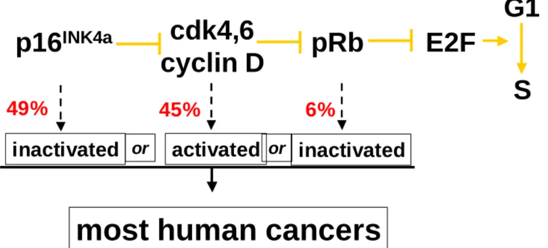
Figure 1.7. Common cancer causing mutations in the pRb pathway are shown here. Mutations in the whole pRb pathway including mutations to inactivate p16, activate cdk4.6 and inactivate pRb are very common in all human cancers. The frequency of these mutations in breast cancer is shown in red (Buckley et al. 1993, Geradts, Wilson 1996). These mutations result in abnormal cell proliferation and are very often coupled with mutations in p53, resulting in loss of cell cycle checkpoint control and decreased apoptosis.
1.10.2 Rb family proteins show functional overlap in multiple cell types.
Two proteins that are similar to pRb in structure and function, retinoblastoma-like 1 (Rbl-1, previously known as p107) and retinoblastoma-like 2 (Rbl-2, previously known as p130), have very similar binding properties to pRb and also bind E2F transcription factors (Mulligan, Jacks 1998, Mulligan, Wong & Jacks 1998). There is significant functional overlap between these three proteins, hence loss of all three of these genes may be required in cell types where all three are expressed, to elicit aberrant cell cycle progression and proliferation. Most human cancers including retinoblastoma do not exhibit mutations or loss in p107 and p130, though mutations in pRb are very common (MacPherson, Dyer 2007,
p16
INK4acdk4,6
cyclin D
pRb
E2F
G1
S
inactivated
oractivated
orinactivated
most human cancers
Scambia, Lovergine & Masciullo 2006). This suggests that the Rb family proteins may have distinct functions in different species. But it has been shown in mouse models that all three of the Rb family genes are expressed in the mammary gland and Rb1 alone is dispensable in normal mammary gland development (Maandag et al. 1994). So loss of pRB alone in the mouse does not result in mammary gland tumors, probably due to functional compensations (Robinson, Wagner & Hennighausen 2001).
1.10.3 pRb mutations in breast cancer
RB1 gene mutations have been reported in about 20-35 % breast cancers (Fung,
Simin et al showed for the first time that Rbf mutation in the mammary gland could induce an
apoptotic response from P53 on day one lactation. Subsequent tumor progression occurs through selective pressure for loss of p53 (Simin et al. 2004). However this model targeted the loss of Rbf in the entire mammary gland and not in a single cell or a subset of cells within the normal mammary gland. This is not similar to how human breast cancer begins in a single cell and progresses stochastically with the accumulation of further co-operating lesions. The possible co-operation between Rbf inactivation and p53 loss could not be studied in this model as most of the p53 germline inactivated mice (p53 knockout mice) developed non-mammary tumors (lymphomas and sarcomas). The tumor latency in this model was extremely long (median of ten months). To overcome these drawbacks we sought to induce somatic and conditional inactivation of both Rb pathway and p53 in the mammary gland.
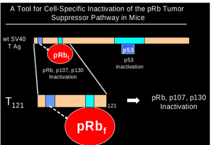
1.10.4 T121 specifically binds to and inactivates pRbf
shared by SV40 T antigen and human papilloma virus E7 (that causes human cervical cancer). This binding results in a conformational change in the pRb protein and prevents its binding to the E2F transcription factors.
Interestingly, SV40 T antigen also has a p53-binding domain and can specifically bind to and inactivate p53. The discovery of the mechanism by which SV40 T antigen functioned had a profound impact in understanding the cancers caused by these viruses. It also provided researchers with a powerful tool that they could now use to their advantage to specifically inactivate RB and P53 in multiple cells in the mouse and ask a variety of questions about their roles in tumorigenesis. We used this tool to our advantage by using a truncated version of the SV40 T antigen that only inactivates the pRBf but not P53. The
truncated T antigen referred to as T121 in the rest of this work, contains the first 121 amino
acid sequence of SV40 T antigen. By leaving an active P53 we were able to specifically study the role of pRBf inactivation in mammary tumor initiation. To specifically target the
mammary gland we used the MMTV promoter that expressed early in development in the mammary luminal epithelial cells (Wagner et al. 2001). To generate an inducible expression of T121 we inserted an eGFP cassette within floxed stop sequences (Figure 1.8). Upon
crossing to mammary specific Cre strains the bi transgenic mice would express T121 and
hence have functional pRBf inactivation in the mammary glands only. We also hoped by
using the MMTV promoter to uncouple T121 expression from regulation by pregnancy
Figure 1.8. The truncated SV40 T antigen binds to pRBf and inactivates it functionally.
The pRBf can be specifically inactivated by T121 that is the truncated (first 121 amino acids)
of the SV40 T Antigen. This molecule was originally reported by (Saenz Robles et al. 1994) and has since then been used to target many cell types in the mouse to study the loss of pRBf
inactivation in a cell specific manner (Hill et al. 2005a, Hill et al. 2005b, Lu et al. 2001, McLear et al. 2006, Simin et al. 2004, Simin et al. 2005, Xiao et al. 2002, Xiao et al. 2005).
1.11 The P53 Tumor Suppressor Gene is Frequently Mutated Concomitant to RB1 Pathway Mutations in Human Breast Cancer
P53 has been in the focus of cancer research ever since its discovery in 1979 (DeLeo
et al. 1979, Kress et al. 1979, Lane, Crawford 1979, Linzer, Levine 1979, Linzer, Maltzman & Levine 1979) as the transcription factor binding to SV40 large T-antigen (DeLeo et al., 1979, Lane and Crawford, 1979) and acting as a cell cycle checkpoint control gene. Almost 50% human cancers show P53 mutations alone or in combination with other gene mutations like RB1 (Soussi 2005). The p53 activating pathways (Figure 1.9) in a cell can be broadly classified under the following categories:
A Tool for Cell-Specific Inactivation of the pRb Tumor Suppressor Pathway in Mice
p53 inactivation pRb, p107, p130
Inactivation
wt SV40 T Ag
pRb, p107, p130 Inactivation
T121 121
pRbf
Figure 1.9. The signals activating p53 and their downstream effects are shown here. A variety of physiologic stress in the cell can induce p53 activation. The resulting p53 undergoes a variety of posttranslational modifications and induces many cellular responses. The response to cell cycle arrest can either be irreversible, called “senescence” or reversible and the cells can start proliferating again. In certain circumstances p53 can trigger apoptosis. (Adapted from Weinberg, 2007)
(i) p53 pathway activating signals, for example, DNA damaging agents like UV radiation, cellular hypoxia, cellular glucose starvation (Feng et al. 2005, Jones et al. 2005)
(ii) mediator signals upstream to p53 that sense the activation signals and as a result can increase or decrease the levels and functional state of the p53 protein in the cell (MDM2, COP-1, and PIRH-2)
(iv) downstream target genes of p53 activation like genes resulting in cell cycle (G1-S and G2-M) arrest (p21, GADD45), genes regulating apoptosis (Fas, Caspase 8,9.3, Bid, Bax, Noxa, Puma, Bcl-2, Bcl-XL and genes regulating cellular senescence
(v) effects of the downstream gene activation resulting in responses like cell cycle arrest, apoptosis and / or senescence (Levine, Hu & Feng 2006).
P53 mutations in cancer are often missense mutations (Jerry et al. 1993, Ozbun et al.
1993, Ozbun, Butel 1995) or loss of heterozygosity in the later more advanced stages of cancer progression. Frameshift mutations resulting in premature stop codons also occur, and more infrequently there occur a separate missense mutation on each allele. Human beings with the Li-Fraumeni syndrome have one copy of mutant P53 and another wild type copy of the gene. These people are predisposed to multiple cancers during their lifetime, including breast and ovarian cancer (Greenblatt et al. 1994, Hollstein et al. 1991, Hollstein et al. 1996, Malkin 1994a, Malkin 1994b, Malkin 1994c, Petitjean et al. 2007).
Several studies using both cell culture and mouse models have been able to recapitulate one or more features of specific types of human breast cancer that show P53 loss or mutation (Attardi, Jacks 1999, Blackburn et al. 2004, Blackburn, Jerry 2002, Derksen et al. 2006, Dittmer et al. 1993, Jerry et al. 1998, Jerry et al. 1999, Jerry et al. 2000, Jonkers et al. 2001, Koch et al. 2007, Kuperwasser et al. 2000, Lin et al. 2004a, Olive et al. 2004, Seluanov et al. 2001, Sigal, Rotter 2000, Zambetti et al. 1991). Studies in knockout mice have shown that mice with one mutant p53 allele have an increased risk of developing spontaneous tumors (Donehower et al. 1992) similar to the Li-Fraumeni patients. But these mice developed breast cancers at a very low frequency. Conditional mouse models were developed to target loss of p53 in the mammary gland alone. While loss of p53 alone pre-disposed to mammary adenocarcinomas, the very long latencies in these models suggested accumulation of additional lesions. But p53 loss in mouse mammary tumor models initiated by the loss or expression of other genes resulted in a drastic reduction in tumor latency. For example, it was shown that loss of p53 in conjunction with loss of E-Cadherin can recapitulate several features of human lobular breast cancer and the combined loss of p53 and
Brca1 can lead to human “basal like” cancers often associated with familial loss of BRCA1
(Liu et al. 2007). This suggested that loss of p53 had a role in the progression rather than initiation of breast cancer.
(Simin et al. 2004). The germline loss of p53 made these mice very prone to lymphomas and sarcomas and hence it was impossible to use this model to study the complete loss of p53 in mammary tumor progression. In this project it was our goal to study the effect of complete loss of p53 in a Rbf inactivated mammary tumor model and also study the co-operating role of Rbf, p53 and Brca1 in mammary tumorigenesis.
1.12 Loss of BRCA1 Combined with Loss of P53 and RBf Pre-disposes to Highly
Malignant Breast Cancers
general patients with this class of breast cancer have a poor diagnosis and rate of survival is low . Long term treatments with tamoxifen fail in these cancers as they are ER negative. Also, breast cancers arising from BRCA1 mutations tend to occur in younger women and early pregnancy is not a good predictor of good prognosis for this type of breast cancers as it is for some other types.
Figure 1.10. BRCA1 protein and its binding partners are shown here. The BRCA1 protein acts as a scaffolding molecule. It binds to and assembles other DNA repair proteins to form large protein complexes. These protein complexes can then help the repair of dsDNA breaks by homology directed repair (HDR). The loss of the binding partners of BRCA1 affects specific cell cycle checkpoint controls and also compromise homologous recombination and HDR. (Adapted from Weinberg, 2007)
Shen et al. 1998). However, the combined loss of Brca1 and p53 resulted in a wide spectrum of mammary tumors with varying latencies (Brodie et al. 2001, Brodie, Deng 2001). It was recently shown by Liu et al., 2007 that tumors with concomitant loss of Brca1 and p53 have much higher levels of genetic instability compared to those with loss of Brca1 or p53 alone. However, studies have shown that embryonic lethality induced by loss of Brca1 in mice is incompletely rescued by the loss of p53 (Xu et al. 2001). This suggests that loss of p53 alone is not sufficient for the survival of Brca1 mutated cells and other genetic alterations are most likely necessary for promoting tumor progression. Additional apoptotic pathway, possibly through the Fas ligand could be playing a role in the early lethality of these mice. We hypothesize that loss of the Rbf pathway that promotes increased proliferation by activation of E2F target genes, along with loss of p53 may be the two other genetic lesions necessary for a synergistic effect on the loss of Brca1 in promoting mammary tumorigenesis.