Development and Testing of a CBCT Educational Module for Dental
Providers in the US Navy
Martin E. Evers, Jr., DDS
A thesis submitted to the faculty of the University of North Carolina at Chapel Hill in partial fulfillment of the requirements for the degree of Master of Science in Oral and
Maxillofacial Radiology at the University of North Carolina School of Dentistry
Chapel Hill 2012
Approved by:
Enrique Platin
John B. Ludlow
ii Abstract
MARTIN E. EVERS, JR.: Development and Testing of a CBCT Educational Module for Dental Providers in the US Navy
(Under the direction of Enrique Platin)
Objectives: This project included the development of an educational module
addressing cone beam computed tomography (CBCT) basics, anatomy, and
interpretation of CBCT reconstructed multiplanar images containing pathology. The
subsequent objective was to test the efficacy of this educational tool.
Methods: 32 Navy dental providers completed a brief online demographic survey
and multiple-choice pretest evaluating their knowledge of CBCT. The web based
educational module was then made available to the cohort. After completing the
module, participants were given the link to the posttest and short feedback
questionnaire.
Results: 24 posttests and feedback questionnaires were completed. The data
demonstrated an increase in the number of correct responses between the pre and
posttests which was not statistically significant when comparing overall scores.
(75.2% (pretest) to 77.5% (posttest) [p=.379]). Interestingly, the results limited to
sectional scores provided different outcomes. The basics/anatomy sections
combined yielded a statistically significant change from 77.4% to 86.6% (p= .010).
iii
(Pretest=69.7% vs. Posttest=58.6% [p=.400] ) 100% of respondents agreed that
the module was helpful and met its objective.
Conclusions: The information presented in the CBCT basics and anatomy sections
of the module resulted in the participants’ increased performance on the posttest.
The decrease in performance on the interpretation section could be attributed to the
skill set required to interpret CBCT multiplanar images. Image interpretation is a skill
set that cannot be developed using a few examples of how disease is manifested
iv Dedication
v
Acknowledgments
A special “Thank You” to the members of my thesis committee: Dr. Platin, Dr.
vi
Table of Contents
Tables/Figures ... vii
Abbreviations ... viii
Introduction/Objectives ... 1
Use of Surveys ... 2
Educational Methods of Presentation ... 3
The Module ... 5
Platform ... 5
Content ... 6
CBCT Basics ... 6
Dental Applications ... 7
Normal Anatomy ... 8
Interpretation ... 8
Methods/Materials ... 10
Results ... 13
Demographic and CBCT Familiarity Survey ... 13
Pre and Posttest ... 13
Feedback ... 17
Discussion ... 18
Conclusions ... 21
Appendix 1-Pretest ... 22
Appendix 2-Posttest ... 32
vii
List of Tables/Figures
Table 1. Number of Participants ... 19
Figure 1. Control panel of WordPress® ... 6
Figure 2. Educational module’s Learning Objectives ... 12
Figure 3. Mean Test Scores ... 14
Figure 4. Posttest-Case 1 responses ... 15
Figure 5. Posttest-Case 1 part 2 responses ... 16
viii
List of Abbreviations
AEGD ... Advanced Education in General Dentistry
CBCT ... Cone Beam Computed Tomography
CT ... Computed Tomography
FOV ... Field of View
GPR ... General Practice Residency
IAN ... Inferior Alveolar Nerve
MPR ... Multi-Planar Reconstructions
MRI ... Magnetic Resonance Imaging
PGY-1 ... Post Graduate Year 1
Introduction/Objectives
In 1998 Mozzo, et al introduced Cone Beam Computed Tomography (CBCT)
for dental use, and in May 2001 CBCT it reached the U.S. market.1 Since that time,
this technology has revolutionized oral and maxillofacial imaging. Three dimensional
anatomical objects would no longer be limited to two dimensional visualization in the
field of dentistry. Previous technologies (e.g. stereoscopy, Tuned Aperture
CT[TACT]) had attempted to address this issue2; however, their popularity was
relatively short lived. Although conventional CT and MRI remain mainstays of
medical imaging, issues of dose (CT), cost, and accessibility limit their application in
dentistry. Still over a decade after its introduction, CBCT’s impact on the field of
dentistry continues to grow.3
The U.S. Navy has a group of general and specialty trained dental providers
educated at various dental schools. Collectively, this group’s familiarity with CBCT
imaging is very diverse. To improve the usefulness of this modality, an educational
module could provide Navy clinicians with some of the necessary basic tools to
understand this technology.
The aims of this project are as follows:
1. Development of an educational module addressing the basic principles of
2
CBCT, and an introduction to the interpretation skills required for differentiating
normal from abnormal findings.
2. Evaluate the usefulness of the module utilizing a pretest and posttest
study design.
A thorough review of the literature revealed only one previous study that
tested a web based CBCT educational module. This previous study concluded that
additional studies would be beneficial to further evaluate the efficacy of web based
modules. However, in contrast to the present study’s module, the module tested in
the previous study was limited to CBCT anatomy.5
The pre and posttest study design was chosen because of its simplicity and
according to the I-tech group from the University of Washington, to understand
exactly what knowledge can be credited to the training itself, using a pre- and
post-test methodology is important. In addition, a well-designed pre- and postpost-test can
help trainers understand which concepts were well taught during the training and
which ones need additional time, or need to be covered using alternative methods.6
It is also anticipated that the module will be modified and retested according to the
results of this study.
Use of Surveys
Surveys are excellent tools for acquiring data. They are inexpensive, useful
in describing the characteristics of a large population, can be administered from
remote locations, can be used on large samples, many questions can be asked
3
standardized questions make measurements more precise by enforcing uniform
definitions upon the participants. Also, standardization ensures that similar data can
be collected from groups then interpreted comparatively. Usually high reliability is
easy to obtain by presenting all subjects with a standardized stimulus and observer
subjectivity is greatly eliminated.
A limitation of utilizing surveys is the lack of control over the response rate by
the investigator. Despite this limitation it was decided to use a survey in data
collection for this study because of its many advantages.
Educational Methods of Presentation
The options considered to convey the desired information for this study were
traditional lecture, classroom computer assisted learning, distance education, and a
combination of the previously mentioned modalities, learning. Advantages to
E-learning include the flexibility of schedules and the E-learning pace. And, there is also
no need to travel (i.e. the classroom is brought to the student). Presently there are
many calls to move away from the traditional lecture to interactive computer learning
systems that allow students access to information when and where they need it.8
Therefore, online training and education (e-learning) based on information
technologies, especially through the Internet, will stimulate the teaching market to
the detriment of traditional teaching methods.9 Due to this trend in educational
methods, convenience for both the presenter and the students, accessibility to the
4
distance from the primary investigator the decision to present the material in a web
5 The Module
Platform
There are multiple software platforms to choose from when considering web
page design. The first attempt for this project was using Dreamweaver by Adobe.
This is an excellent product, but requires technical expertise beyond the scope of
this project’s designer. Alternatives include Joomla®, Drupal®, and WordPress® to
name a few.
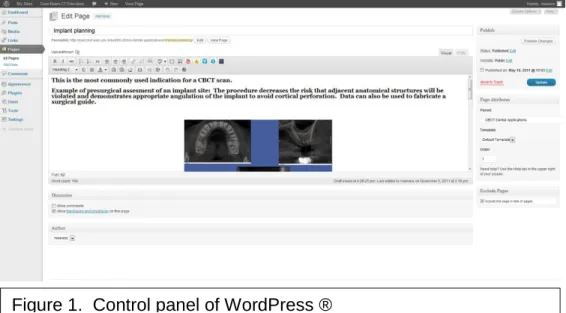
WordPress® is a free, user friendly web publishing software. Using this
technology one simply has to cut and paste desired content and the software applies
the prescribed themes with the click of a button. Plugins expand the functionality of
a variety of thematic templates. The control panel is straightforward and easy to
use. (Figure 1) Changes to the developing website are not executed by the software
until a proof is reviewed by the designer. The bottom line about the utility of this
6
Content
Determining what to include in the module was an issue of concern. If the
topic list for the CBCT module was all-inclusive, the model might be considered too
lengthy and participants in the study of the module’s efficacy would be less likely to
participate due to time constraints. After a review of the literature and consideration
of the purpose of the module, four topic areas were selected—CBCT Basics, Dental
Applications, Normal Anatomy, and Interpretation.
CBCT Basics
When introducing CBCT technology much of the literature compares this
modality to that of conventional CT. This gives the reader a fixed reference with
which he/she can compare and contrast CBCT. Conventional CT is a more familiar
modality due to the relative age of the technology. Patient positioning during image
7
market-- each constructed with one of the various patient position options—seated,
standing, or supine. Each of these positions has its advantages and disadvantages.
Also specific to the manufacturer’s and model’s specifications is the field of view or
the area of image visualization. These range in size from <5 cm to 22 cm,2,4 and
some models are capable of multiple FOVs with collimation. Different diagnostic
tasks require distinct FOVs. The basic views presented in image viewing software
are the MPR or Multi-Planar Reconstructions. These include an axial, coronal, and
sagittal planar images.
Dental Applications
The purpose of including the dental applications section was to inform the
target audience of the many capabilities of the technology. So often we focus on the
areas of our specialty. This section demonstrates the versatility of CBCT. From
implant treatment planning to TMJ evaluation to orthodontic evaluation, CBCT
images can benefit each of these disciplines and many more. This section
contained case examples of all the common dental applications for this modality,
specifically, implant imaging and treatment planning, evaluation of developmental
abnormalities, evaluation of 3rd molar/IAN canal relationship, pathology, trauma,
TMJ evaluation, orthodontic evaluation, airway analysis, and periapical and
periodontal findings. It should be noted that there are other applications not listed.
8
Normal Anatomy
As our ability to recognize abnormal findings in diagnostic imaging is
dependent upon our knowledge of normal anatomy, this section was emphasized in
both the educational module and the tools used to evaluate it. Without this basic
knowledge of normal anatomy, interpretation of the images is not possible. While
this section did not contain an all-inclusive list of anatomical features of the
craniofacial region, the structures presented can serve as landmarks in navigating
through a CBCT volume. While viewing each image’s highlighted structure, one can
also visualize the adjacent anatomy and get an overall sense of normal anatomy as
it appears in CBCT volumetric images. In future versions of the module, it is
planned to make this section more interactive. This upgrade requires further
development.
Interpretation
The basic principles for interpretation were presented in this section of the module with the following guides:
Know normal anatomy- remembering that normal is a range.
Symmetry- comparing structures bilaterally
Radiographic Signs- density, shape, borders, etc.
Independence of radiographic signs regardless of imaging modality- a
well-defined radiolucency in a panoramic radiograph will present similarly in CBCT images.
The systematic image/lesion description methods were also discussed. Being able
9
terminology is an asset of great value. This can provide a significant service to the
patient and facilitate treatment decisions by the providers.
Also included in this portion of the module were examples of each of the following pathological categories:
Cyst
Benign Neoplasia
Malignant Neoplasia
Inflammatory Lesion
Benign Fibro-Osseous Lesion
Vascular Anomaly
Systemic Disease
Trauma
Developmental Abnormality4
Knowledge of specific pathological entities within these categories is also helpful, but
10
Methods/Materials
For the initial testing of the educational module thirty three individuals
associated with various Navy Post Graduate Year (PGY-1) programs were invited to
participate. Email was the communication medium selected to correspond with the
invitees. This group of potential participants included residents and directors from
both the Advanced Education in General Dentistry/General Practice Residency
(AEGD/GPR) programs. It was decided to invite these individuals because of their
previously expressed interest in the study. Prior to publishing the survey, the pretest
was submitted for review to three imaging educational experts. Their feedback
resulted in modification of the survey/pretest document. Shortly thereafter, an
additional email communication to the study participants provided a link to the
demographic survey/pretest. The participating locations included Portsmouth, VA;
Great Lakes, IL; and San Diego, CA. Thirty two of the thirty three Navy dental
providers completed the brief online demographic survey and multiple-choice
pre-test evaluating their knowledge of CBCT. The web based educational module was
subsequently made available to them. After completing the module, participants
were given the link to the post-test and short feedback questionnaire. After three
reminder emails spanning a two month period, the final tally revealed that twenty
11
All the data collection steps utilized Qualtrics (Provo, UT) software. The
demographic survey consisted of ten multiple choice questions encompassing topics
of gender, age, previous dental and CBCT training, and CBCT utilization. The
pretest contained twenty multiple choice questions regarding CBCT basics, common
dental applications, normal anatomy as seen in CBCT images, and interpretation of
CBCT images. Cases for interpretation for both the pre and posttest were selected
from the teaching files at the University of North Carolina at Chapel Hill School of
Dentistry. The posttest included twenty multiple choice questions, which like the
pretest questions, were focused on testing the participants’ knowledge of the topics
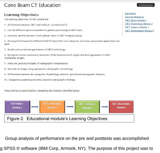
presented in the “Learning Objectives” of the module. (Figure 2) While the posttest
questions were similar in content and form to the pretest questions, they were a
different question set designed to be comparable in level of difficulty. The feedback
questionnaire consisted of two 5 point scale questions and two open answer/text
input questions. These questions were designed to solicit feedback which will aid in
12
Group analysis of performance on the pre and posttests was accomplished
using SPSS ® software (IBM Corp, Armonk, NY). The purpose of this project was to
evaluate the educational module, and according to findings presented at the
National Health Science Curriculum Conference, group analysis is able to
appropriately identify the strengths and weaknesses of instruction, targeting areas
for improvement.10,11 The paired t-test was used to analyze the data. Significance
level was set at .05.
13 Results
Demographic and CBCT Familiarity Survey
There was a nearly 4:1 male to female ratio of participants in this study. 70%
of those surveyed were under the age of 30, and 85% graduated from dental school
less than one year prior to participating in the study. The respondents received their
dental school training at 22 different dental schools. It was reported that 8 of the 22
dental schools offered some level of CBCT training as part of the dental school
curriculum. 100% of those participating are general dentists. 85% of those
surveyed currently do not view any CBCT images in their practice/educational
program. For those viewing CBCT scans the most reported indication for the
imaging procedure was to evaluate pathology. The most reported frequency of
viewing scans was 1-5 per week.
Pre and Posttests
The mean pretest score was 75.2. The posttest scores were slightly
improved with a mean of 77.5. A paired t-test was used to compare the mean
values. While the scores trended upward, the overall difference between the pre
14 Figure 3. Mean Test Scores
Sorting the results by question type yielded significant results. The CBCT
basics and anatomy sections resulted in a statistically significant increase in correct
responses comparing the pre vs. posttest scores (77.4% to 86.6% [p= .010]).
However, in the interpretation section there was a decrease in performance (p=
.400). One possible explanation for the decrease in performance is that the cases
selected for the posttest were more difficult.
It was also anticipated that those who reported having received previous
training would have scored better on the pretest than their colleagues that had not
received previous training in the CBCT imaging modality. While they did score
slightly higher on the pretest (76.8 vs. 74.2), this increased level of performance was
not statistically significant (p=.305). 0 10 20 30 40 50 60 70 80 90 100
Basics/Anatomy Interpretation Overall
Pretest
15
As previously noted, the most difficult area of the tests for the study
population was the interpretation section. Multiple questions on the posttest
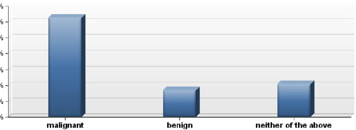
exemplified this trend. In Case 1 for example, where the participants were asked to
distinguish between neoplasia (both malignant and benign) and another possible
etiology for the patient’s condition. The majority of respondents concluded that it
was a malignancy, although the correct cause was trauma. (Figures 4 and 5)
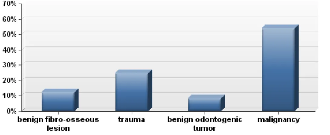
Another example that proved difficult for this cohort is Case 2, also of the posttest,
were only 17 percent of the participants answered correctly when asked to identify
the most likely pathological category based on the radiographic signs. The majority
responded that it was a benign neoplasia. (Figure 6) The correct response was
inflammation.
16 Figure 5. Posttest-Case 1 part 2 responses
17 Feedback
The responses from the participants were unanimously positive. Using a five
point scale ( 1=strongly agree, 2=agree, 3=neither agree or disagree, 4=disagree,
5=strongly disagree), 50% of the participants responded “1” and the other 50%
responded “2” when asked if the module was helpful to them. When queried if the
module adequately addressed all the learning objectives, the responses were 27%
18 Discussion
When asked about additional information to add to the module, responses
included more information on anatomy and interpretation and specific dental
applications, specifically implant treatment planning, endodontic treatment planning
and caries diagnosis. One respondent desired additional information on current
CBCT manufacturers and models. Suggestions for improvement of the module were
also well received. These responses included such topics as more in depth
information and making the module more interactive. There was also a request for a
database to include normal and pathologic findings from different imaging modalities
to be used as a reference for Navy dentists. While incorporating these ideas into the
educational module would be useful, one must consider the resources (e.g. server
space) and the purpose of the module which is not to train radiologists but to provide
basic information regarding CBCT to Navy providers and give them tools to
recognize abnormalities in these images and make the appropriate treatment
decisions and/or referrals.
The attrition levels or non-completion rate by location (Table 1) reveal
interesting findings. The total (pre and posttest) completion rate for the cohort was
75%. The Midwest group had the highest number of participants completing the
pretest (twelve), but only seven individuals completed the posttest. The east coast
group had only one individual not complete both tests, and the west coast group had
19 Table 1: Number of Participants
Location Pretest Posttest Change
Portsmouth, VA 11 10 -1
Great Lakes, IL 12 7 -5
San Diego, CA 9 7 -2
Total 32 24 -8
Prior to the last 3 respondents of the posttest, in the anatomy section for
example, the entire cohort had responded correctly to 5 of the 8 questions—an
overall correct response average of 94%. After the results of the last 3 participants
were tabulated, the overall anatomy score dropped to 90%. A possible cause for the
abrupt decline in performance by the last of the respondents is that when repeatedly
encouraged to complete the posttests, they did so hastily just to be finished with the
task. Additional evidence that supports this theory is the fact that in the open
ended/text input portion of the feedback survey none of the last 3 respondents had
input and their surveys were completed 2 weeks after the rest of the group.
Some observers may conclude that because of the limited variability of the
study population’s demographics, they do not represent an appropriate sample.
That is, all of the participants were general dentists with the majority lacking
experience and training, having recently graduated from dental school. An
alternative perspective and the position held by the primary investigator of this study
20
module. The majority of participants were either AEGD or GPR residents currently
involved in the academic process--eager and ready to learn.
Although proven useful, there are limitations to the pre and posttest study
design, especially when the tests are administered remotely online and at the leisure
of the study population. As the participants are not isolated during the time between
tests, there is no guarantee that the intervention is solely responsible for the
21 Conclusions
While it would be difficult to generalize conclusions for this study with such a
small sample size, we consider meaningful the following:
The module did prove useful in the fact that the overall mean scores for the
cohort trended upward.
The CBCT basics and anatomy sections of the module effectively imparted
knowledge to the participants.
According to the feedback of the group, the module was helpful and
adequately addressed the learning objectives.
The Navy will have an educational tool that interested providers will be able to
access as a reference. This will allow them to improve their knowledge of this useful imaging modality--CBCT.
Although not specifically tested, we can infer that the interpretation of CBCT
images is not mastered after a single educational intervention.
We look forward to further development and testing of the module. Future
experiments will include testing the module--not only comparing the results of pre
and posttests but also comparing the test results between groups—traditional lecture
vs. web based instruction. This continued effort will ultimately result in an improved
22
Appendix 1-Pretest
Please answer the following survey questions A - J, followed by the Pretest questions 1 - 20. A. By answering yes you are consenting to participate in this study. The module, pre and post tests will be accessible online. The only
identifiable information collected will be an email address and will only be viewed by the PI and the electronic survey provider.
A.
Yes No
B. Gender
Male Female
C. Age
<26 26-30 31-35 36-40 >40
D. In what year did you graduate from dental school?
Prior to 1985 1986-1995 1996-2005 2006-2010 2011
23
F. If you received post doctoral/specialty training, please indicate your specialty/program. Please enter none if appropriate.
G. Have you received previous CBCT education/training?
Yes No
H. In what manner did you receive this education/training?
Dental School Curriculum CE course
Personal Study Residency Not applicable
I. How many CBCT scans do you view per week?
None 1-5 6-10
More than 10
J. For what purpose are the scans ordered? (check all that apply)
Pathology
Implant Planning Trauma
Pre-surgical extractions Orthodontics
Periodontics Endodontics
24
PRE TEST:1. Read the following two statements and select the best
answer. Statement a: A small field of view CBCT scan is adequate for all patients’ comprehensive orthodontic treatment planning. Statement b: A CBCT scan must be acquired on all patients prior to orthodontic treatment planning.
Both statements are true Both statements are false
Statement a is true; statement b is false Statement b is true; statement a is false
2. Which of the following includes the patient positions utilized in CBCT imaging?
supine, prone, seated prone, seated, standing supine, seated, standing prone and seated only
3. Which of the following imaging techniques is most useful in detecting interproximal caries?
CBCT panoramic bitewing occlusal
4. Read the following two statements and select the best answer. Statement a: CBCT is utilized to evaluate impacted teeth. Statement b: CBCT is the imaging modality of choice to distinguish between the various soft tissues of the maxillofacial area.
25
5. All of the following are components of CBCT multi-planar views except
26
Normal Anatomy- Use the images above to answer questions #6 - 13. Please select the anatomic structure indicated by the arrow.
6.
ethmoid air cell frontal sinus nasal cavity nasolacrimal duct
7.
nasal septum pterygoid plate hard palate vomer
8.
maxillary sinus sphenoid sinus oral cavity frontal sinus
9.
condyle
malar process coronoid process
27 10.
cerebellum pons orbit
pineal gland
11.
maxillary sinus sphenoid sinus ethmoid sinus frontal sinus
12.
maxillary sinus sphenoid sinus ethmoid sinus frontal sinus
13.
nasopharyngeal tonsil adenoid
28
Case 1- Use the images above to answer questions #14 and 15. 54 y/o asymptomatic male, teeth #21 and 22 are vital.
14. The lesion above is most consistent with a ___________________ process.
malignant benign
none of the above
15. Which is the category of disease most suggested by the radiographic and clinical findings?
benign fibro-osseous lesion cyst
29
Case 2 - Use the image above to answer questions #16 and 17.
16. The lesion indicated be the arrows is found in which bone?
maxilla hyoid mandible
none of the above
17. This lesion is most likely a ________________ process.
30
Case 3 - Use the image above to answer questions #18 - 20.
18. This lesion can be described as a _______________.
well defined, partially corticated radiolucency ill defined, non corticated radiolucency
a mixed density lesion none of the above
19. Read the following two statements and select the best answer.Statement a: In accordance with the radiographic findings above, a malignancy would not be
expected. Statement b: Inflammation and malignancy often have similar radiographic findings.
31
20. One would anticipate that this disease process would present _____________.
bilaterally unilaterally
32
Appendix 2-Posttest
1. A small field of view CBCT scan may be appropriate for all of the following applications except:
endodontics periodontics
orthodontic treatment planning
single unit implant treatment planning
2. Which of the following is not a patient position used in CBCT image acquisition?
supine prone seated standing
3. Read the following two statements and select the best answer: a. CBCT is the modality of choice for detecting interproximal caries. b. CBCT scans are frequently utilized for implant treatment planning.
Both statements are true Statement a is true, b is false Statement b is true, a is false Both statements are false
4. Read the following two statements and select the best answer: a. Medical CT produces more scatter than CBCT. b. CBCT scans are higher dose than
conventional CT.
33
5. Read the following two statements and select the best answer: a. The ethmoid sinuses are located posterior to the sphenoid sinus. b. As evident in traditional panoramic imaging, a patient has two hyoid bones.
34
Normal Anatomy- Use the images above to answer questions #6 - 13. Please select the anatomic structure indicated by the arrow.
6.
nasal septum inferior turbinate nasolacrimal gland none of the above
7.
condyle
malar process coronoid process
ossified stylohyoid ligament
8.
mastoid process vomer
cruciform ligament pterygoid plate
9.
mandibular condyle malar process coronoid process
ossified stylohyoid ligament
10.
35 11.
sublingual gland submandibular fossa pterygoid plate lingual foramen
12.
maxillary sinus sphenoid sinus orbit
foramen magnum
13.
36
Case 1- Use the image above to answer questions #14 and 15.
14. The lesion above is most consistent with a __________________ tumor.
malignant benign
37
15. Which is the category of disease most suggested by the radiographic and clinical findings?
benign fibro-osseous lesion trauma
benign odontogenic tumor malignancy
Case 2 - Use the image above to answer questions #16 and 17.
16. The lesion indicated be the arrow is found in which bone?
maxilla hyoid mandible
38 17. This most appropriate pathologic category is:.
benign tumor inflammation cyst
39
Case 3 - Use the images above to answer questions #18 - 20.
18. This lesion can be described as a _______________.
well defined, partially corticated radiolucency ill defined, non corticated radiolucency
40
19. Based on the radiographic signs, which pathological category can be ruled out?
cyst
benign tumor systemic condition all of the above
20. One would anticipate that this disease process would present _____________.
bilaterally unilaterally
as a generalized condition throughout both jaws none of the above
Feedback questions:
A. The CBCT module is helpful.
Strongly Agree Agree
Neither Agree nor Disagree Disagree
Strongly Disagree
B. The module appropriately addressed all of the learning objectives.
Strongly Agree Agree
Neither Agree nor Disagree Disagree
Strongly Disagree
Please list any CBCT subtopics in which you desire additional information:
41 References
1. Mozzo P, Procacci C, Tacconi A, Martini PT, Andreis IA. A new volumetric CT machine for dental imaging based on the cone-beam technique: preliminary results. Eur Radiol 1998;8(9):1558-64.
2. Scarfe W,C. What is cone-beam CT and how does it work? Dent Clin N Am 2008;52:707-730.
3. Miles D. The future of dental and maxillofacial imaging. Dent Clin N Am 2008;52:917-28.
4. White S, Pharoah M. Oral Radilogy: Principles and Interpretation. Sixth ed. St. Louis, MO:Mosby Elseviewr: 2009.
5. Al-Rawi WT, Jacobs R, Hassan BA, Sanderink G, Scarfe WC. Evaluation of web-based instruction for anatomical interpretation in maxillofacial cone beam
computed tomography. Dentomaxillofac Radiol 2007; 36:459-464.
6. I-TECH. Guidelines for pre- and post-testing: a technical implementation guide. University of Washington, 2008.
7. Advantages and Disadvantages of the Survey Method. [Internet]. Available from: http://writing.colostate.edu/guides/research/survey/com2d1.cfm
8. Edlich RF. My last lecture. J Emer Med 1993; 11(6):771-774.
9. Pinto A, Selvaggi S, Sicignano G, Vollono E, Iervolino L, Amato F, et al. E-learning tools for education: regulatory aspects, current applications in radiology and future prospects. Radiol Med 2008; 113:144-157.
10. Metz K. Benefits of online courses. Techniques 2010;9:20-23.
11. National Health Science Curriculum Conference. 2010 [Internet] Available from: http://www.healthscienceconsortium.org/docs/assessment_success.pdf
12. Asadoorian J, Batty HP. An evidence-based model of effective self-assessment for directing professional learning. J Dent Educ 2005; 69:1315-1323.
13. Eaton KA, Reynolds PA. Continuing professional development and ICT: target practice. Br Dent J 2008; 205:89-93.
42
15. Goldman S. The Educational Kanban: promoting effective self-directed adult learning in medical education. Acad Med 2009; 84:927-934.
16. Johnson LA. Continuing dental education on the World Wide Web. Dent Clin North Am 2002; 46:589-604
17. Perryer G, Walmsley AD, Barclay CW, Shaw L, Smith AJ. Development and evaluation of a stand-alone web-based CAL program. A case study. Eur J Dent Educ 2000; 4:118-123.
18. Ryder MI, Sargent P, Perry D. Evolution and revolution: the curriculum reform process at UCSF. J Dent Educ 2008; 72:1516-1530.
19. Welk A, Splieth C, Wierinck E, Gilpatrick RO, Meyer G. Computer-assisted learning and simulation systems in dentistry--a challenge to society. Int J Comput Dent 2006; 9:253-265.
20. Wenzel A, Gotfredsen E. Students' attitudes towards and use of
computer-assisted learning in oral radiology over a 10-year period. Dentomaxillofac Radiol 1997; 26:132-136.