Single-cell transcriptomics reveals gene
expression dynamics of human fetal kidney
development
Mazène Hochane1, Patrick R. van den BergID1, Xueying Fan2☯, Noe´mie Be´renger-CurriasID1☯, Esme´e AdegeestID1☯, Monika Bialecka2, Maaike Nieveen2,
Maarten MenschaartID1, Susana M. Chuva de Sousa LopesID2,3‡*, Stefan SemrauID1‡*
1 Leiden Institute of Physics, Leiden University, Leiden, The Netherlands, 2 Department of Anatomy and Embryology, Leiden University Medical Center, Leiden, The Netherlands, 3 Department of Reproductive Medicine, Ghent University Hospital, Ghent, Belgium
☯These authors contributed equally to this work.
‡ These authors share equal senior authorship on this work.
*[email protected](SMCSL);[email protected](SS)
Abstract
The current understanding of mammalian kidney development is largely based on mouse models. Recent landmark studies revealed pervasive differences in renal embryogenesis between mouse and human. The scarcity of detailed gene expression data in humans there-fore hampers a thorough understanding of human kidney development and the possible developmental origin of kidney diseases. In this paper, we present a single-cell transcrip-tomics study of the human fetal kidney. We identified 22 cell types and a host of marker genes. Comparison of samples from different developmental ages revealed continuous gene expression changes in podocytes. To demonstrate the usefulness of our data set, we explored the heterogeneity of the nephrogenic niche, localized podocyte precursors, and confirmed disease-associated marker genes. With close to 18,000 renal cells from five dif-ferent developmental ages, this study provides a rich resource for the elucidation of human kidney development, easily accessible through an interactive web application.
Author summary
Regenerative medicine offers an exciting avenue to potential cures of kidney disease. However, a detailed knowledge of the structure and embryonic development of the kidney is crucial, both for stimulating regeneration in the body and for growing healthy kidney tissue in a dish. Most of such knowledge has been obtained from mice, whose develop-ment differs in crucial ways from that of humans. We therefore studied the composition of human fetal kidney tissue from five developmental ages, using a technique that can measure gene expression in individual cells. Our measurements revealed 22 distinguish-able cell types, some of which we localized in the tissue by fluorescence microscopy. We found several subpopulations of nephron progenitors—cells that give rise to the nephron, the functional unit of the kidney. Our study also focused on the development of
a1111111111 a1111111111 a1111111111 a1111111111 a1111111111 OPEN ACCESS
Citation: Hochane M, van den Berg PR, Fan X,
Be´renger-Currias N, Adegeest E, Bialecka M, et al. (2019) Single-cell transcriptomics reveals gene expression dynamics of human fetal kidney development. PLoS Biol 17(2): e3000152.https:// doi.org/10.1371/journal.pbio.3000152
Academic Editor: Emma Rawlins, University of
Cambridge, UNITED KINGDOM
Published: February 21, 2019
Copyright:©2019 Hochane et al. This is an open access article distributed under the terms of the Creative Commons Attribution License, which permits unrestricted use, distribution, and reproduction in any medium, provided the original author and source are credited.
Data Availability Statement: The single-cell
RNA-seq data has been deposited in the GEO database under accession number GSE114530. An interactive web application accompanying this paper, which provides convenient access to the data, can be found here:http://www.semraulab. com/kidney.
Funding: M. H., P. v.d. B., N. B.-C. and S.S. were
podocytes, a cell type that is crucial for the filtration function of the kidney, and our results might inform attempts to recreate these cells in a dish. We hope that our data set, made conveniently accessible through a web application, will help scientists develop new regen-erative medicine approaches to kidney disease.
Introduction
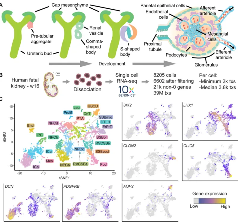
Mammalian kidney development initiates in the intermediate mesoderm through crosstalk between the metanephric mesenchyme (MM) and the ureteric bud (UB). The UB originates from the nephric duct, invades the MM, and starts to subdivide progressively into multiple ramifications. The UB tip cells, which make the first contact with the MM, become enveloped by an assembly of mesenchymal cells, the cap mesenchyme (CM) (Fig 1AandS1 Fig). The CM contains nephron progenitor cells (NPCs), which give rise to the whole nephron epithelium through tightly regulated morphogenic transformations [1]. Self-renewal of (mouse) NPCs is governed by key transcription factors, such asSix2andMeox1, which mark the nephrogenic zone of the kidney [2]. Signaling between UB tip cells and NPCs regulates the balance between self-renewal and differentiation of NPCs [3]. In humans, about 1 million nephrons are pro-duced before the NPC population is irrevocably exhausted a few weeks before birth [4]. During nephrogenesis, NPCs undergo mesenchymal-epithelial transition and differentiate into a suc-cession of intermediate structures: the pretubular aggregate (PTA), renal vesicle (RV), and comma- and s-shaped body (CSB and SSB, respectively). Then, via the capillary loop stage, mature and functional glomerular and tubular structures are eventually formed. In contrast to the nephron epithelium, the collecting duct system originates from the UB. Glial cell-derived neurotrophic factor/ RET proto-oncogene (GDNF/RET) signaling between the UB and CM critically regulates proliferation of UB tip cells and branching morphogenesis of the UB [5]. Stromal cells—such as interstitial cells (ICs), mesangial cells, juxtaglomerular cells, smooth muscle cells, fibroblasts, and pericytes—derive from a common interstitial progenitor [6,7]. Finally, vascular endothelial cells and the highly specified glomerular endothelium originate from the MM [8], and leukocytes and erythrocytes enter with the blood stream. The current understanding of mammalian kidney development is largely based on mouse studies, although it is clear that human and mouse kidneys are morphologically different. Three recent land-mark studies have revealed, in great detail, a significant divergence between mouse and human renal embryogenesis in terms of morphology as well as gene expression [9–11]. These studies underline that the prevailing lack of data on human kidney development severely hinders the detailed understanding of human kidney development and possible developmental origins of kidney disease.
In the study described here, we used single-cell RNA sequencing (scRNA-seq) to study gene expression dynamics in human fetal kidney development. Analysis of a fetal kidney from week 16 (w16) of gestation revealed 22 cell types, which we identified by known marker genes. Pseudotime analysis clarified their temporal relationship. We further defined specifi-cally expressed cell type marker genes of which many have not been implied in kidney devel-opment. Comparison to four additional samples (from w9, w11, w13, and w18) suggested that most cell types have a constant expression pattern, with the notable exception of podo-cytes. To highlight two ways in which our data set can be interrogated, we then explored the nephrogenic niche and the development of podocytes. Gene expression differences between four NPC clusters were related to spatial heterogeneity by immunostaining and single-mole-cule fluorescence in-situ hybridization (smFISH). Expression of the disease-associated gene was supported by the China Scholarship Council
(www.cscscholarships.net, 201307040026). M.B. and S.M.C.S.L. were supported by an European Research council Consolidator Grant (https://erc. europa.eu/, ERC-CoG-2016-725722). We further acknowledge support from Generade (https://www. hsleiden.nl/generade, project number G2016-14). This work was carried out on the Dutch national e-infrastructure with the support of SURF Foundation. The funders had no role in study design, data collection and analysis, decision to publish, or preparation of the manuscript.
Competing interests: The authors have declared
that no competing interests exist.
Abbreviations: AUROC, area under the ROC; BMP,
bone morphogenetic protein; CM, cap
UNCXwas localized to NPCs and their early derivatives. Finally, we focused on podocyte development, which proceeds via a distinct precursor state. By immunostaining and smFISH,
Fig 1. Single-cell transcriptomics identified 22 unique cell types in the human fetal kidney. (A) Schematic of kidney epithelium development. (B) Overview of the
scRNAseq experiment. (C) Top left: 2D tSNE map of 6,602 human fetal kidney cells. Colors and labels indicate the assigned cell type. (Other panels) tSNE maps
indicating expression ofSIX2,LHX1,CLDN2,CLIC5,DCN,PDGFRB, andAQP2. Expression is indicated by color; expression values of 1 are plotted in gray. The
numerical data underlying this figure can be found inS1 Data. CnT, connecting tubule; DTLH, distal tubule/loop of Henle; End, endothelial cells; ErPrT, early proximal
tubule; ICa, interstitial cells a; ICb, interstitial cells b; IPC, interstitial progenitor cell; Leu, leukocyte; Mes, mesangial cell; NPCa, nephron progenitor cells a; NPCb, nephron progenitor cells b; NPCc, nephron progenitor cells c; NPCd, nephron progenitor cells d; Pod, podocyte; Prolif, proliferating cells; PTA, pretubular aggregate; RVCSBa, renal vesicle/comma-shaped body a; RVCSBb, renal vesicle/comma-shaped body b; scRNA seq, single-cell RNA sequencing; SSBm/d, s-shaped body medial/ distal; SSBpod, s-shaped body podocyte precursor cells; SSBpr, s-shaped body proximal precursor cells; tSNE, t-distributed stochastic neighbor embedding; tx, transcript; UBCD, ureteric bud/collecting duct; w16, week 16.
we localized these precursors in situ and confirmed the disease-associated geneOLFM3as a marker.
Results
Clustering and identification of cell types
We performed single-cell transcriptomics on a human fetal kidney from w16 of gestation, equivalent to 14 weeks of development (Fig 1B). After data pruning and stringent removal of cells affected by stress (S2 Fig, Methods), 6,602 cells were retained for further analysis. Clusters of cells were identified by hierarchical clustering after k-nearest neighbor smoothing [12]. We assigned cell types to these clusters by expression of marker genes from the literature on mouse kidney development. The studies that linked the genes of this literature set to particular cell types are referenced inS1 Table. After merging similar clusters (S3 Fig, Methods), we obtained 22 cell types (S4 Fig) and visualized the single-cell transcriptomes in a two-dimen-sional t-distributed stochastic neighbor embedding (tSNE) map [13] (Fig 1C).
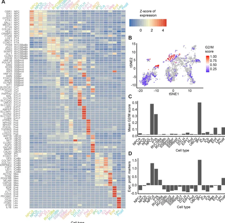
The mean expression levels of the literature set genes showed clear differences between cell types (Fig 2A). NPCs, which were distributed over four distinct clusters (NPCa–d), were marked by the established markersSIX2(Fig 1C),CITED1,MEOX1, andEYA1. Expression of these progenitor markers was highest in NPCa, which we hence considered “bona fide” self-renewing NPCs. NPCb showed lower levels ofCITED1andSALL1and higher levels ofGDNF
andHES1compared to the other NPC clusters.HES1, a transcription factor downstream of NOTCH signaling, is important for further renal cell differentiation. Compared to the other NPC clusters, NPCc showed higher expression ofCRABP2, which is related to retinoic acid signaling [14]. NPCd exhibited lowOSR1,CITED1, andMEOX1expression and increased lev-els ofLEF1, a known indicator of NPC induction towards differentiation. Compared to the other NPC subtypes, NPCd were also marked by a larger fraction of cells in G2/M-phase of the cell cycle (Fig 2B and 2C) and a higher expression of proliferation markers (Fig 2D), which indicated faster proliferation. We will discuss the relationship between the various NPC clus-ters in more detail below (see Heterogeneity in the nephrogenic niche).
Nephrogenesis continues with the creation of the pre-tubular aggregate (PTA) cells, which in turn develops into the RV and CSB. In our data, PTA cells were identified based on high expression ofLHX1(Fig 1C),JAG1,WNT4, andCCND1. Because RV and CSB are mainly dis-tinguishable by morphology, cells belonging to these two structures were grouped in our anal-ysis (RVCSB). RVCSB cells were marked by the same genes as PTA cells, but they appeared to proliferate less (Fig 2B–2D). Furthermore, they expressed markers reflecting more advanced regional patterning, which allowed us to discriminate between two subtypes (a and b). RVCSBa had a higher expression of genes that were recently associated with the distal RV (SFRP2,DLL1,LHX1), whereas RVCSBb expressed genes that indicate the proximal RV (CDH6,FOXC2,MAFB,CLDN1,WT1).
Fig 2. Known markers elucidated the cell types corresponding to each cluster. (A) Heatmap of literature set gene expression in the 22 identified cell types. Expression
was Freeman-Tukey transformed, averaged over all cells in a cluster and standardized gene-wise. (B) tSNE map of all cells with color indicating the G2/M score (calculated by the Cyclone tool [15]). This score reflects the likelihood that a cell is in G2/M phase. (C) G2/M scores from panel B averaged over the cells in each cell
type. (D) Mean expression of proliferation markers [16] (Z-scores) per cell type. The numerical data underlying this figure can be found inS1 Data. CnT, connecting
tubule; DTLH, distal tubule/loop of Henle; End, endothelial cells; ErPrT, early proximal tubule; G2/M, cell cycle phase G2/M; ICa, interstitial cells a; ICb, interstitial cells b; IPC, interstitial progenitor cells; Leu, leukocytes; Mes, mesangial cells; NPCa, nephron progenitor cells a; NPCb, nephron progenitor cells b; NPCc, nephron progenior cells c; NPCd, nephron progenitor cells d; Pod, podocyte; Prolif, proliferating cells; PTA, pretubular aggregate; RVCSBa, renal vesicle/comma-shaped body a; RVCSBb, renal vesicle/comma-shaped body b; scRNAseq, single cell RNA sequencing; SSBm/d, s-shaped body medial/distal; SSBpod, s-shaped body podocyte precursor cells; SSBpr, s-shaped body proximal precursor cells; tSNE, t-distributed stochastic neighbor embedding; UBCD, ureteric bud/collecting duct.
low expression ofPAPPA2andMALand the absence ofCDH6andHNF1A. Cell types that are known to develop from the SSBm/d were found together in one cluster (distal tubule/loop of Henle [DTLH]). This cluster showed high expression of the distal markersMAL,CLCN5,
SLC12A3, andPOU3F3, which are specific to the distal tubule, as well asSLC12A1,PAPPA2, andUMOD, which are found in the loop of Henle. Finally, cells that likely gave rise to podo-cytes, SSBpod, clustered separately. These cells showed high expression ofMAFBandFOXC2, both transcription factors necessary for the development of podocyte identity, and low levels of the mature podocytes markersCLIC5(Fig 1C),PTPRO,NPHS1, andNPHS2. This cluster also showed the highest expression ofOLFM3, previously identified as a specific marker of podocyte precursors residing in the visceral part of the proximal segment of the SSB. In con-trast to SSBpod, podocytes (Pods) showed higher expression of mature podocyte markers and lower levels ofMAFB. Differences between SSBpod and Pods will be studied in more detail below (see Podocyte development). Because of the high similarity in gene expression between SSB and capillary loop stage, we could not exclude that the SSB clusters also contained cells from the capillary loop stage.
Cells of the connecting tubule (CnT), which connects the distal tubule to the collecting duct, could also be identified in the data. They shared markers with the collecting duct (such asALDH1A1,TACSTD2, andCDH1), distal tubule (SOX9,POU3F3), and UB (RET,KRT8,
KRT18,MMP7). Cells of the UB and collecting duct (UBCD) were strongly marked by well-known genes likeAQP2(Fig 1C),CALB1,KRT8,KRT18,RET, andGATA2, found in the col-lecting duct as well as the stalk and tip of the UB.
The developing nephrons are surrounded by interstitial tissue, a separate lineage that origi-nates in interstitial progenitor cells (IPCs). We identified IPCs by coexpression ofFOXD1and
GDNF. These cells also expressed lower levels of markers known to be found in more mature cells likePDGFRAfor ICs orPDGFRB(Fig 1C) andACTA2for mesangial cells. We identified two subtypes of ICs (a and b), which were similar in their marker gene profile. Compared to IPCs, they lackedFOXD1and expressed less (ICa) or no (ICb)GDNF. ICa showed high levels ofFGF7, which has been localized to the renal fibroblasts or stroma surrounding the ureter and the collecting system. ICa also showed high levels ofTPM2andACTA2, markers of smooth muscle-like cells. ICb, on the other hand, expressed genes likeDCN(Fig 1C),DES,
SERPINE2, andCOL3A1, which are known to mark cortical stromal cells. Endothelial cells were identified by markers such asKDRandTEK, whereas leukocytes showed many specifi-cally expressed genes, such asCD37orCD48. Finally, one cluster of cells (Prolif) had a higher expression of proliferation markers compared to most other cell types (Fig 2D) but lacked dis-cernible cell type markers.
Developmental flow
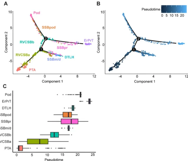
distal cell fates (Fig 3A). This might indicate that some of these cells preceded the RVCSBb, whereas others were primed to develop into distal fates. RVCSBb cells, however, only appeared after branch point 1, which confirmed that they were likely progenitors of proximal cell fates. On three separate branches, SSBm/d preceded DTLH, SSBpr preceded ErPrT, and SSBpod preceded Pods, which confirmed the identity of the SSB clusters. The temporal relationship of the NPC subtypes will be discussed in detail below (see Heterogeneity in the nephrogenic niche).
Comparison with existing single-cell transcriptomics data
To further confirm the interpretation of the cell clusters, we wanted to compare our data with an existing single-cell transcriptomics study of a w17 fetal kidney by Lindstro¨m and colleagues [19]. To that end, we first corrected for batch effects, using a method based on matching mutual nearest neighbors in the two data sets [20]. After correction, the two data sets showed a
Fig 3. Pseudotime analysis clarified the developmental relationship of the cell clusters. (A) Two-dimensional embedding (with the
DDRTreealgorithm [18]) of all w16 kidney cells, calculated byMonocle 2[17]. The graph learned by the algorithm is shown as a black
line. Colors and labels indicate cell types. (B) Same embedding as in panel A. Color indicates pseudotime calculated byMonocle 2. (C)
Box plots of cell type distribution over pseudotime. The numerical data underlying this figure can be found inS1 Data. DTLH, distal
tubule/loop of Henle; ErPrT, early proximal tubule; Pod, podocyte; PTA, pretubular aggregate; RVCSBa, renal vesicle/comma-shaped body a; RVCSBb, renal vesicle/comma-shaped body b; SSBm/d, s-shaped body medial/distal; SSBpod, s-shaped body podocyte precursor cells; SSBpr, s-shaped body proximal precursor cells; w16, week 16.
large degree of overlap (S5A Fig). This allowed us to use the cell types found by Lindstro¨m and colleagues to classify the cell clusters found here, using a k-nearest neighbors approach (see Methods). NPCa–c were also classified as NPC by Lindstro¨m and colleagues, whereas NPCb were considered “primed NPC,” which supports the notion that NPCb were primed to differ-entiate. The NPCd cluster was classified as “proliferating cells.” This classification is in agree-ment with our observation that NPCd seemed to proliferate more than other NPC subtypes (Fig 2B–2D). Because NPCd expressed low levels of NPC markers (such asSIX2andCITED1), these cells were likely in a transition state between NPCs and PTA cells. Whereas the majority of PTA cells identified here were considered “PTA/RV I” by Lindstro¨m and colleagues, RVCSBa cells were spread over multiple cell types. This spread was likely due to the fact that transitory cell types are transcriptionally similar, and their clustering is therefore less robust. Nevertheless, the “PTA/RV II” cluster received most of the RVCSBa cells. RVCSBb cells were called “podocyte precursors” in the Lindstro¨m data set, whereas SSBpod as well as Pods were classified as “podocytes.” In our data set, RVCSBb directly preceded SSBpod (Fig 3A), so they could indeed be considered podocyte progenitors. Below, we will show that SSBpod did form a cell state separate from Pods and should not be grouped with them (see Podocyte develop-ment). In agreement with our analysis, the majority of SSBpr were classified as “proximal pre-cursor” or “proximal tubule,” and all ErPrT were considered “proximal tubule” by Lindstro¨m and colleagues. CnT and DTLH were both classified as “distal/loop of Henle (LOH) precur-sor.” The fact that two cell types in the study by Lindstro¨m and colleagues (“podocytes” and “distal/LOH precursor”) were split in multiple subclusters in our study likely reflects differ-ences in sample preparation. Whereas Lindstro¨m and colleagues preferentially released single cells from the nephrogenic niche, here, the whole kidney was used. Consequently, the Lind-stro¨m data set has a finer resolution of NPCs and early, proliferating cell types, whereas our data set allowed us to resolve more mature cell types. The two data sets therefore complement each other.
Marker identification
To confirm the inferred cell types and also identify novel markers, we pursued two comple-mentary strategies. First, we determined a set of marker genes based on their usefulness as clas-sifiers for individual cell types: for each gene, the performance of a binary classifier was evaluated by the area under the receiver operating characteristic (AUROC) and combined with expression level filtering (seeMethods). This resulted in 88 marker genes (S6A Fig, marker set,S3 Table). Only 11 of these markers overlapped with the 89 genes in the literature set (S6C Fig). To our knowledge, many of the remaining markers had not been associated with kidney development in previous studies. As an independent approach, we used theKeyGenes
algorithm [21] to identify classifier genes among the 500 most highly variable genes (HVGs), using two-thirds of all cells as a training set. Based on the classifier genes determined by Key-Genes, we next predicted the cell types of the remaining one-third of the cells (test set). Cell types could be predicted with an average certainty (id score) of 0.59; 24% of the cells in the test set obtained an id score higher than 0.8. Of the 95 classifier genes (S6B Fig,KeyGenes set,S3 Table), 24 were the same as in the marker set, and 14 were common with the literature set (S6C Fig).
label a particular cell type in the human fetal kidney were coexpressed in the same subset of mouse cells. A few markers, however, were either ubiquitously expressed or almost completely absent. This might be due to interspecies differences.
Comparison of different developmental ages
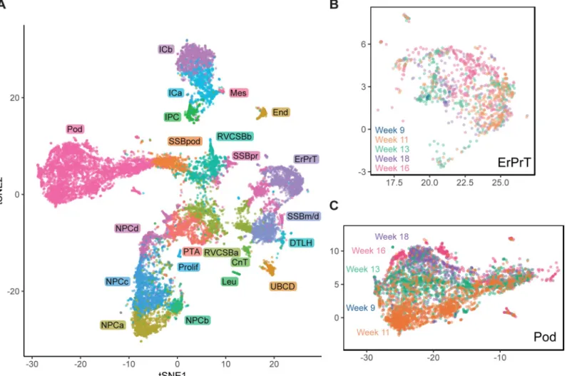
By establishing the identity of cell clusters at w16, we obtained a snapshot of cell type diversity in the fetal kidney. To explore whether the identified expression patterns change dynamically throughout development, we analyzed four additional samples from different developmental ages (w9, w11, w13, and w18), which together contained 11,359 usable cells. Using, again, batch correction based on mutual nearest neighbors [20], we visualized all samples in a com-mon tSNE map (Fig 4,S8 Fig). Overall, gene expression in the different samples was largely
Fig 4. Comparison of different developmental ages suggested continued expression changes in podocytes. (A) tSNE map combining all five samples (w9, w11, w13,
w16, w18). Samples were corrected for batch effects by matching mutual nearest neighbors [20]. Cells in the w9, w11, w13, and w18 samples were classified by
comparing to the w16 sample using a k-nearest neighbors-based approach (seeMethods). (B) tSNE map of all ages restricted to ErPrT. Labels and colors indicate ages.
Six outlier cells were omitted from this plot to improve visualization. (C) tSNE map of all ages restricted to Pods. Labels and colors indicate ages. The numerical data
underlying this figure can be found inS1 Data. CnT, connecting tubule; DTLH, distal tubule/loop of Henle; End, endothelial cells; ErPrT, early proximal tubule; ICa,
interstitial cells a; ICb, interstitial cells b; IPC, interstitial progenitor cell; Leu, leukocyte; Mes, mesangial cells; NPCa, nephron progenitor cells a; NPCb, nephron progenitor cells b; NPCc, nephron progenitor cells c; NPCd, nephron progenitor cells d; Pod, podocyte; Prolif, proliferating cells; PTA, pretubular aggregate; RVCSBa, renal vesicle/comma-shaped body a; RVCSBb, renal vesicle/comma-shaped body b; scRNAseq, single-cell RNA sequencing; SSBm/d, s-shaped body medial/distal; SSBpod, s-shaped body podocyte precursor cells; SSBpr, s-shaped body proximal precursor cells; tSNE, t-distributed stochastic neighbor embedding; tx, transcript; UBCD, ureteric bud/collecting duct; w16, week 16.
overlapping for the majority of cell types. For example, proximal tubules cells (ErPrT) appeared at the same positions in the tSNE map in all samples (Fig 4B). The position of Pods, however, shifted systematically across different ages, which corresponds to a continuing change in expression pattern (Fig 4C). This observation might suggest that Pods further matured in terms of their expression pattern after being specified.
Differential expression analysis of Pods of different ages revealed 109 differentially expressed genes (fold change>2 in any comparison, false discovery rate (FDR)<0.05,S4 Table). Func-tional annotation analysis of these genes showed significant enrichment of two gene ontology (GO) terms—“proteinaceous extracellular matrix” (adjustedp-value = 1.9×10⁻³, including
SPON2,BGN,COL1A2, andCTGF) and “extracellular exosomes” (adjustedp-value = 1.4×
10−3, includingNPNT,S100A10,ANXA1, andEPCAM). Some of the differentially expressed genes have been shown to be important for kidney development. For example,NPNTandDCN
showed increasing expression from w11 to w18. Knockout of the extracellular matrix protein NPNT in mice decreases the invasion of the UB and causes agenesis or hypoplasia [23].NPNT
was further shown to be expressed in the glomerular basement membrane and to be necessary for podocyte adhesion in mice [24]. Ablation of this gene in mice causes podocyte effacement. As in the case ofNPNT,DCNhas been reported to be part of the glomerular basement mem-brane proteins [25]. This gene appeared strongly up-regulated in podocytes between w11 and w13 or w18 (fold changes of 3.25 and 4.6, respectively). The increase ofNPNTandDCN expres-sion over time in our data set could reflect an increase in adheexpres-sion between podocytes and glo-merular basement membrane. Pods further showed significant differential expressions of genes related to stress, likeHSPA1Aand HSPA1BorNFKBgenes (NFKB2,NFKBIA, andREL), with the highest levels at w18. This might suggest that dissociation-related stress increases with age for podocytes, maybe related to stronger adhesion of the cells, or that stress-related genes have another, physiological role in development.
We would like to emphasize that the observed gene expression changes with age should be considered with caution because they might be related to the differences in genotype between the samples. A much larger number of samples would be necessary to rule out such interindi-vidual differences as a cause.
Having established the identity of the cell clusters, we next wanted to demonstrate how the data set can be used to explore different aspects of kidney development. We specifically focused on the nephrogenic niche, which showed pronounced heterogeneity, and the develop-ment of podocytes, which progressed via a distinct, intermediate cell state (SSBpod).
Heterogeneity in the nephrogenic niche
The formation of the nephron epithelium starts with the NPCs that differentiate and form the PTA, RV, and CSB. Studies in the mouse suggest that cells in the NPC compartment are not biased towards a particular lineage and patterning is first detectable in the PTA [26]. Neverthe-less, the w16 scRNA-seq data indicated the presence of several nephron progenitor subpopula-tions, NPCa–d. To clarify the temporal relationship of these clusters, we employedMonocle 2
again to arrange them together with the PTA cells on a pseudotime scale (Fig 5A). NPCa clearly preceded NPCb and c, which seemed to appear around the same pseudotime. NPCd cells followed NPCb and c and preceded PTA. This analysis suggested that NPCa are the “bona fide” NPCs and give rise to NPCb and c. NPCd, which were likely more proliferative than the other NPCs (Fig 2B–2D), seemed to be an intermediate state between (slowly cycling) NPCa–c and the PTA.
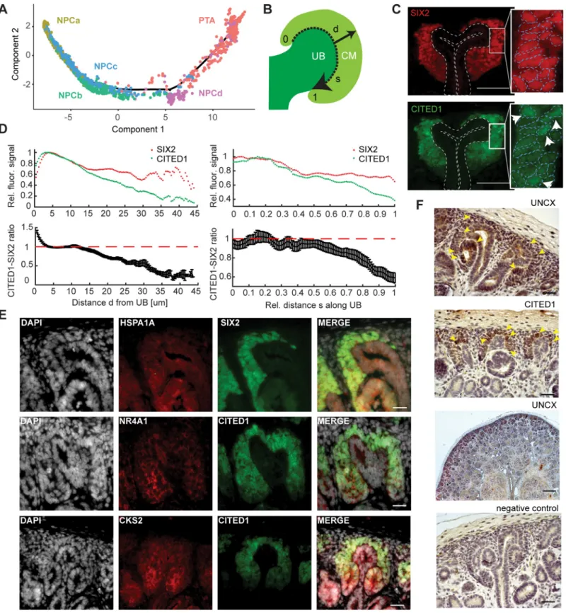
Fig 5. The nephrogenic niche exhibited a complex spatial organization. (A) Pseudotime analysis of the nephrogenic niche (NPC) and the PTA. Two-dimensional
DDRTree[18] embedding and the learned graph (shown as a black line) were calculated withMonocle 2[17]. Labels and colors indicate cell types. (B) Schematic sketch
of the CM indicating the distancedfrom the UB to the edge of the CM (solid arrow) and the relative distancesalong the UB (dashed arrow), in which 0 and 1 represent
the top and bottom of the CM, respectively. (C) Representative image of SIX2 and CITED1 immunostaining in a w15 human fetal kidney. Dashed lines in the insets
indicate the outline of the nuclei, based on DAPI signal. Arrows in the inset point to cells in which CITED1 is concentrated in the nucleus. Scale bar = 50μm. (D)
levels of these markers, NPCb and NPCd had lostCITED1almost completely, while retaining someSIX2expression. In an immunostaining of a w15 kidney, CITED1 and SIX2 appeared overlapping in a subset of cells (Fig 5B–5D). Quantification of the fluorescence signal (Meth-ods) revealed clear differences between their expression patterns. Whereas SIX2 expression was approximately constant throughout the CM, CITED1 expression decreased, relative to SIX2, with increasing (radial) distance from the UB (Fig 5D). A marked drop of CITED1 was visible between 10 and 20μm from the UB, which approximately corresponds to the first layer of cells. To exclude that the observed difference between SIX2 and CITED1 expression was due to the different fluorophores on the secondary antibodies, we repeated the experiment with swapped fluorophores. This measurement produced a very similar expression gradient (S9A Fig). To exclude that the observed effect was influenced by PTA found in the CM towards the stalk of the UB, the analysis was repeated, taking only the 20% of CM cells closest to the edge of the cortex into account. A similar expression gradient was observed (S9C Fig). This result implies the existence of a CITED1 low/SIX2 high subpopulation of cells, which are not in contact with the UB. Secondly, we observed that CITED1 decreased relative to SIX2 towards the interface with the PTA and the stalk of the UB (Fig 5D). A similar observation was made when the experiment was repeated with swapped fluorophores (S9B Fig). Taken together, these results suggested that NPCa and NPCc were located closer to the surface of the UB and closer to the tip of the UB compared to the other NPC subtypes. Additionally, we also observed differences in subcellular localization of CITED1 protein within the CITED1 high compart-ment. Whereas for the majority of cells CITED1 was found in the cytoplasm, in several cells it was concentrated in the nucleus (right inset inFig 5C). In contrast, SIX2 was always found restricted to the nucleus (left inset inFig 5C). This observation might indicate that CITED1 was only active in a small population of cells, which would constitute another layer of cell–cell heterogeneity.
In addition to the observed heterogeneity inCITED1andSIX2, differences between the NPC clusters could also be gleaned from the set of novel markers (marker set,S3 Table).
TMEM100andRSPO3specifically marked NPCa.RSPO3is an activator of the canonical WNT signaling pathway [27], suggesting a role of WNT either in NPC self-renewal or UB branching morphogenesis. Notably, all markers of NPCb (CACYBP,MRPL18,ZFAND2A,
DNAJB1) were related to the stress response in some form. The markers of NPCc (CRABP2,
HAS2,MDK,HOXC6), which were also expressed in the other NPC types, are all either targets of retinoic acid or binding it [14,28,29].MDKhas been shown to be expressed in the CM of the developing rat kidney, and its neutralization reduced the number of formed nephrons in vitro [30,31]. Finally, the NPCd markers CENPF, HMGB2, CCNB2, and NUSAP1 all have a role in cell cycle regulation or proliferation [16]. HMGB2 was recently implied in the activa-tion of quiescent adult neural stem cells [32].
The observation that markers of NPCb were related to the stress response seemed to suggest that this cluster was created as an artefact of cell dissociation [33], despite our best efforts to remove stressed cells (seeMethods). On the other hand, the vast majority of NPCb cells were classified as “primed NPC II” in the Lindstro¨m data set (S5 Fig). The fact that NPCb cells were only detected in the w16 and w18 kidneys is consistent with single-cell dissociation becoming increasingly difficult with fetal age, or alternatively, with a progenitor cell aging phenomenon.
calculated over all evaluated profiles (n= 24). (E) Representative image of HSPA1A, NR4A1, and CKS2 immunostaining in a w15 human fetal kidney. Scale
bar = 20μm. (F) Representative image of UNCX and CITED1 immunostaining. Arrowheads indicate the presence of immunostaining signal. Scale bar = 100μm. The
numerical data underlying this figure can be found inS1 Data. CM, cap mesenchyme; NPC, nephron progenitor cell; PTA, pretubular aggregate; SEM, standard error of
the mean; UB, ureteric bud; w15, week 15.
To explore the differences between NPCb and the other NPC clusters further, we immunos-tained HSPA1A and NR4A1, both known stress-response genes, in w15 kidney sections (Fig 5E).HSPA1Awas identified as a marker of NPCb (S9D Fig,S3 Table), whereasNR4A1was expressed in multiple NPC clusters but highest in NPCb (S9D Fig). Furthermore,NR4A1was also identified in the study by Adam and colleagues [22] to be up-regulated in response to ele-vated temperatures during enzymatic dissociation. HSPA1A and NR4A1 were both observable in the nephrogenic niche at the level of the stalk of the UB and at the transition to the PTA or RV. Additionally, we studied the expression ofEGR1, another stress-related gene that marked NPCb, with smFISH (S9E Fig).EGR1was mainly found toward the stalk of the UB and in a few cells around the tip of the UB, whereasSIX2andCITED1transcripts were visible through-out the CM (S9F Fig). Because results obtained in fixed tissue sections are not confounded by dissociation-related artifacts, these immunostainings and smFISH measurements supported the existence of NPCb cells in the fetal kidney.
The fourth NPC cluster, NPCd, was clearly distinguished by proliferation markers (Fig 2B– 2D). To locate NPCd in the tissue, we immunostained the cell cycle regulator and NPCd markerCKS2(S2G Fig). CKS2 signal could be observed around the stalk of the UB and in RVs (Fig 5E). This result supported the interpretation that NPCd were a proliferating transitory state between NPCa–c and PTA.
Given the crucial role of the nephrogenic niche in the development of nephrons, it is likely that misexpression or mutation of genes that are specifically expressed in NPCs affect kidney function. Mining a database of genome wide association studies (GWAS) revealed that genes that were differentially expressed in NPCs were significantly enriched for association with kid-ney disease (p= 1.7×10⁻³, one-sided Fisher’s exact test). No enrichment was found for lung diseases (p= 0.21, one-sided Fisher’s exact test) (seeMethods,S10 Fig,S4 Table). Unsurpris-ingly, several of the disease-associated genes are known regulators of kidney development, such asSALL1,SOX11, andHAS2[34–37]. The other identified genes had not been previously associated with kidney development. For example,DDX1, which was differentially expressed in NPCs as well as SSBpod, is an RNA helicase that promotes micro-RNA maturation [38].
UNCX, which was broadly expressed in all NPC clusters, is a homeobox transcription factor involved in somitogenesis and neurogenesis [39] and has also been found to be up-regulated in the induced mouse nephrogenic mesenchyme in culture [40]. It was recently associated with renal-function–related traits [41] as well as glomerular filtration rate [42–44]. In our data, the expression profile ofUNCXwas similar to that ofCITED1(S9H Fig). Immunostaining of UNCX confirmed the scRNA-seq results and showed expression of UNCX in the nephrogenic zone, as marked by CITED1 (Fig 5FandS9I Fig). These findings suggestedUNCXas a novel potential regulator of early nephrogenesis.
Podocyte development
Table). Compared to RVCSBb, SSBpod showed higher expression ofMAFBandFOXC2, which are necessary for the determination of podocyte identity [47,48]. On the other hand, compared to Pods, they exhibited lower expression of genes typically associated with more mature podocytes, likeCLIC5,PODXL, andPTPRO. Filtration function-related genes like
NPHS1,NPHS2, andPTPROwere expressed at intermediate levels in SSBpod compared to RVCSBb, where they were absent, and Pods, where they are highly expressed. A similar pattern could be observed for genes associated with podocyte polarization or structural organization as well as pedicel growth and patterning. Finally, Pods showed the expression of genes that negatively regulate the cell cycle and support long term survival, consistent with their postmi-totic nature [49]. In contrast, SSBpod specifically expressedORC4, which has a function in DNA replication. However, proliferation markers were lowly expressed in both SSBpod and Pods (Fig 2D), which suggested low proliferative potential in both cell types. In contrast to NPCs, association with kidney disease was not significantly enriched among genes differen-tially expressed in SSBpod (p-value = 0.1, one-sided Fisher’s exact test). One of the disease-associated genes wasOLFM3, which has been associated with glomerular filtration rate [50]. OLFM3, a secreted glycoprotein, has a known function in brain and retina development [51] and has been identified as a marker for podocyte precursors in two independent studies [19,46]. In our data set, it was specifically expressed in SSBpod (Fig 6C) and was a marker for this cell type in the marker set and KeyGenes set (S3 Table).
In order to localize SSBpod, Pods and mesangial cells in situ, we immunostained w15 kid-ney sections with antibodies for MAFB, PODXL, and ACTA2 (Fig 6D). As expected, PODXL and MAFB were found in Pods at the capillary loop stage and in more mature glomeruli. MAFB staining extended to the proximal segment of the SSB, which indicated that SSBpod may be part of this structure. To locate the SSBpod cells more precisely, we performed smFISH onCLIC5andMAFB, expressed both in Pods and SSBpod (Fig 6E). We observed a subpopula-tion ofMAFB+/CLIC5−cells outside the glomeruli, which we identified as the SSBpod. These cells could be found predominantly in the visceral part of the proximal segment of the SSB but also at the capillary loop stage. This result supported the notion that SSBpod were transient cells that preceded (mature) Pods. Having localized the SSBpod, we next wanted to confirm OLFM3 as a marker of this cell type. smFISH ofOLFM3,MAFB, andCLIC5showedOLFM3
to be coexpressed withMAFBbut absent in cells that were positive forCLIC5(Fig 6E), a marker that persists in podocytes in the adult kidney. Quantification of the density of smFISH signals (Fig 6F) showed thatOLFM3was absent in glomeruli but could be detected in the sub-population we identified as SSBpod (MAFB+/CLIC5−). In summary, these results supported
OLFM3as a robust marker of podocyte precursors.
Fig 6. Podocytes developed via a precursor state localized in the visceral proximal SSB. (A) Schematic of development from the SSB to the glomerulus. Regional
patterning is shown in color—proximal (purple), medial (green), distal (orange). (B) Expression heat map of the subset of marker set genes that are markers for SSBpod and Pods. Expression was Freeman-Tukey transformed, averaged over all cells in a cluster and standardized gene-wise. (C) Two-dimensional tSNE map
showing the expression ofOLFM3. Expression is indicated by color; expression values of 1 are plotted in gray. (D) Typical images of MAFB, PODXL, and ACTA2
immunostaining in SSB and glomeruli. w15 female kidney. Scale bar = 50μm. (E) Representative images of smFISH ofOLFM3,MAFB, andCLIC5in SSBpod and
Pods. w15 female kidney. Scale bar = 10μm. (F) Box plots of smFISH signal densities in SSB (n= 10), capillary loop (n= 4), and glomeruli (n= 8), forOLFM3,
MAFBandCLIC5(�adjustedp<0.05,��adjustedp<0.0005). w15 female kidney. (G) Volcano plot of differential gene expression between SSBpod and Pod. L2FC
Pod over SSBpod versus−log10 (adjustedp-value). Genes with an adjustedp<0.05 and L2FC>1 were considered significant (colored data points). Genes with an
AP-1 binding site are shown in red. The numerical data underlying this figure can be found inS1 Data. CnT, connecting tubule; DE, differentially expressed; DTLH,
distal tubule/loop of Henle; End, endothelial cells; ErPrT, early proximal tubule; ICa, interstitial cells a; ICb, interstitial cells b; IPC, interstitial progenitor cell; Leu, leukocyte; L2FC, log2 fold change; Mes, mesangial cells; NPCa, nephron progenitor cells a; NPCb, nephron progenitor cells b; NPCc, nephron progenitor cells c; NPCd, nephron progenitor cells d; Pod, podocyte; Prolif, proliferating cells; PTA, pretubular aggregate; RVCSBa, renal vesicle/comma-shaped body a; RVCSBb, renal vesicle/comma-shaped body b; smFISH, single molecule fluorescence in situ hybridization; SSBm/d, s-shaped body medial/distal; SSBpod, s-shaped body podocyte precursor cells; SSBpr, s-shaped body proximal precursor cells; tSNE, t-distributed stochastic neighbor embedding; tx, transcript; UBCD, ureteric bud/ collecting duct; w15, week 15.
Finally, we were wondering whether our data set would also allow us to identify candidate mechanisms that drive development from SSBpod to Pods. Differential expression analysis revealed 228 genes that had a significant, bigger than 2-fold changes between SSBpod and Pods (Fig 6G,S4 Table). Among these we found factors belonging to multiple signaling path-ways, such asFGF1,VEGFA,HES1, andEGF1.VegfaandFgf1are known to have a homeo-static function in podocytes [52–54], whereasHes1, a target of the NOTCH signaling pathway, seems to be necessary for the synthesis of extracellular matrix proteins in these cells [55]. Bind-ing sites for the transcription factor AP-1 were strongly enriched in this set of genes (145 out of 228 genes, adjustedp-value = 1.3×10⁻⁵). AP-1 would therefore be an interesting target for perturbation studies in mouse models.
All in all, the results presented here complement other, recent, single-cell transcriptomics studies of the fetal kidney. We demonstrated how the data can be interrogated to find expres-sion patterns that will improve our understanding of human kidney development.
Discussion
Heterogeneity in the nephrogenic niche
Building an organ during development requires the careful balance between two fundamental processes—growth and the creation of structure. In many organs, these two functions are rec-onciled by self-renewing progenitor cells that can be induced to differentiate. In kidney devel-opment, NPCs give rise to the epithelium of the nephron, the functional unit of the kidney. To balance growth with patterning, self-renewal and differentiation of NPCs have to be tightly controlled. It is well established that the niche of the NPC plays an important role in this con-trol, but the precise mechanisms are not well understood. In particular, it is not clear how the position and movement of NPCs in the niche might impact the induction towards differentiation.
Heterogeneity in the nephrogenic niche was brought to light first by Mugford and colleagues in 2009 [26] and has been confirmed by multiple recent studies [10,19,46,56,57]. Mugford and colleagues used in situ hybridization to study the localization of transcriptional regulators in E15.5 mouse kidney. Three distinct compartments were defined in the CM—inner capping mesenchyme (which lies closest to the cleft of the UB), outer capping mesenchyme (at the tip of the UB), and induced mesenchyme (at the level of the stalk of the UB). Although all compart-ments expressedSix2, only inner and outer capping mesenchyme expressedCited1. The induced CM was distinguished by Wnt pathway activity, as evidenced byWnt4expression. Sev-eral recent scRNA-seq studies confirmed heterogeneity in the nephrogenic niche. Brunskill and colleagues studied an E12.5 mouse kidney and found two subpopulations in the CM, which they classified as uninduced (Six2positive,Cited1positive) and induced (Six2positive,Cited1
negative) [56]. Among the hundreds of genes that were differentially expressed between these two populations, they found genes related to the Wnt signaling pathway as well as protein vesic-ular trafficking and degradation. Wang and colleagues also found two subclusters in the CM of the human fetal kidney [57]. They interpreted one subcluster as the self-renewing compartment due to higher expression of markers for cell division. The other subcluster, which showed gene expression related to NOTCH signaling (HES1,HEY1), was considered induced. Two studies by Lindstro¨m and colleagues [10,19] also explored NPC heterogeneity. The first study [10] iden-tified four NPC clusters (self-renewing, primed, differentiating, and proliferating), whereas the second [19] revealed four clusters of NPCs (I–IV), two clusters of primed NPCs (I–II), as well as several clusters of proliferating cells.
colleagues [10,19], they expressed the highest levels ofCITED1andTMEM100compared with the other NPCs. Furthermore, they preceded all other NPC clusters in pseudotime analysis. NPCb showed expression of several genes that modulate NOTCH, bone morphogenetic pro-tein (BMP), and transforming growth factor beta (TGF-beta) pathway activity, as well as low levels ofLEF1, which has been shown to indicate induction towards differentiation [10,19]. The classification of NPCb as “primed NPC” by comparison to the Lindstro¨m data set sup-ported the interpretation of NPCb as a state distinct from NPCa that is primed to differentiate. The fact that we detected NPCb only at w16 and w18 leads us to speculate that NPCb could be the result of continuous changes in the nephrogenic niche over the course of development. The third NPC cluster, NPCc, appeared together with NPCb in pseudotime and was distin-guished from the other NPCs by higher expression of genes involved in or regulated by reti-noic acid signaling. The retireti-noic-acid binding protein CRABP2 has been identified as an NPC marker in other reports [10,57]. We speculate that NPCc are the result of spatially varying con-centrations of retinoic acid, which is produced in the cortical interstitium [58]. Finally, NPCd appeared between NPCb–c and PTA in pseudotime and were clearly distinguished from the other NPC clusters by increased proliferation, at least as far as that can be inferred from gene expression data. In agreement with our analysis, NPCd were classified as “proliferating cells” by comparison with the Lindstro¨m data set [19]. NPCd cells also lowly expressed markers of induction towards differentiation (such asLEF1,LHX1,WNT4), which indicates a transitory state between induced and/or primed NPCs and PTA. The suggested developmental flow from NPCa via NPCb–c to NPCd was supported by a gradual decrease ofOSR1, which is a well-known marker of the early CM.
By in situ detection of CITED1, SIX2, and other genes, we also explored the spatial localiza-tion of the different NPC clusters. NPCa seemed to reside closest to the tip of the UB, the induced and/or primed NPC b and c were situated closer to the stalk, and NPCd were closest to the PTA. This finding is consistent with the recent report of NPCs streaming from their niche at the UB tip towards the UB branch point to form the PTA and RV [19]. On this path, the cells gradually lose the NPC transcriptional program, and differentiation is induced. In the mouse, trajectories of NPCs also seem to have a large stochastic component: NPCs repeatedly detach from the UB and attach again and also shuttle back and forth between the “uninduced region” at the UB tip and the “committed region” around the stalk of the UB [59]. This obser-vation could indicate that varying expression levels of genes such asCITED1occur as a conse-quence of cell migration and are not necessarily functionally relevant. Indeed,Cited1
knockout has no adverse effects on kidney development in the mouse [60]. Taken together, our results support a model in which (self-renewing) NPCa reside at the tip of the UB, proba-bly in close proximity or even in contact with the UB. Movement away from the UB tip, toward the stalk, is accompanied by decreasedCITED1expression and transformation to the (induced and/or primed) NPCb–c states. Arrival at the stalk of the UB is characterized by the NPCd expression state, increased proliferation, and eventually transformation to the PTA. It is conceivable that cells sometimes visit the different NPC states in reverse order, which would reconcile this model with the observed high, multidirectional motility of NPCs [59].
Proximal–distal patterning
temporal and spatial cues: whereas NPCs that are recruited to the PTA early develop into distal cell types, NPCs that are integrated later contribute to the proximal compartment. In our study, we identified the PTA by known marker genes (CCND1,LHX1, andWNT4) and high proliferation. We were unable to detect any substructure within the PTA, which might be due to the limited resolution of our scRNA-seq method. RV/CSB, the next developmental stage, however, was split in two clusters (a and b). Pseudotime analysis suggested that RVCSBa was a heterogeneous cluster comprising early RV cells (which appeared before RVCSBb) and the dis-tal segment of the RVCSB. This observation is consistent with the time-dependent recruitment model by Lindstro¨m and colleagues [19] in the sense that in that model, distal specification precedes proximal patterning.
Podocyte development
Single-cell transcriptomics studies of various organs have brought to light many new, interme-diate cell states. This has provoked the question of whether we should consider gene expres-sion in complex tissues as a continuum rather than a collection of distinct expresexpres-sion profiles. In developmental systems, it is certainly useful to think about gene expression change as a con-tinuous process. Nevertheless, there are clearly distinct intermediate cell states even within lin-ear developmental paths. In our study, we observed that RVCSBb gave rise to Pods via an intermediate state, the SSBpod, which directly preceded the Pods in pseudotime. Specific expression ofOLFM3made it likely that this cluster is identical to previously identified podo-cyte precursors, which were marked by this gene [19,46]. Menon and colleagues defined a clus-ter of “immature” or “early” podocytes, characclus-terized by highOLFM3and lowMAFB
expression. In that study, podocytes showed increasedMAFBexpression but loss ofOLFM3
[46]. Lindstro¨m and colleagues locatedOLFM3positive cells to the proximal part of the SSB [19]. In our study, we confirmed all of these observations:OLFM3was localized to the visceral part of the proximal segment of the SSB, andOLFM3negative Pods showed higher expression of mature podocyte markers compared to theOLFM3positive SSBpod. Functional annotation analysis of genes that were differentially expressed between SSBpod and Pods revealed enrich-ment of a binding site for AP-1. This transcription factor has been found to be important for the development of the skin [61], neural precursor cells [62], and the heart valve [63] in mice. A role of AP-1 in kidney development has not been described yet, and further research is needed to elucidate its potential function. The analysis of the kidneys from different gestational ages showed high similarity of cell types across different ages with the exception of Pods. These displayed a systematic change in expression pattern, which might indicate the continued mat-uration of Pods over time. This observation is in agreement with a study by Brunskill and col-leagues [64] in the mouse, which compared embryonic (E13.5 and E15.5) with adult podocytes (defined asMafBpositive cells). That study found hundreds of genes that were differentially expressed between embryonic and adult podocytes. Furthermore, targeted experiments are needed to demonstrate the possible maturation of podocytes in human kidney development.
In summary, we have leveraged a combination of single-cell transcriptomics and in situ imaging to study the intricate structure of the developing human kidney. The transcriptomics data, accessible via a web application (http://www.semraulab.com/kidney), will be a valuable starting point for discovering gene regulatory mechanisms or finding new disease
Materials and methods
Ethics statement
The collection and use of human material in this study was approved by the Medical Ethics Committee from the Leiden University Medical Center (P08.087). The gestational age was determined by ultrasonography, and the tissue was obtained by vacuum aspiration from women undergoing elective abortion. The material from six embryos (w9, male; w11, male; w13, female; w15, female; w16, male; and w18, female) was donated with written informed consent. Questions about the human material should be directed to S. M. Chuva de Sousa Lopes ([email protected]).
Experimental methods
Single-cell dissociation of human fetal kidney. One human embryo of w16 (male) was
isolated and the kidney dissected in cold saline solution (0.9% NaCl, Versylene Fresenius, Almere, Netherlands). For sex genotyping, polymerase chain reaction (PCR) for AMELX/Y was used as previously described [65]. The obtained kidney was decapsulated and kept on ice in dissociation buffer (DPBS + Penicillin 100 U/mL + Streptomycin 0.1 mg/mL; all from Life Technologies) before cutting it into 1–2 mm pieces. The pieces were washed three times with washing solution (Advanced DMEM F12 supplemented with ITS commercial solution [Insu-lin–Transferrin–Selenium; Thermofisher], Glutamax, Penicillin 100 U/mL, and Streptomycin 0.1 mg/mL) with brief centrifugation (160 g) in order to remove as many red blood cells as possible. The washed kidney tissue was then incubated with digestion solution (Trypsin/ EDTA solution 0.25% and Collagenase-II 280 U/ml) and incubated overnight at 4˚C. The next day, the digestion solution was removed, and the kidney was rinsed with washing solution and incubated with washing solution for 30 min at 37˚C with agitation. Subsequently, the sample was sequentially passed through sterile cell strainers of 100, 70, and 40μm pore size with the help of washing solution. The cells were then centrifuged and counted, and viability was mea-sured to be 78% (trypan blue assay) before proceeding with scRNA sequencing library prepa-ration. Four additional human fetal kidneys (w9, male; w11, male; w13, female; and w18, female) were dissected as described above, but, additionally, live cells were purified by fluores-cence activated cell sorting (FACS) before library preparation [66].
scRNA-seq library preparation and sequencing. scRNA-seq libraries were prepared
using the Chromium Single Cell 3’ Reagent Kit, Version 2 Chemistry (10×Genomics) accord-ing to the manufacturer’s protocol. Libraries were sequenced on a NextSeq500 in Mid Output mode using a version 2, 150-cycle kit (Illumina).
Immunostaining. A paraffin-embedded w15 human kidney (female) was sectioned
(5μm) using a RM2255 microtome (Leica Microsystems GmbH) and mounted on StarFrost slides (Waldemar Knittel).
anti-UNCX (1:10, PA5-69485; Thermo Fisher Scientific), rabbit anti-CKS2 (HPA003424, 1:100; Sigma Aldrich), rabbit anti-NURR77 (NR4A1) (ab13851, 1:50; Abcam Biochemicals), and mouse HSP70 (HSPA1A) (ab2787, 1:50; Abcam Biochemicals). The secondary anti-bodies were diluted in blocking buffer and applied at room temperature for 1 h followed by nuclear counterstaining with 40,6-diamidino-2-phenylindole (DAPI; Life Technologies). The
secondary antibodies used were Alexa Fluor 647 donkey anti-rabbit (1:500, A-31573; Life Technologies), Alexa Fluor 594 donkey anti-mouse (1:500, A-21203; Life Technologies), and Alexa Fluor 555 donkey anti-goat (1:500, A32727; Life Technologies). The sections were then mounted using ProLong Gold (Life Technologies).
For immunohistochemistry, sections were deparaffinized and blocked as above. After over-night incubation with primary antibodies rabbit anti-UNCX (1:10, PA5-69485; Thermo Fisher Scientific) and mouse anti-CITED1 (1:500, H00004435-M03; Novus Biologicals) in blocking buffer, 0.3% H2O2was used to quench endogenous peroxidase activity for 20 min. Next, the
sections were incubated with biotin-labeled goat anti-rabbit IgG (1:200, BA-1000; Vector Lab-oratories) diluted in normal goat serum (1:66, S-1000; Vector LabLab-oratories) or biotin-labeled horse anti-mouse (1:200, BA-2000; Vector Laboratories) diluted in normal horse serum (1:66, S-2000; Vector Laboratories) for 40 min. Sections were then treated for 40 min with avidin-biotin-peroxidase complex (VECTASTAIN Elite ABC HRP Kit, #PK-6100; Vector Laborato-ries) following the manufacturer’s instructions, followed by DAB (D5637; Sigma-Aldrich) and hematoxylin (1043020025; Merck) and were mounted with Entellan (1079610100; Merck).
Single-molecule FISH. Paraffin embedded sections from the w15 human fetal kidney
(female) used for immunostaining were also used for smFISH experiments. Paraffin was removed by immersion in xylene twice for 10 min at room temperature. The sections were then rehydrated by sequential immersion in ethanol solutions—100% (2×, 10 min), 85% (2×, 5 min), and 70% (2×, 3 min). Subsequently, sections were permeabilized in 70% ethanol for 5 h before incubation with proteinase-K (P4850; Sigma Aldrich) for 15 min at 37˚C (23μg/mL in TE buffer at pH = 8) and a wash in RNAse-free water (3×, 5 min). smFISH was performed as described previously [67]. Briefly, custom designed smFISH probes (BioCat,S5 Table), labeled with Quasar 570, CAL FLuor Red 610, or Quasar 670, were incubated with the samples for 16 h at 30˚C in hybridization buffer (100 mg/mL dextran sulfate, 25% formamide, 2X SSC, 1 mg/ mL E.coli tRNA, 1 mM vanadyl ribonucleoside complex, 0.25 mg/mL BSA). Samples were washed twice for 30 min at 30˚C with wash buffer (25% formamide, 2X SSC) containing DAPI (1μg/mL, D9542; Sigma). All solutions were prepared with RNAse-free water. Finally, the sec-tions were mounted using ProlongGold (P36930; Life Technologies) and imaged the next day.
Imaging. Immunostained and smFISH-treated kidney sections were imaged on a Nikon
Ti-Eclipse epifluorescence microscope equipped with an Andor iXON Ultra 888 EMCCD camera, using a 100×/1.45 Plan Apo Lambda oil objective (Nikon, Tokyo, Japan) and dedi-cated, custom-made fluorescence filter sets (Nikon). To cover large areas of the sectioned kid-ney, images of multiple adjacent areas were taken and combined using the tiling feature of the NIS Elements software (Nikon). For imaging of smFISH signals, z-stacks were collected with distances of 0.3–0.5μm between planes in four fluorescence channels (GFP, Quasar 570, CAL FLuor Red 610, Quasar 670).
Quantification and statistical analysis
scRNA-seq data pruning and normalization. Single-cell expression for the w16 sample
transcripts per cell (S2A Fig). Given the recent report that dissociation can have a significant influence on the single-cell transcriptome [33] and that the kidney is notoriously difficult to dissociate, special attention was paid to dissociation-related artifacts. A group of 1,859 cells with signs of stress were removed from the data set (S2B Fig). These cells had more than 10% of their expression come from mitochondrial genes (MT-ND1,MT-ND2,MT-CO1,MT-CO2,
MT-ATP8,MT-ATP6,MT-CO3,MT-ND3,MT-ND4L,MT-ND4,MT-ND5,MT-ND6, MT-CYB) or more than 5% from stress markers. Stress markers were defined as those genes that were significantly up-regulated upon prolonged enzymatic incubation of mouse kidney tissue in the study by Adam and colleagues [22] (S2 Table,S2B Fig). Mouse genes from this list were converted to human genes using biomart [68]. Genes of the literature set (S1 Table) only showed small differences between stressed and nonstressed cells (S2C and S2D Fig), and stressed cells did not form a separate cluster (S2E and S2F Fig). Therefore, removing stressed cells did not reduce the cell type diversity in the sample. Additionally, 42 cells had more than 1% of their expression coming fromHBB,HBA1, andHBA2and were therefore classified as red blood cells and discarded from any further analysis (S2E Fig). Sporadic expression of hemoglobin genes in other cells was likely due to red blood cells that burst before isolation. The same filtering approach was applied to the samples from the other developmental ages as well as the data from Lindstro¨m and colleagues [19]. Raw UMI counts were smoothened by k-nearest neighbors smoothing version 2.1 [12]. This procedure reduces technical noise by shar-ing information between transcriptionally similar cells, which likely belong to the same cell type. Briefly, the expression profiles of each cell and its k-nearest neighbors were summed (k = 10; distance metric: Euclidean distance of the first 10 PCs with a dither of 0.05). The resulting smoothened count matrix had a higher total count than the original and was there-fore scaled back to the original matrix by a global factor. Expression was normalized by the method developed by Lun and colleages [69] (as implemented in the scran [version 1.10.1] R package using the functions quickCluster and computeSumFactors). Normalized gene expres-sion was Freeman-Tukey transformed in further analyses unless stated otherwise.
Reduction of dimensionality. Variability of gene expression was calculated using the
improvedCV2-function from the scran R package. Intercell distances were calculated using the 5% most HVGs excluding stress markers [22] (S2 Table) and ribosomal genes (obtained from the HGNC website) without any filter for minimum mean expression. For maps of individual samples, we used (1–Pearson correlation) as distance measure. For maps of combined samples, we used Euclidean distance in the MNN-corrected principal component space. All tSNE maps used a perplexity setting of 500. For the DDRTree embedding used with pseudotime analysis, see section Pseudotime analysis.
Clustering. Hierarchical cluster analysis was performed using Ward linkage and the same
dendrogram as well as in tSNE space (S3B Fig). Consequently, these clusters were merged. For example, clusters 4 and 5 had similar expression of genes known to be expressed in mature podocytes (NPHS2,PTPRO,PODXL) compared to cluster 6, which showed very weak expres-sion of these genes and had distinctive expresexpres-sion ofOLFM3, which has been shown to be spe-cifically expressed in podocytes precursors [19,46]. Furthermore, we also merged clusters 7 and 25, which were more distant in the dendrogram of the hierarchical clustering but had very similar literature marker profiles (e.g.,LHX1,WNT4,CCND1,JAG1,PAX2, andPAX8) and appeared in close proximity in tSNE space. Finally, clusters 26 and 29 were also merged. Clus-ter 26 was a heClus-terogeneous clusClus-ter of only 56 cells that were spread in tSNE space between mul-tiple other clusters. This cluster was closest to PTA (cluster 29) in terms of literature marker expression (WNT4,LHX1, andCCND1) and differed from it with respect to proliferative state, which may account for the heterogeneous distribution.
Combining different data sets. To compare cells from multiple scRNA-seq data sets, we
used the fastMNN function [20] implemented in scran (version 1.10.1) on the first 50 principal components of the 5% HVGs without stress markers or ribosomal genes. We used a k-nearest neighbor approach to infer the cell types of unclassified cells from already classified cells. For each unclassified cell, the 20 nearest neighbors in batch-corrected principal component space (Euclidean distance) were determined. The most common cell type among these neighbors was then assigned to the unclassified cell. For the comparison with the data set from Lindstro¨m and colleagues [19], we restricted our data set to the nephrogenic niche. The cluster identities for the Lindstro¨m and colleagues data set were kindly provided to us by the group of Andrew D. Smith.
Cell cycle and proliferation. Cell cycle scores were calculated using the Cyclone tool [15] from the scran (version 1.10.1) R package. A list of proliferation markers was adopted from a publication by Whitfield and colleagues [16].
Pseudotime analysis. We used the Monocle 2 algorithm [17] to perform embedding and pseudotime analyses on the 2,594 cells of the nephron epithelium, starting from the PTA (cells classified as PTA, RVCSBa, RVCSBb, SSBm/d, SSBpr, SSBPod, DTLH, ErPrT, or Pods), and separately on the 2,153 cells of the nephrogenic niche (NPC) and the PTA. The 5% HVGs (without stress or ribosomal genes) were used as input to the algorithm. We used the reduce-Dimension function (max_components = 3 forFig 3; max_components = 2 forFig 5A) to run the DDRTree algorithm [18]. The root of the graph learned by DDRTree was placed on the branch that starts with the PTA to obtain the pseudotime shown inFig 3B.
Marker genes and KeyGenes. For each gene, the cluster of interest (COI) was defined as
the cluster that had the highest mean expression of the gene. Then, a binary classifier based on an expression threshold was defined: cells with expression above that threshold were consid-ered to be part of the COI. We systematically varied this threshold to create a receiver operat-ing characteristic (ROC) based on the cells’ true cluster identities. The AUROC (area under the ROC) was then used to determine the usefulness of this gene as a marker (rather than the specificity or sensitivity at a specific threshold). We defined genes as marker set candidates if they satisfy all of the following four criteria: (1) they have an AUROC exceeding 0.8, (2) they are detected in at least 80% of the cells in the COI, (3) they have a minimum mean expression of 1.5 in the COI, and (4) they are significantly expressed in at most 25% of the cells outside the COI (S3 Table). Significant expression was defined here as an expression level higher than the 25th percentile of expression in the COI. Subsequently, the top four candidate marker genes per cluster, as ranked by the AUROC, resulted in a final set of 88 marker genes (marker set,S3 Table).
trained on the training set with LASSO shrinkage using the 500 most HVGs, filtered for stress markers and ribosomal genes. The shrinkage parameter was determined by 20-fold cross vali-dation. To apply the KeyGenes method to single cells, each cell was treated as a sample, and cross validation was used to control for overfitting. The model obtained a list of 95 classifier genes with nonzero weights (KeyGenes set,S3 Table). Thereafter, the cells in the test set were assigned to the cell type with the highest identity (id) score; 84% of the cells in the test set were classified correctly (16% test error). On average, the id score was 0.59, and 24% of the cells in the test set obtained an id score higher than 0.8.
Differential expression analysis. For all differential expression analyses, we used EdgeR
(version 3.24.0) [71] on raw counts. Normalization and dispersion estimates were calculated by calcNormFactors and estimateDisp, respectively. We modeled gene expression with a nega-tive binomial generalized linear model with glmQLFit. Besides the conditions to be compared, a detection rate for each gene was added to the design matrix. The detection rate is defined as the fraction of cells with nonzero expression. In the comparison of different ages, we excluded the w9 and w16 samples. The w9 sample contained only a few cells, which results in high uncertainty for average gene expression levels. The w16 sample was created separately from the other samples. Therefore, to avoid batch effects, which are not corrected for in the differen-tial expression analysis, we therefore also excluded the w16 sample.
GWAS analysis. The NHGRI-EBI GWAS catalog was used to retrieve genes associated
with traits related to kidney diseases. Specifically, we selected the following traits: kidney stone, kidney disease, rapid kidney function decline, chronic kidney disease, kidney amyloid deposi-tion measurement, acute kidney injury, type 1 diabetes nephropathy, nephrolithiasis, diabetic nephropathy, proteinuria, GFR change measurement, renal cell carcinoma, serum creatinine measurement, cystatin C measurement, type 2 diabetes nephropathy, immunosuppressive agent, tacrolimus measurement, focal segmental glomerulosclerosis, nephrotic syndrome, membranous glomerulonephritis, lupus nephritis, IgA glomerulonephritis, renal system mea-surement, and Wegener’s granulomatosis. This selection resulted in a list of 560 genes (S4 Table, Kidney GWAS genes). As a negative control, we also obtained a list of 1,508 genes asso-ciated with lung diseases (S4 Table, Lung GWAS genes) by selecting the following traits: lung adenocarcinoma, lung carcinoma, interstitial lung disease, squamous cell lung carcinoma, lung disease severity measurement, family history of lung cancer, non-small cell lung carci-noma, diffusing capacity of the lung for carbon monoxide, pulmonary function measurement, vital capacity, emphysema, idiopathic pulmonary fibrosis, chronic bronchitis, chronic obstruc-tive pulmonary disease, pneumonia, and asthma. We performed a one-sided Fisher’s exact test to determine whether the genes in the GWAS lists were significantly enriched in the genes that were differentially expressed in our clusters of interest.
Multiple hypothesis testing. In all cases in which significance is reported,p-values were adjusted for multiple hypothesis testing using the Benjamini-Hochberg method.
Functional annotation enrichment. To look for enrichment of GO terms or
transcrip-tion factor binding sites we use the DAVID Functranscrip-tional Annotatranscrip-tion tool [72], version 6.8 (https://david.ncifcrf.gov/) with all genes in the human genome as background gene set. For enrichment of transcription factor binding sites, we used the “UCSC_TFBS” category.
Image analysis. smFISH image stacks were processed with 3D deconvolution and