Journal of Dental Research 2017, Vol. 96(3) 292 –299
© International & American Associations for Dental Research 2016
Reprints and permissions: sagepub.com/journalsPermissions.nav DOI: 10.1177/0022034516678169 journals.sagepub.com/home/jdr
Research Reports: Clinical
Introduction
Fanconi anemia (FA) is a rare genetic disease characterized by chromosomal instability and impaired DNA damage repair (Alter 2014). Patients show progressive pancytopenia and are at a high risk of developing malignant solid tumors, particularly head and neck squamous cell carcinomas (HNSCC) (Alter 2014). FA patients develop HNSCC much earlier and at a higher frequency than the general population—700 times more likely—even in the absence of known risks for cancer, such as tobacco and alcohol (Romick-Rosendale et al. 2013; Alter 2014). Cancer rates increase significantly after hematopoietic stem cell transplantation (HSCT), which is the main treatment to restore normal hematopoiesis (Alter 2014), and among patients with a history of graft-versus-host disease (GVHD).
The causes of the high incidence of oral squamous cell car-cinoma (OSCC) in FA patients remain unclear. Human papil-lomavirus (HPV) has been identified as an etiological agent for oropharyngeal cancer (van Monsjou et al. 2013). However, the contribution of HPV to oral carcinogenesis in FA patients is controversial and published reports seem contradictory (Alter et al. 2013).
There is increasing evidence for an etiological role of bac-teria in cancer development. Elevated levels of certain species/
genera such as Helicobacter pylori, Neisseria and Veilonella, and Fusobacterium nucleatum have been associated with
1Graduate Program in Dentistry, Federal University of Paraná, Curitiba,
PR, Brazil
2Department of Stomatology, Federal University of Paraná, Curitiba, PR,
Brazil
3Bone Marrow Transplantation Unit, Hospital de Clínicas, Federal
University of Paraná, Curitiba, PR, Brazil
4Department of Cell Biology and Physiology, School of Medicine,
University of North Carolina at Chapel Hill, Chapel Hill, NC, USA
5Department of Research Computing, University of North Carolina,
Chapel Hill, NC, USA
6Department of Dental Ecology, School of Dentistry, University of
North Carolina at Chapel Hill, Chapel Hill, NC, USA
7Department of Periodontology, University of North Carolina at Chapel
Hill, School of Dentistry, Chapel Hill, NC, USA
A supplemental appendix to this article is available online.
Corresponding Author:
F.R.F. Teles, Department of Periodontology, University of North Carolina at Chapel Hill, School of Dentistry, Chapel Hill, Koury Health Sciences Building, 385 S. Columbia Street, Room 3406, Chapel Hill, NC 27515, USA.
Email: [email protected]
The Salivary Microbiome and Oral Cancer
Risk: A Pilot Study in Fanconi Anemia
C.P. Furquim
1, G.M.S. Soares
2, L.L. Ribeiro
3, M.A. Azcarate-Peril
4,
N. Butz
4, J. Roach
5, K. Moss
6, C. Bonfim
3, C.C. Torres-Pereira
2,
and F.R.F. Teles
7Abstract
Fanconi anemia (FA) is a rare genetic disease characterized by chromosomal instability and impaired DNA damage repair. FA patients develop oral squamous cell carcinoma (OSCC) earlier and more frequently than the general population, especially after hematopoietic stem cell transplantation (HSCT). Although evidence of an etiological role of the local microbiome and carcinogenesis has been mounting, no information exists regarding the oral microbiome of FA patients. The aim of this study was to explore the salivary microbiome of 61 FA patients regarding their oral health status and OSCC risk factors. After answering a questionnaire and receiving clinical examination, saliva samples were collected and analyzed using 16S rRNA sequencing of the V3-V4 hypervariable region. The microbial profiles associated with medical and clinical parameters were analyzed using general linear models. Patients were young (mean age, 22 y) and most had received HSCT (n = 53). The most abundant phyla were Firmicutes [mean relative abundance (SD), 42.1% (10.1%)] and Bacteroidetes [(25.4% (11.4%)]. A history of graft-versus-host disease (GVHD) (n = 27) was associated with higher proportions of Firmicutes (43.8% × 38.5%, P = 0.05). High levels of gingival bleeding were associated with the genera Prevotella (22.25% × 20%), Streptococcus (19.83% × 17.61%), Porphyromonas (3.63% × 1.42%, P = 0.03), Treponema (1.02% × 0.28%, P = 0.009), Parvimonas (0.28% × 0.07%, P = 0.02) and Dialister (0.27% × 0.10%, P = 0.04). Finally, participants transplanted over 11 y ago showed the highest levels of Streptococcus (18.4%), Haemophilus (12.7%) and Neisseria (6.8%). In conclusion, FA patients that showed poor oral hygiene harbored higher proportions of the genera of bacteria compatible with gingival disease. Specific microbial differences were associated with a history of oral GVHD and a history of oral mucositis.
gastric adenocarcinoma, esophageal carcinoma and colorectal carcinoma, respectively (Correa and Houghton 2007; Mima et al. 2015; Pilato et al. 2016). Mager et al. (2005) showed that certain oral species had diagnostic properties in identifying OSCC patients, even after controlling for smoking and alcohol consumption. Although association does not imply causation, several mechanisms have been postulated by which the local microbiome can contribute to carcinogenesis, including epi-genetic DNA modifications, direct DNA damage, inflamma-tion and synthesis of carcinogenic metabolites (Schwabe and Jobin 2013). Collectively, these studies provide a compelling conceptual framework to explore the associations between the oral microbiome and carcinogenesis in FA.
Due to the complexity of the oral microbiome, which encompasses more than 700 bacterial species, 35% of which have not yet been cultivated (Dewhirst et al. 2010), the com-prehensive analysis afforded by sequencing of the 16S rRNA gene is rather appealing. Its open-ended character allows for the discovery of new taxa potentially related to disease, and its culture-independent nature does not exclude species and phy-lotypes that are difficult to grow or currently uncultivated but that might have a role in disease initiation. The most recent studies of the association between the oral microbiome and OSCC have used 454 pyrosequencing (Schmidt et al 2014; Guerrero-Preston et al. 2016). However, this form of sequenc-ing has recently been outperformed by MiSeq Illumina sequencing (Caporaso et al. 2012), which has a lower error rate and generates over 10 times more reads (Nelson et al. 2014) and has thus become the leading sequencing platform for human microbiome studies (Amarasekara et al. 2015).
The growing evidence for the role of infection and inflamma-tion in carcinogenesis in the general populainflamma-tion has made it rather compelling to evaluate the microbial challenges sustained by a population genetically predisposed to DNA damage and cancer development, such as the FA population. Despite their extreme risk for OSCC, there are no comprehensive microbio-logical assessments of the FA oral cavity. Therefore, the aim of this study was to explore the microbial salivary profiles of FA patients considering their oral health status and OSCC risk fac-tors using 16S rRNA MiSeq Illumina sequencing.
Materials and Methods
Study Population
This study was approved by the Brazilian National Research Ethics Committee (Approval number 1.219.800) and the Office of Human Research Ethics from North Carolina at Chapel Hill University (Study #15-2381). The methodology is in accord with Strengthening the Reporting of Observational Studies in Epidemiology (STROBE) guidelines. All patients who participated in the IV Brazilian FA Family Meeting (Curitiba, Brazil) were invited to participate in this study.
Increasing age, history of HSCT, oral GVHD and oral mucositis, and the presence of potentially malignant oral lesions (PMOL) are proposed risk factors for OSSC develop-ment in FA (Bonfim et al. 2016). Thus, the microbial profile of
each parameter was analyzed as a composite, as follows: high risk, HSCT for more than 5 y before the current examination or
≥18 y and presenting with PMOL; moderate risk, <18 y and presenting with PMOL or >18 y without PMOL; low risk, <18 y without PMOL and HSCT within the last 5 y.
After signing an informed consent, participants responded to a questionnaire including socio-demographic and behavioral information and modification of the instrument validated by Eke et al. (2013) for self-reporting periodontitis. Blood test results, time of transplantation, GVHD history, and oral muco-sitis were obtained from clinical charts.
Saliva Collection and Clinical Monitoring
Patients were asked to refrain from eating or tooth brushing for 1 h before sample collection. For each patient, non-stimulated saliva was collected into a sterile Falcon tube on ice. The tube was vortexed and 500 µL aliquots of saliva were frozen in ster-ile cryogenic tubes (−80 ºC) and stored until analysis.
Clinical Measurements
All measurements were performed by one trained dentist using an artificial light, gauze, and a mouth mirror. The oral health status was determined by the Visual Plaque Index (VPI), using the dichotomous plaque score of 6 sites for each natural tooth selected (16, 11, 26, 36, 31, and 46) (Ainamo and Bay 1975). The number of decayed, missing, and filled teeth (DMFT) was determined according WHO guidelines, and full mouth Gingival Bleeding Index (GBI) was determined using dental floss (Carter and Barnes 1974). The dentist then performed a clinical exami-nation for the presence of leukoplakia, erythroplakia, erythro-leukoplakia and oral GVHD lesions (collectively classified as PMOL to distinguish them from benign oral conditions). PMOLs were classified per the WHO and NIH criteria for leukoplakia and GVHD, respectively (Warnakulasuriya et al. 2007). The history of oral mucositis was stratified into levels I (Level 0 and 1), II (Level 2) and III (3 and 4), according to increasing severity.
DNA Extraction, Library Preparation
and 16S rRNA Sequencing
dsDNA Reagent, Molecular Probes). Equimolar amounts of each library were pooled, gel-purified, analyzed and quantified (2100 Bioanalyzer, DNA High Sensitivity chip; Agilent Technologies). The library mixture (6 picomolars spiked with 30% PhiX) were run on a MiSeq instrument (Illumina). Automated cluster genera-tion and paired–end sequencing using a 500-cycle reagent kit were performed per the manufacturer’s instructions. The entire protocol is described in the Supplemental Appendix.
Data Analysis
After removal of chimeric sequences and sequences that failed quality control, sequencing reads were evaluated using the QIIME analysis pipeline (Quantitative Insights Into Microbial Ecology, qiime.org, version 1.8). Reads were grouped into Operational Taxonomic Units (OTUs) using UCLUST and the bacterial taxonomy was determined using Greengenes. After taxonomic assignment, OTUs were combined with sample metadata. The relative abundance of the taxa present in each sample was computed at the phylum and genus levels. The microbial profile variables of interest (i.e., potentially associ-ated with increased risk to OSCC), including age, time since bone marrow transplantation, history of GVHD and oral health status, were evaluated using general linear models, correlation analyses and t and x2 tests. The false discovery rate (FDR) was
used to adjust for multiple comparisons.
Results
Sociodemographic, Behavioral and Oral Health
Characteristics of the Study Population
The study population was composed of 61 FA patients, most of whom were young [mean (SD), 22 y (7.6 y)] and male (57.4%). Clinical examination indicated that the mean VPI and GBI were 31.3% (27.1%) and 34.2% (25.9%), respectively, and that the mean DMFT was 5.3 (5.6). PMOL was identified in 38 partici-pants (62.3%), primarily in the palate (n = 21), tongue (n = 19) and retrocomissural mucosa (n = 19) (Appendix Table 3); leuko-plakia was identified in 81.5% of these participants with PMOL (Appendix Table 4). PMOLSs were more frequent in older patients (PMOL presence x absence; 24 years old x 19 years old, P = 0.007) and in participants with longer time since HSCT (12 years x 8 years, P = 0.025.
Prevalence of Potential Risk Factors for OSCC in FA
On average, the study participants had been diagnosed with FA for 13 y (SD, 6 y; range, 2 to 28 y), and 86.9% of these partici-pants had received HSCT. Among these, the mean time since HSCT was 11 y (6 y; 1 to 27 y). Several patients had experi-enced systemic (56.6%) or oral (50.9%) GVHD or mucositis (92.4%) post-HSCT. Further, a few participants described themselves as current (n = 1) or former (n = 6) smokers, or cur-rent (n = 11) or former (n = 2) alcohol drinkers. Regarding the OSSC risk-based grouping, participants in the high-risk cate-gory reported more gingival bleeding (32.8% v. 4.9% in the
medium- to low-risk category) and loose bone around their teeth (18% v. 3.3%); although, those differences did not reach statistical significance. In addition, the 2 groups had similar scores for DMFT, VPI and GBI (Table).
Overall Sequencing Data and Microbial Profile
All samples were sequenced in the same run, which generated a total of 1,208,196 reads, and yielded, on average, 20,137 (24,731) reads (median, 17,184), and a range of 3,298 to 203,958 reads (all but one sample were in the 3,298 to 30,393 range). One sample was excluded from the analysis due to low read numbers. Sequencing revealed 13 different phyla, where Firmicutes [42.16% (10.17%)] and Bacteroidetes [25.7% (11.16%)] predominated, and 77 genera were present, led by Prevotella [20.61% (11.28%)], Veillonella [(18.28% (9.56%)] and Streptococcus [18.06% (9.41%)]. (Appendix Tables 1 and 2).
Microbial Profiles Associated with Oral Health
Parameters and Measurements
DMFT, VPI and GBI were dichotomized according to the upper quartile of their distribution. Participants with a DMFT greater than or equal to 8 had higher levels of Streptococcus (23.48% v. 16.26%; P = 0.007) and Treponema (0.99% v. 0.32%, P = 0.01) and those with DMFT < 8 had higher propor-tions of Prevotella (21.26% v. 17.83%), Veillonella (18.42% v. 17.51%) and Actinomyces (2.83% v. 1.61%, P = 0.04) (Appendix Fig. 1). Participants at 75th percentile of the VPI distribution harbored greater levels of Veillonella (19.26% v. 17.80%, P = 0.05), Streptococcus (18.91% v. 17.88%), Porphyromonas (2.86% v. 1.59%), Selenomonas (1.36% v. 0.53%) and Treponema (0.96% v. 0.33%) (Appendix Fig. 2). Participants at the 75th percentile of the GBI distribution showed higher levels of Prevotella (22.25% v. 20%), Streptococcus (19.83% v. 17.61%), Porphyromonas (3.63% v. 1.42%; P = 0.03), Treponema (1.02% v. 0.28%; P = 0.009), Parvimonas (0.28% v. 0.07%, P = 0.02) and Dialister (0.27% v. 0.10%, P = 0.04) as compared with participants in the lower GBI, who, comparatively, presented with higher levels of Veillonella (19.65% v. 12.94%, P = 0.02), Haemophilus (12.83% v. 8.48%, P = 0.05), Actinomyces (2.85% v. 1.90%) and Rothia (2.36% v. 1.55%) (Appendix Fig. 3). Patients with PMOL had higher proportions of Haemophilus (12.24% v. 11.03%), Neisseria (6.67% v. 4.07%), Porphyromonas (2.37% v. 1.18%), Actinobacillus (1.65% v. 0.71%; P = 0.04), Selenomonas (0.96% v. 0.40%; P = 0.047), Capnocytophaga (0.66% × 0.28%; P = 0.009) and Treponema (0.59% v. 0.33%), as compared with those without PMOL (Appendix Fig. 4).
Microbial Profiles Associated with Potential
Risk Factors for OSCC in FA
Table. Participants’ Sociodemographic Characteristics (n = 61).
High Risk
83.6% (n = 51) Moderate and Low Risk 16.4% (n = 10) P Value 100% (n = 61)
Sociodemographic characteristicsa
Age, yb Median (range) 22 (11 to 44) 16 (11 to 40) 0.010*** 100
Gender Male 47.5 (29) 9.8 (6) 0.57** 57.4 (35)
Female 36.1 (22) 6.6 (4) 42.6 (26)
Marital status Married 60.7 (37) 13.1 (8) 0.479** 73.8 (45)
Not married 23.0 (14) 3.3 (2) 26.2 (16)
Ethnicity White 41 (25) 8.2 (5) 0.955* 49.2 (30)
Not White 42.6 (26) 8.2 (5) 50.8 (31)
Level education Elementary school 37.7 (23) 11.5 (7) 0.38** 49.2 (30)
High school 36.1 (22) 3.3 (2) 39.3 (24)
Graduate 9.8 (6) 1.6 (1) 11.5 (7)
Profession Student 31.1 (19) 8.2 (5) 0.51** 39.3 (24)
Working 24.6 (15) 1.6 (1) 26.2 (16)
Not working 27.9 (17) 6.6 (4) 34.4 (21)
Family income monthly ≤$438.25 53.3 (32) 10 (6) 0.539** 63.3 (38)
>$438.25 30 (18) 6.7 (94) 36.7 (22)
Per-capita income monthlyb Median (range) $109.56
($9.73 to $834.24) ($43.83 to $834.24)$129.77 0.758*** 98.3 (60) Oral cancer risk factor
HSCT Yes 80.3 (49) 6.6 (4) 0.0001** 86.9 (53)
No 3.3 (2) 9.8 (6) 13.1 (8)
HSCT time Median (range) 11 (1 to 27) (n = 49) 3 (2 to 4) (n = 4) 0.003*** 100 (53) Mucositis level Level I (Level 0 and 1) 28.6 (14) 2.4 (1) 0.99** 31 (15)
Level II (Level 2) 49.0 (24) 4 (2) 53 (26)
Level III (Level 3 and 4) 16 (8) 0 16 (8)
Systemic GVHD Yes 50.9 (27) 5.7 (3) 0.412** 56.6 (30)
No 41.5 (22) 1.9 (1) 43.4 (23)
Oral GVHD Yes 45.3 (24) 5.7 (3) 0.320** 50.9 (27)
No 47.2 (25) 1.9 (1) 49.1 (26)
PMOL Yes 60.7 (37) 1.6 (1) 0.0001** 62.3 (38)
No 23 (14) 14.8 (9) 37.7 (23)
Neutrophilsb Median (range) 3,024/mm3
(460 to 7,560/mm3) 1,349/mm 3 (593 to
4,972/mm3) 0.017*** 100 (61) Plateletsb Median (range) 217,000/mm3
(66,000 to 468,000/mm3) 91,500/mm
3 (24,000 to
247,000/mm3) 0.002*** 100 (61)
Tobacco use Yes 1.6 (1) 0 0.646** 1.6 (1)
No 72.1 (44) 16.4 (10) 88.5 (54)
Former smoker 9.8 (6) 0 9.8 (6)
Alcohol consumption Yes 18 (11) 0 0.249** 18 (11)
No 62.3 (38) 16.4 (10) 78.7 (48)
Former drinker 3.3 (2) 0 3.3 (2)
Oral status
Gingival bleeding Yes 32.8 (20) 4.9 (3) 0.432** 37.3 (23)
No 50.8 (31) 11.5 (7) 62.3 (38)
When gingiva bleeds Tooth brushing 73.9 (17) 8.7 (2) 0.16** 82.6 (19)
Spontaneously 0 4.3 (1) 4.3 (1)
Other 13 (3) 0 13 (3)
Gum disease Yes 14.8 (9) 0 0.259** 14.8 (9)
No 60.7 (37) 16.4 (10) 77 (47)
I don’t know 8.2 (5) 0 8.2 (5)
Oral health reported Excellent, Very good or
Good 50.8 (31) 11.5 (7) 1.0** 62.3 (38)
Regular 27.9 (17) 4.9 (3) 32.8 (20)
Poor 4.9 (3) 0 4.9 (3)
Teeth became loose Yes 11.55 (7) 0 0.265** 11.55 (7)
No 72.1 (44) 16.4 (10) 88.5 (54)
Lost bone around teeth Yes 18 (11) 3.3 (2) 1.0** 21.3 (13)
No 62.3 (38) 13.1 (8) 75.4 (46)
I don’t know 3.3 (2) 0 3.3 (2)
DMFTb Median (range) 4 (0 to 25) 4 (0 to 11) 0.543*** 100 (61) VPIb Median (range) 25 (0 to 100) 19.45 (2.8 to 47.20) 0.227*** 100 (61) GBIb Median (range) 27.4 (0 to 100) 20.4 (3.80 to 50) 0.270*** 91.8 (56)
DMFT, decayed, missing, and filled teeth; GVHD, graft-versus host disease; GBI, gingival bleeding index; HSCT, hematopoietic stem cell transplantation; PMOL, potentially malignant oral lesions; VPI, visual plaque index.
*Chi-square test; **Fisher exact test; ***Mann–Whitney U test; P values in bold are P < 0.05.
aData are presented as % (n), unless otherwise indicated.
bMedian (all participants) values for: Age, 21 y (range, 11 to 44 y); Per-capita Income Monthly, $113.14 ($9.73 to $834.24); Neutrophils, 3,134/mm3
(Appendix Fig. 5). When risk factors were analyzed individually, history of oral GVHD (n = 27) was associated with higher pro-portions of Firmicutes (43.8% v. 38.5%; P = 0.05). Levels of Veillonella (20.2% v. 15.7%), Streptococcus (18.7% v. 16%) and Haemophilus (13.0% v. 10.7%) were also higher in this group as compared with participants who did not have oral GVHD (Fig. 1).
Regarding history of oral mucositis, more severe cases (III: Levels 3 and 4) harbored higher levels of Streptococcus (19.25%), Haemophilus (12.8%), Aggregatibacter (3.6%, P =
0.009), as well as Selenomonas (1.9%, P = 0.007), Capnocytophaga (1.02%, P = 0.02) and Corynebacterium (0.5%, P = 0.01) as compared with the less severe groups (Fig. 2).
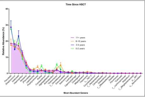
The time since HSCT was stratified into 4 levels: 0 to 2 y, 3 to 5 y, 6 to 10 y and 11 y and over. First, we assessed HSCT using princi-pal coordinate analysis based on unweighted Unifrac distances (Fig. 3). This analysis sug-gested a tendency for there to be data parti-tioning, as samples from participants who had HSCT in recent years appeared to cluster together and away from the samples pro-vided by participants who had HSCT in ear-lier years. The genus-level analysis of the data indicated that the group whose time since HSCT was 11 y or more had the highest lev-els of Streptococcus (18.4%), Haemophilus (12.7%) and Neisseria (6.8%) as compared with other groups (Fig. 4).
Discussion
Susceptibility to OSCC is not fully explained in FA or in the general population. Due in part to their young age and awareness of their cancer susceptibility, FA patients do not typi-cally smoke or consume alcohol. Within the general population, even among smokers and drinkers, only a small proportion of individu-als develop OSCC. Therefore, other environ-mental factors may synergize with those exposures. Thus, this study aimed to explore the microbial salivary profiles of FA patients about OSCC risk factors. The inherent genomic instability of FA patients may coop-erate with other environmental exposures (such as the local microbiome) aside from the classical risk factors, and this would add to the multitude of elements that can collec-tively promote carcinogenesis.
Infection and inflammation are important mediators of cancer development, particu-larly for OSCC (Meurman and Bascones-Martinez 2011). The mouth is heavily colonized by bacteria (Dewhirst et al. 2010), and exposure stemming from the microbi-ome, induced local inflammation, or metabolites produced by the host or microbiota, may contribute to OSCC development. Several oral species have been associated with OSSC (Mager et al. 2005) and a link between plaque-induced periodontal dis-eases and oral cancer has been proposed (Meurman and Bascones-Martinez 2011).
In the present study, Firmicutes and Bacteriodetes were the most common phyla, which is in accord with studies of the oral microbiome in health and disease (Guerrero-Preston et al. 2016). In addition, Prevotella, Veillonella, and Streptococcus VeillonellaPrevotella Streptococcu s HaemophilusNeisseri a [Prevotella ] ActinomycesLeptotrichia FusobacteriumPorphyromona s Rothia
AggregatibacterActinobacillusf__Gemellacea e GranulicatellaSelenomona s CapnocytophagaMegasphaer a Unassigne d LautropiaTreponemac__TM7-3
f__NeisseriaceaeCampylobacterCorynebacteriumOribacterium f__Lachnospiracea e f__[Weeksellaceae ] CatonellaDialiste r Parvimona s
*Atopobiump__SR1Moryella
f__[Mogibacteriaceae ] 0 1 0 2 0 3 0 4 0 5 0
Most Abundant Genera
Relative Abundance (%
)
Absence Presence
History of Oral Graft Versus Host Disease (GVHD)
Figure 1. Mean relative abundance of the most prevalent genera in saliva samples from patients with (red, n = 27) and without (blue, n = 26) a history of oral graft-versus-host disease (GVHD). When a genus-level resolution could not be achieved, the next taxonomic level was presented. p, phylum; c, class; o, order; f, family. *P < 0.05 (Differences did not reach false discovery rate [FDR] adjusted statistical significance).
Strep tococ cus Veillo nella Haem ophil us *Prev otella Neiss eria *Agg regati bacte r Actin obac illus [Prev otella ] Actin omyc es Rothi a *Sele nomo nas Lepto trich ia Porp hyro mona s Fuso bacte rium *Cap nocy topha ga f__Ge mella ceae *Gran ulica tella Trepo nema Lautr opia *f__N eisse riace ae Coryn ebac terium Mega spha era Camp yloba cter Unas signe d c__T M7-3 *f__[W eeks ellac eae] Oriba cteriu m Caton ella f__La chno spira ceae Card iobac terium Morax ella g__T G5 o__Bacteroidales King ella p__S R1 0 10 20 30
Level of Mucositis
Most Common Genera
Level I Level II Level III
Relative Abundance (%
)
were the most frequently detected genera, which is in line with the literature (Al-Hebshi et al. 2015; Guerrero-Preston et al. 2016). FA patients showed VPI (31.3%) and GBI (34.2%) lev-els that are higher than the recommended levlev-els. However, FA patients do not present inherently poorer oral health. Lyko et al. (2016) found no differences in DMFT and oral hygiene index between FA individuals and a paired control group. Our DMFT results were in line with the findings of Yalman et al. (2001), who reported a mean DMFT of 5 in transplanted FA patients. Using culture, the authors compared the microbiota and DMFT of HSCT and non-HSCT FA patients, and found no differences in the levels of Lactobacilli and Streptococcus mutans. In the present study, we found a trend for increased levels of Streptococcus ssp. in participants with DMFT equal to or above 8. Among the oral parameters examined, we focused on GBI because of its association with inflammatory responses to the local microbial insult and because it is a more objective metric than VPI. Participants with a higher GBI (Appendix Fig. 3) showed elevated levels of Porphyromonas, Treponema, Prevotella, Parvimonas and Dialister spp. Interestingly, those genera include members of the red (Porphyromonas gingivalis, Treponema denticola) and orange (Prevotella intermedia and Parvimonas micra) complexes (Socransky and Haffajee 2005) as well as newly proposed periodontal pathogens (Dialister pneumosintes and Dialister invisus) (Pérez-Chaparro et al. 2014), all of which are associated with the inflammation of periodontal tissues (Socransky and Haffajee 2005). Only one previous study investigated the presence of periodontal patho-gens (Aggregatibacter actinomycetemcomitans, Porphyromonas gingivalis, Fusobacterium nucleatum, and Treponema dentic-ola) in the oral cavity of subjects with FA and found no differ-ences in their levels as compared with matched controls (Lyko et al. 2013).
Based on the literature and our clinical experience, we pro-pose a set of risk factors and categories. When comparing microbial profiles, we found a recurring theme in terms of the elevated presence of Streptococcus, Neisseria, Veillonella and Haemophilus concomitant with oral GVHD (Streptococcus, Veillonella, Haemophilus; Fig. 1), severe mucositis (Streptococcus, Haemophilus; Fig. 2), longer time since HSCT (Streptococcus, Neisseria, Haemophilus; Fig. 4), PMOL (Neisseria, Haemophilus; Supplemental Fig. 4) and high risk (i.e., patients that received HSCT more than 5 years prior to the present examination or subjects ≥18 years old presenting an oral lesion) (Neisseria, Haemophilus; Appendix Fig. 5). Our results are in accord with recently reported associations between these taxa and cancer. Guerrero-Preston et al. (2016) demonstrated that Streptococcus Haemophilus and Veillonella were the most abundant taxa in the saliva of OSCC patients, and Pustelny et al. (2015) showed that Veillonella parvula preferentially colo-nizes neoplastic tissues. Higher proportions of Haemophilus and Neisseria have been detected in patients with oral leuko-plakia (Hu et al. 2016) and oral erosive lichen planus (Wang et al. 2016), respectively. Further, Al-Hebshi et al. (2015) found that Neisseria, Haemophilus and Streptococcus were among the most abundant genera in OSCC. Also, Neisseria
accounted for 8%, 4%, and 5% of the microbiota of laryngeal squamous cell carcinoma, adjacent normal tissue and control tissues, respectively (Gong et al. 2013). Finally, esophageal precancerous lesions are enriched for Neisseria and Veillonella (Pilato et al. 2016).
The relevance of our findings is further supported by an earlier study that reported high salivary levels of Streptococcus mitis in patients with OSCC (Mager et al. 2005), and recent investigations showing that Streptococcus spp. (including S. mitis) and Neisseria can synthesize acetaldehyde (Moritani et al. 2015), a well-known carcinogen. Indeed, the oral microbiota seems to be a major determinant of salivary acetaldehyde lev-els (Marttila et al. 2013), which can be decreased using an antimicrobial rinse (Homann et al. 1997). Moreover, the syner-gism between conventional risks factors and microorganisms via aldehyde production has been described for esophageal cancer (Peng et al. 2016). Although a few studies have found similar levels of Streptococcus and Neisseria (Hu et al. 2016) in healthy subjects and OSCC patients, we hypothesize that their levels are a bigger challenge for FA patients, considering their deficiency in DNA repair mechanisms.
Our results are particularly relevant for the FA population because of the toxicity of aldehydes noted in these patients (Garaycoechea et al. 2012). Indeed, higher aldehyde concentra-tions seem to interact directly with lipid peroxidation of the cell and induce DNA damage (Garcia et al. 2011). Ethanol exposure can also lead to acetaldehyde accumulation, causing bone mar-row dysfunction in FA patients (Ghosh et al. 2014). We found that some participants classified as a high risk in our study were current or former smokers (n = 11) or alcohol drinkers (n = 13), and these insults might have influenced the profiles of these patients. Further, both habits enhance acetaldehyde production by the oral microbiome (Marttila et al. 2013).
There are limitations in the present study. The first is its sample size. However, because FA is estimated to occur in 1:250,000 births (Dong et al. 2015), the present study population is of a
representative size for an FA study. Still, the smaller sample size presents many challenges, as conventional sample size calculations are difficult to achieve, and the heterogeneity of the group hampers the utilization of strict inclusion and exclusion criteria. A second limitation of the present study is its cross-sectional design, which precludes any inference on a causal role of the oral microbiome in OSCC susceptibility. However, despite these considerations, our results represent a significant first step in understanding the FA oral microbiome and its potential contribution to OSCC development in FA and, possibly, in the general population.
Author Contributions
C.P. Furquim, contributed to conception, design, data acquisition, analysis, and interpretation of data, drafted and critically revised the manuscript; G.M.S. Soares, contributed to design, data acquisi-tion, analysis, and interpretaacquisi-tion, drafted and critically revised the manuscript; L.L. Ribeiro, M.A. Azcarate-Peril, and N. Butz, con-tributed to data acquistion, critically revised the manuscript; J. Roach and K. Moss, contributed to data analysis, critically revised the manuscript; C. Bonfim, contributed to data acquisition, criti-cally revised the manuscript; C.C. Torres-Pereira and F.R.F. Teles, contributed to conception, design, data acquisition, analysis, and interpretation, drafted and critically revised the manuscript. All authors gave final approval and agree to be accountable for all aspects of the work.
Acknowledgments
This study was supported by research grants and from the Brazilian Support Agency for Superior Education (CAPES) and in part by the National Institutes of Health / National Institute of Dental and Craniofacial Research (R01-DE024767 to F.R.F.T. and P30DK 34987 to the UNC Microbiome Core) and the Fanconi Anemia
Figure 4. Mean relative abundance of the most prevalent genera in saliva samples according to time since hematopoietic stem cell transplantation (HSCT): 0 to 2 y (green, n = 5); 3 to 5 y (blue, n = 8); 6 to 10 y (orange, n = 9); 11 y and over (purple, n = 31). When genus level resolution could not be achieved, the next taxonomic level was presented. p, phylum; c, class; o, order; f, family. *P < 0.05 (Differences did not reach FDR-adjusted statistical significance).
Research Fund (FARF to F.R.F.T.). The authors also thank the staff of the Bone Marrow Transplant Unit, Federal University of Paraná Hospital, who were involved in the Brazilian Fanconi Anemia Patient and Family Meeting. The authors declare no potential conflicts of interest with respect to the author-ship and/or publication of this article.
References
Ainamo J, Bay I. 1975. Problems and proposals for recording gingivitis and plaque. Int Dent J. 25(4):229–235.
Al-Hebshi NN, Nasher AT, Idris AM, Chen T. 2015. Robust species taxonomy assignment algorithm for 16S rRNA NGS reads: application to oral carcinoma samples. J Oral Microbiol. 7:28934. eCollection 2015. Alter BP. 2014. Fanconi anemia and the development
of leukemia. Best Pract Res Clin Haematol. 27(3– 4):214–221.
Alter BP, Giri N, Savage SA, Quint WG, Koning MN, Schiffman M. 2013. Squamous cell carcinomas in patients with Fanconi anemia and dyskeratosis congenita: a search for human papillomavirus. Int J Cancer. 133(6):1513–1515.
Amarasekara R, Jayasekara RW, Senanayake H, Dissanayake VH (2015). Microbiome of the pla-centa in pre-eclampsia supports the role of bacteria in the multifactorial cause of pre-eclampsia. J Obstet Gynaecol Res. 41(5):662–669.
Bonfim C, Ribeiro L, Nichele S, Bitencourt M, Loth G, Koliski A, Funke VA, Pilonetto DV, Pereira NF, Flowers ME, et al. 2016. Long-term survival, organ function, and malignancy after hematopoietic stem cell transplanta-tion for fanconi anemia. Biol Blood Marrow Transplant. 22(7):1257–1263. Caporaso JG, Lauber CL, Walters W, Berg-Lyons D, Huntley J, Fierer N,
Owens SM, Betley J, Fraser L, Bauer M, et al. 2012. Ultra-high-throughput microbial community analysis on the Illumina HiSeq and MiSeq platforms. ISME J. 6(8):1621–1624.
Caporaso JG, Lauber CL, Walters WA, Berg-Lyons D, Lozupone CA, Turnbaugh PJ, Fierer N, Knight R. 2011. Global patterns of 16S rRNA diversity at a depth of millions of sequences per sample. Proc Natl Acad Sci U S A. 108(Suppl 1):4516–4522.
Carter HG, Barnes GP. 1974. The gingival bleeding index. J Periodontol. 45(11):801–805.
Correa P, Houghton J. 2007. Carcinogenesis of helicobacter pylori. Gastro-enterology. 133(2):659–672.
Dewhirst FE, Chen T, Izard J, Paster BJ, Tanner AC, Yu WH, Lakshmanan A, Wade WG. 2010. The human oral microbiome. J Bacteriol. 192(19):5002–5017. Dong H, Nebert DW, Bruford EA, Thompson DC, Joenje H, Vasiliou V. 2015.
Update of the human and mouse Fanconi anemia genes. Hum Genomics. 9:32. Eke PI, Dye BA, Wei L, Slade GD, Thornton-Evans GO, Beck JD, Taylor GW, Borgnakke WS, Page RC, Genco RJ. 2013. Self-reported measures for sur-veillance of periodontitis. J Dent Res. 92(11):1041–1047.
Garaycoechea JI, Crossan GP, Langevin F, Daly M, Arends MJ, Patel KJ. 2012. Genotoxic consequences of endogenous aldehydes on mouse haematopoi-etic stem cell function. Nature. 489(7417):571–575.
Garcia CC, Angeli JP, Freitas FP, Gomes OF, Oliveira TF, Loureiro AP, Mascio PD, Medeiros MH. 2011. [13C2]-Acetaldehyde promotes unequivocal for-mation of 1,N2-propano-2’-deoxyguanosine in human cells. J Am Chem Soc. 133(24):9140–9143.
Ghosh S, Sur S, Yerram SR, Rago C, Bhunia AK, Hossain MZ, Paun BC, Paun BC, Ren YR, Iacobuzio-Donahue CA, et al. 2014. Hypersensitivities for acetaldehyde and other agents among cancer cells null for clinically rel-evant Fanconi anemia genes. Am J Pathol. 184(1):260–270.
Gong HL, Shi Y, Zhou L, Wu CP, Cao PY, Tao L, Xu C, Hou DS, Wang YZ. 2013. The composition of microbiome in larynx and the throat biodiversity between laryngeal squamous cell carcinoma patients and control popula-tion. PLoS One. 8(6):e66476.
Homann N, Jousimies-Somer H, Jokelainen K, Heine R, Salaspuro M. 1997. High acetaldehyde levels in saliva after ethanol consumption: methodologi-cal aspects and pathogenetic implications. Carcinogenesis. 18(9):1739–1743. Hu X, Zhang Q, Hua H, Chen F. 2016. Changes in the salivary microbiota of
oral leukoplakia and oral cancer. Oral Oncol. 56:e6–e8
Lyko K, Bonfim C, Benelli EM, Torres-Pereira CC, Amenabar JM. 2013. Salivary detection of periodontopathic bacteria in Fanconi’s anemia patients. Anaerobe. 24:32–35.
Lyko K, Lemes AL, Bonfim C, Torres-Pereira CC, Amenábar JM. 2016. Oral health status in children and adolescents with Fanconi anemia. Spec Care Dentist. 36(2):71–74.
Mager DL, Haffajee AD, Devlin PM, Norris CM, Posner MR, Goodson JM. 2005. The salivary microbiota as a diagnostic indicator of oral cancer: a descriptive, non-randomized study of cancer-free and oral squamous cell carcinoma subjects. J Transl Med. 3:27.
Marttila E, Uittamo J, Rusanen P, Lindqvist C, Salaspuro M, Rautemaa R. 2013. Acetaldehyde production and microbial colonization in oral squa-mous cell carcinoma and oral lichenoid disease. Oral Surg Oral Med Oral Pathol Oral Radiol. 116(1):61–68.
Meurman JH, Bascones-Martinez A. 2011. Are oral and dental diseases linked to cancer? Oral Dis. 17(8):779–784.
Mima K, Nishihara R, Qian ZR, Cao Y, Sukawa Y, Nowak JA, Yang J, Dou R, Masugi Y, Song M, et al. 2015. Fusobacterium nucleatum in colorec-tal carcinoma tissue and patient prognosis. Gut. 65(12):1973–1980. Moritani K, Takeshita T, Shibata Y, Ninomiya T, Kiyohara Y, Yamashita
Y. 2015. Acetaldehyde production by major oral microbes. Oral Dis. 21(6):748–754.
Nelson MC, Morrison HG, Benjamino J, Grim SL, Graf J. (2014). Analysis, optimization and verification of Illumina-generated 16S rRNA gene ampli-con surveys. PLoS One 9:e94249.
Peng Q, Chen H, Huo JR. 2016. Alcohol consumption and corresponding fac-tors: a novel perspective on the risk factors of esophageal cancer (Review). Oncol Lett. 11(5):3231–3239.
Pérez-Chaparro PJ, Gonçalves C, Figueiredo LC, Faveri M, Lobão E, Tamashiro N, Duarte P, Feres M. 2014. Newly identified pathogens associated with periodontitis: a systematic review. J Dent Res. 93(9):846–858.
Pilato V, Freschi G, Ringressi MN, Pallecchi L, Rossolini GM, Bechi P. 2016. The esophageal microbiota in health and disease. Ann N Y Acad Sci. 1381(1):21–33.
Pustelny C, Komor U, Pawar V, Lorenz A, Bielecka A, Moter A, Gocht B, Eckweiler D, Müsken M, Grothe C, et al. 2015. Contribution of Veillonella parvula to Pseudomonas aeruginosa-mediated pathogenicity in a murine tumor model system. Infect Immun. 83(1):417–429.
Romick-Rosendale LE, Lui VWY, Grandis JR, Wells SI. 2013. The Fanconi anemia pathway: repairing the link between DNA damage and squamous cell carcinoma. Mutation Research. Mar-Apr:743–744:78–88.
Schmidt BL, Kuczynski J, Bhattacharya A, Huey B, Corby PM, Queiroz ELS, Nightingale K, Kerr AR, DeLacure MK, Veeramachaneni R, et al. 2014. Changes in abundance of oral microbiota associated with oral cancer. PLoS One. 9(6):e98741.
Schwabe RF, Jobin C. 2013. The microbiome and cancer. Nat Rev Cancer. 13(11):800–812.
Socransky SS, Haffajee AD. 2005. Periodontal microbial ecology. Periodontol 2000. 38:135–187.
van Monsjou HS, Wreesmann VB, van den Brekel MW, Balm AJ. 2013. Head and neck squamous cell carcinoma in young patients. Oral Oncol. 49(12):1097–1102.
Wang K, Lu W, Tu Q, Ge Y, He J, Zhou Y, Gou Y, Nostrand JDV, Qin Y, Li J, et al. 2016. Preliminary analysis of salivary microbiome and their potential roles in oral lichen planus. Sci Rep. 6:22943.
Warnakulasuriya S, Johnson NW, van der Waal I. 2007. Nomenclature and classification of potentially malignant disorders of the oral mucosa. J Oral Pathol Med. 36(10):575–580.