Structure-Function Studies of Essential Centrosomal Proteins Associated with Primordial Dwarfism and Microcephaly
By Sarah Speed
Senior Honors Thesis Department of Biology
University of North Carolina at Chapel Hill
6 April 2015
Approved:
1 Abstract:
2 Introduction:
In eukaryotes, the main microtubule-organizing center in the cell is the centrosome, a non-membrane-bound organelle involved in such essential cell processes as organization of the microtubule cytoskeleton and mitotic spindle, regulation of protein trafficking and cell division, and establishment and maintenance of cell polarity (1, 2). Centrosomes are composed of two structural elements: a centriole, shown to be an electron-dense cylinder of proteins that exhibits distinctive nine-fold symmetry, and surrounding pericentriolar material (PCM) (3). The PCM was previously thought to be an amorphous cloud of proteins; however, recent super-resolution microscopy studies have shown the PCM in Drosophila to contain distinct structural layers. The innermost layers closest to the centrioles contain primarily scaffolding proteins such as Sas-4, Asl, and PLP. The outermost layers primarily contain microtubule recruiting and organizing proteins such as Spd-2, γ-TURC, and CNN (4 - 6). Several hundred other proteins are concentrated within the PCM, a fact that highlights the centrosome’s role as a coordination center of the cell (7). The recruitment and subsequent organization of these proteins is a complex process that strictly controls the regulation of centrosome function. However, explicit details on how they are organized to form a functional centrosome are currently poorly understood.
3
9, reviewed in 10). It is during this time that centrosomes nucleate the microtubules that will form the mitotic spindle, a crucial step for proper cell division. In organizing the mitotic spindle, centrosomes facilitate accurate segregation of chromosomes. Therefore, tight regulation of centrosome composition and number is imperative to ensure mitotic defects do not occur. This concept is corroborated by a correlation between supernumerary centrosomes arising due to deregulation of centrosome biogenesis and aneuploidy, polyploidy, and chromosome fragmentation (11, 12). These abnormal cellular conditions are implicated in tumorigenesis, although whether these conditions are corollary or causative has yet to be established (13). Studies have also shown a link between heritable point mutations in centrosomal proteins and developmental diseases, including primordial dwarfism and microcephaly (14, 15, 16).
Among the proteins implicated in the development of these diseases are Asterless (Asl), Centrosomin (CNN), Pericentrin-Like Protein (PLP), and Spindle Assembly defective-4 (Sas-4), each of which have identified human orthologues CEP152 (14), CDK5RAP2 (17), Pericentrin (18), and CPAP (19), respectively. Each of these proteins plays crucial roles either in centriole duplication, PCM recruitment or both, in flies as well as in humans. For example, Sas-4 and its human orthologue CPAP have been shown to be essential for centriole replication. Cells treated with sas-4 RNAi and thus lacking Sas-4 function were observed to contain only one centrosome and were defective in S-phase progression (20). Reduced levels of Sas-4 result in centrosomes with significantly less PCM, while in contrast, overexpression of Sas-4 leads to elongated centrioles with associated PCM as well as acentriolar centrosomes (21 - 24). Clearly, Sas-4 is involved in both centriole duplication and the recruitment of essential PCM components; however, the mechanistic details behind these roles remain unclear.
4
CNN, and PLP near the centriole (25 - 30). In flies mutant for Asl, the stabilization and maintenance of the PCM is severely disrupted and the centrioles are often absent altogether, indicating a lack of structural integrity at the centrosome (28, 29). Both Asl and PLP exist at the centrosome in an extended conformation with their carboxyl-termini facing inwards towards the center and their amino-termini facing outwards, extending from the centriole’s outer wall into the PCM (5). This organization emphasizes their potential roles as scaffolding proteins and raises the possibility that they form a physical bridge linking the centrioles with the PCM and thus contribute to a structurally sound centrosome. However, it is not known exactly how Asl and PLP bind and interact with their partners to form such a bridge due to a lack of structural studies.
Other proteins essential for proper PCM formation are CNN and PLP. Centrioles are able to control the total amount of PCM they organize throughout the cell cycle by regulating the rate of CNN incorporation into existing PCM (31). The human orthologue of CNN, CDK5RAP2, interacts with the human orthologue of PLP, Pericentrin. This interaction is necessary for correct centrosome maturation and bipolar spindle formation (32). Interestingly, a conserved domain known as the PACT domain is located within PLP, Pericentrin, and a similar protein in humans, AKAP450 (33, 34). This domain is sufficient for centrosomal targeting and contains specific Calmodulin (CaM) binding sites (33). In fact, interaction of CaM with the PACT domain is required for proper targeting of PLP to the centrosome. A point mutation disrupting the PACT-CaM interaction within PLP causes inefficient centrosomal targeting and highly disorganized PCM (35), likely due to the loss of the scaffolding function of PLP. Thus, CaM also plays a crucial role in the organization of PCM and regulation of the centrosome.
5
which also contains CP-190 and 𝛾-tubulin. It has been proposed that this S-CAP complex forms as a result of Sas-4 acting as a scaffold for pre-assembled cytoplasmic complexes before tethering them at the centrosome (36). The S-CAP complex is held at the centrosome via Sas-4 during centrosome maturation, providing an initial layer of PCM upon which more PCM is added later (36). In cells mutant for sas-4 that are unable to properly form S-CAP complexes, centrosomes are still present but with greatly reduced levels of PCM (36). Needless to say, without any one of the proteins present in the S-CAP complex, centrosomes would fail to form properly, leading to microtubule-associated mitotic defects such as aneuploidy and chromosome fragmentation. Thus, by studying the structural and biochemical properties of these centrosomal proteins both independently and in complex with one another, we can more deeply understand how centrosome duplication and maturation is regulated in vivo.
6 Materials and Methods:
Cloning, Expression, and Purification of Protein Fragments
7
0.1% BME). In the case of PACTF, cells expressing either HIS-CaM or GST-PACTF were sonicated together, and the clarified supernatant was purified first over a Ni-NTA column, then over a GST-column, in the same manner as previously described. Fractions containing protein were pooled and dialyzed overnight into appropriate storage buffers containing PreScisson protease to cleave the HIS-tag and/or GST-tag. All dialyzed protein fractions were run through a Glutathione Sepharose column to remove HIS-PreScission Protease, free GST-tag, and/or uncleaved protein, then concentrated, flash frozen in liquid nitrogen, and stored at -80°C.
Transfection and Fixation of S2 Cells
Drosophila melanogaster S2 cells were resuspended in Schneider’s media (5% FBS,
anti/anti). One milliliter of resuspended cells was centrifuged for 3 minutes at 200 g to pellet the cells. The pellet was resuspended in 100 μL Amaxa solution with 1 μg of vector containing the protein of interest fused to an N-terminal GFP-tag under an actin promoter. This solution was placed in a cuvette and cells were electroporated. The cells were then resuspended in 2 mL of Schneider’s media and plated in a 6-well culture dish. Forty-eight hours post-transfection, cells were plated on Concanavalin A-coated coverslips and allowed to adhere for 30 minutes. The media was aspirated from the dish and the cells were washed with 1X PBS. The cells were then washed with ice cold (-20⁰C) 100% dry MeOH. The dish was again washed with ice cold 100% dry MeOH and incubated with the MeOH at -20⁰C for 20 minutes. After aspirating away the MeOH, the dish was washed three times with 1X PBS.
Staining of S2 Cells
8
fragment, the primary antibody used was Guinea Pig anti-Asl at a 1:30,000 dilution, the secondary antibody used was Rabbit anti-Guinea Pig 568, and Rabbit:Phosphohistone 3 was used in the same manner. All antibodies were diluted in a 1X PBS/5% Normal Goat Serum solution. The prepared coverslips were incubated with primary antibody for 30 minutes and then washed three times with 1X PBS. These steps were repeated with the secondary antibody and with PH3. The coverslips were then mounted on slides.
Circular Dichroism (CD) Analysis
CD spectra were recorded on a Cirascan Plus: Steady State Circular Dichroism/ Fluorescence spectrometer with titration and automated temperature ramping capabilities at 20°C using a 0.1 cm path-length cuvette. Asl 842-961 was incubated in several buffers of varying pH (20mM sodium citrate for pH 3, 20mM sodium acetate for pH 5, and 20mM sodium phosphate for pH 7) at a concentration of 0.1 mg/mL. In order to compare CD spectra recorded at different pH values, the concentration of each sample was kept constant and the CD spectra were recorded after buffer exchanging to each pH.
Isothermal Titration Calorimetry (ITC) Analysis
9 Crystallization Trials
Trials began for purified proteins using broad screening kits HR2-110 and HR2-126 commercially available from Hampton Research. Broad screens were set-up using the hanging-drop vapor diffusion method in 24-well Linbro trays (Hampton) for Asl 842-961, CNN F1, Asl 626-719+CNN F1, Asl 842-961+CNN F1, and CaM+PACTP (see Figure 1). Drops were set by adding 2 µL of protein to 2 µL of well solution. Trays were incubated at 20°C. Proteins used in cocrystallization trials were incubated together at molar ratios on ice for one hour before setting the drops. Once possible crystallization conditions were identified in the broad screens, narrower optimization screening conditions were employed.
Optimization conditions for Asl 842-961 used a screen of Ammonium fluoride that varied down the tray in 0.05 M increments from 0.05 M to 0.2 M and varied Polyethylene glycol (PEG) 3350 across in 5% increments from 5% to 30% (weight/volume). The second optimization crystal tray varied the percentage of PEG across in 5% increments from 5% to 30%, and varied the molecular weight of the PEG (PEG-1500, -4000, -6000, and -8000) down while maintaining a constant concentration of CAPS buffer (stock 1M CAPS pH 10.00, 0.1% 2-mercaptoethanol) of either 0.1 M or 0.3 M. After the hanging drops equilibrated over the course of seven days, a potential crystallization hit was identified, and a second optimization crystal tray was set up varying PEG 8000 across in 2.5% increments from 17.5% to 30% (weight/volume) and a varying CAPS buffer concentration down in 0.05 M increments from 0.05 M to 0.2 M.
10
were streak-seeded with microseeds from the original condition. A second optimization tray lowered PEG 8000 to 14% to 20%, and the tray was incubated at 4°C.
Co-crystallization trials for Asl 842-961+CNN 1-365 were screened using various molar ratios of Asl and CNN created by mixing fixed amounts of the two components from the following individual stocks: 4 mM of Asl 842-961 and 0.16 mM CNN 1-365 both in a CAPS buffer. Drops were streak-seeded with carbon nanotubes at a concentration of 0.1 mg/mL. Possible crystal conditions were identified in condition #47 of HR2-110. Two optimizations of this condition were tested. The first optimization used a screen that varied Ammonium sulfate across in 0.5 M increments from 0.5 M to 3.0 M and varied Sodium acetate pH 4.6 down in 0.05 M increments from 0.1 M to 0.25 M. A second optimization tray was set-up identical to the first, and this tray was incubated at 4°C.
Co-crystallization trials of Asl 626-719+CNN 1-365 were conducted in the same manner as the broad screening trials of Asl 842-961+CNN 1-365, however at ratios of 1.06 mM Asl:0.276 mM CNN and 2.12 mM Asl:0.276 mM CNN. Rod-shaped crystals appeared in condition #44 of HR2-126. An optimization screen was set-up by varying Ammonium phosphate across in 0.05 M increments from 0.1 M to 0.35 M and varying PEG 3350 down in 2% increments from 14% to 20%. Asl and CNN were incubated at a ratio of 0.708 mM Asl:0.184 mM CNN for the optimization. This optimization tray was repeated using molar ratios of 0.185 mM Asl:0.185 mM CNN and 0.370 mM Asl:0.185 mM CNN.
11
Sodium acetate pH 4.5 in 0.2 M increments across from 0.6 M to 0.16 M and varying Ammonium sulfate down in 0.1 M increments from 0.5 M to 0.8 M with a constant concentration of 0.5 M Guanidine hydrochloride throughout the tray.
Expression and Solubility Tests
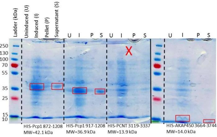
S. pombe Pcp1 872-1208, S. pombe Pcp1 917-1208, H. sapiens PCNT 3119-3337, and H. sapiens AKAP450 3664-3744 were transformed into BL21 E. coli cells, grown at 37°C to OD600 of 0.6 and induced with 0.1 mM IPTG. After induction, cells were shifted to 18°C for 18 hours. Cell pellets were collected by centrifugation and stored in Buffer A at -20°C. Cells were lysed by sonication and then centrifuged for 45 minutes at 36,000 g to obtain clarified lysate supernatant. PMSF was used during sonication to limit protease activity.
Results:
Bacterial expression and column chromatography yielded pure protein for all fragments of interest
All protein fragments were derived from full-length Drosophila melanogaster proteins (Fig. 1). Asl and CNN full-length proteins were divided into smaller fragments in order to isolate as much predicted secondary structure as possible, with the expectation of obtaining more stable fragments during expression and purification. All Asl and CNN fragments as well as full-length CaM were purified for biochemical and structural analysis (Fig. 3, Fig. 4). They expressed well in BL21 E. coli cells induced with IPTG and remained soluble during the purification process.
12
4). However, we only observed GST-PACTF to be soluble when copurified with CaM, prior to cleavage of the GST-tag. Due to PACTF having a similar molecular weight to CaM, its presence as a soluble band after GST-tag cleavage cannot be determined by our SDS-PAGE gel (Fig. 4). A better resolved SDS-PAGE gel will determine if PACTF continues to remain soluble after cleavage. Due to the limited solubility of PACTF, a smaller region of the conserved PACT domain, labeled as PACTP in Figure 1, was synthesized as a low molecular weight peptide in order to aid in biochemical and structural analyses.
Asterless and Centrosomin fragments exhibit robust localization to centrosomes
In order to assess the ability of Asl F1, F2, F3 and CNN F1 to localize to the centrosome
in vivo, we transfected Drosophila melanogaster S2 cells with GFP-tagged versions of these fragments. Endogenous PLP and Asl were tagged with red fluorescent antibodies and served as centrosomal markers within GFP-Asl and GFP-CNN transfected cells, respectively. As can be seen in Figure 5 by the presence of green fluorescence at the red fluorescently marked centrosome, all three Asl fragments and CNN F1 robustly localized to the centrosome. Phosphohistone 3 was also used to stain mitotic chromatin, however no Phosphohistone 3 staining was observed on the slides. These localization studies demonstrated the ability of each fragment to independently localize to the centrosome, likely mediated by interactions with other centrosomal proteins.
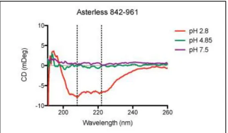
Circular dichroism analysis reveals helical structure of Asl 842-961 at an acidic pH
13
structure begins to emerge at pH 2.8 as shown by the characteristic peaks at 208 nm and 222 nm (Fig. 6). This result is in agreement with a previous study on the emergence of structure in centrosomal proteins through protein-protein interactions and pH changes at the centrosome (reference #38; see discussion).
Isothermal titration calorimetry reveals a strong binding association between CaM and PACTP In order to determine if further structural studies with our synthesized peptide PACTP were warranted, we wished to establish evidence of a binding association between CaM and PACTP. Using isothermal titration calorimetry (ITC), we quantified several variables involved in the association between CaM and PACTP. The raw data as well as a fitted curve to the titration of PACTP into CaM can be seen in Figure 7. The fitted curve indicates an exothermic reaction and favorable binding interactions. The stoichiometry between CaM and PACTP is approximately one (N = 1.33 ± 0.0164 sites). As determined from an association constant of 7.71 x 10⁷ (K = 7.71 x 10⁷ ± 7.68 x 10⁷ M-1), the dissociation constant, Kd, is approximately 13.0 nM, indicating a high affinity between PACTP and CaM. It is interesting to note that this association was observed in a buffer solution lacking the addition of free calcium, which also contained a chelating agent (EGTA) to bind any Ca2+ ions present in normally distilled water. This means that CaM was in an apo state (not bound to Ca2+ ions) when bound to PACTP. No conclusive results have yet been recorded for the affinity between CaM and PACTP when Ca2+ ions are present in the buffer solution.
Protein fragments were screened independently and in various combinations for diffraction-quality crystal growth
14
crystallographic analysis. We were unable to use the entire third fragment of Asl due to the presence of multiple species (i.e. degradation products) during purification (Fig. 3). Asl F3 formed higher order species that maintained association even after being run through a denaturing gel. Therefore, a smaller fragment from amino acids 842-961 that lacks cysteine residues and eliminates predicted unstructured regions was chosen for crystallization trials. Exposed cysteine residues are capable of interacting with one another to form di-sulfide bonds (either within the same protein chain or between adjacent chains). The lack of cysteine residues aids in crystallization by preventing the formation of aberrant disulfide bonds. Disulfide bond formation could lead to incorrect or multiple folding configurations and thus folded protein fragments that are unable to pack into a uniform crystal structure.
Crystallization trials with CNN F1 alone as well as cocrystallization trials of CNN F1 with Asl 626-719 and Asl 842-961 were attempted. We predicted that binding interactions between Asl and CNN would help stabilize their respective structures and thus increase the likelihood of uniform crystal packing. Various molar ratios of Asl:CNN were screened due to a lack of stoichiometric binding information between Asl and CNN. It is necessary to create as homogenous a solution as possible for crystallization trials in order to eliminate any unbound protein molecules that may interfere with a uniform crystal arrangement. Therefore, by screening various molar ratios, we hoped to find a stoichiometric ratio between Asl and CNN that would remove excess unbound protein molecules from solution.
15
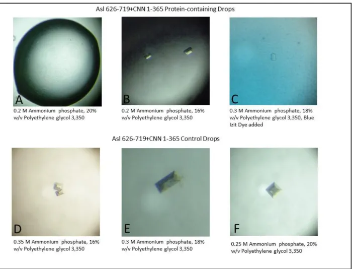
To date, crystal growth of our protein fragments has yet to be observed. This lack of crystal growth could be due to a number of confounding variables such as poor binding interactions or the possible use of heterogeneous solutions in which unbound protein effectively blocks crystal packing. Another problem could be a lack of protein stability in solution, as precipitate was observed for most drops containing Asl, CNN, or Asl+CNN fragments indicating non-uniform packing of proteins. False positives can also lead to unfounded optimization of conditions. For example, Figure 8A-C shows crystal growth in drops containing Asl 626-719 and CNN F1. These crystals appeared several days prior to the appearance of similar crystals in control drops containing only buffer and well solution (Fig. 8 D-F). Figure 8C shows a crystal subjected to Blue Izit dye (Hampton) which protein crystals readily absorb while salt crystals exclude. Both the growth of crystals in the control drops as well as the exclusion of the blue dye by the crystal in the protein-containing drop indicate that these crystals are composed of salt ions, not proteins.
Other PACT domains in yeast and human proteins show solubility
16 Discussion:
The main goal of our structure-function studies was to elucidate high-resolution structural models of interacting centrosomal proteins through X-ray crystallography in order to more fully understand their function in centrosome regulation. We were able to confirm that our chosen fragments were suitable for crystallization trials through in vivo localization trials. We found robust centrosomal localization of Asl and CNN fragments during interphase demonstrating their ability to independently localize, most likely through interactions with other centrosomal proteins. This robust localization indicates that the protein fragments likely contain important binding and/or targeting domains, and thus these results warranted further crystallization studies. It should be noted, however, that these localization trials were not performed in a knock-down background, and therefore there remains the possibility of dimerization between the fragments and their respective full-length endogenously expressed proteins.
17
Another important consideration about Asl and CNN function lies in the fact that their localization was observed in interphase cells. We did not observe any Phosphohistone 3 staining used to stain mitotic chromatin for our cells, and therefore we were unable to observe if localization changes during mitosis. Asl and CNN are essential for proper centrosome regulation (28, 29, 31), and as the centrosome matures and expands its PCM throughout the cell cycle, it is likely that Asl and CNN are spatially controlled in order to temporally regulate their functional roles. It will be of great interest to determine if Asl and CNN levels change during different stages of the cell cycle.
Due to the presence of unstructured regions within Asl F3, we chose to purify a smaller fragment, Asl 842-961, which eliminated these unstructured regions in order to aid in biochemical and structural analysis. CD analysis of Asl 842-961 revealed an intriguing structural feature of this fragment, which begins to exhibit its predicted helical structure at the acidic pH of 2.8 while remaining disordered at higher pH values (Fig. 3). This result is in agreement with a study by Trevino et al. (38) in which helical content for several centrosomal proteins was explored as a function of pH. They similarly found that all of the peptides tested became more structured at a lower pH. The study also found that this result held true regardless of the isoelectric point of the peptide, even for those with isoelectric points above 7 (predicted pI of Asl 842-961 = 8.23). The dependence of structural changes on pH could be due to an underlying mechanism of global regulation at the centrosome. The roles of centrosomal proteins remain dynamic throughout the cell cycle. The use of a centrosome wide pH variance during the cell cycle could result in more or less structured proteins, offering an attractive way to temporally regulate their functions.
18
centrosome (35). We were unable to purify Drosophila PACT (PACTF) alone due to its limited solubility, and therefore we had a small portion of the CaM-binding region within the PACT domain synthesized as a peptide (PACTP). Our ITC analysis indicated a high affinity between CaM and PACTP (Kd ~13 nM). We wish to replicate these results in order to reduce error. However, this initial result provided us with sufficient evidence to continue with crystallization trials. Interestingly, this interaction was observed in a calcium-free buffer with CaM in an apo state. Further ITC analysis in which Ca2+ ions are present and bound to CaM may provide insightful details concerning the effects of calcium binding on the affinity of CaM for the PACT domain of PLP. If future results indicate a weakened or ablated affinity between CaM and PACTP in the presence of calcium, it could indicate a potential regulatory mechanism in vivo in which CaM-PLP binding and thus PLP localization is dependent on changing calcium concentrations within the cell.
19
interactions, long-term instability in solution, blocking of crystal packing from unbound protein molecules in our cocrystallization trials, improper concentrations, and the limited availability of broad screening buffer conditions.
Despite not yet being able to crystallize Asl or CNN, our ITC analysis of PACTP has provided strong evidence that crystals can be produced for CaM and PACTP, especially considering the large number of solved CaM structures to date. A solved structure of this PACT peptide would indeed prove useful, however having a structure of a portion of endogenously expressed PACT would provide even more compelling evidence as to how CaM is targeting PLP to the centrosome. We will therefore continue forward with further solubility analysis and crystallization trials of CaM and PACTF. The solubility of the other PACT domains from yeast and humans (see Fig. 9) also allows us to continue with purification and crystallization trials of these PACT fragments independent of CaM, which can eventually afford the opportunity to compare the structures of CaM-bound vs. unbound PACT.
20 References:
1. Strnad, P., Gönczy, P. Mechanisms of procentriole formation. Trends Cell Biol (2008):18, 38996.
2. Doxsey, Stephen, Dannel Mccollum, and William Theurkauf. "Centrosomes In Cellular Regulation." Annual Review of Cell and Developmental Biology (2005): 411-34.
3. Kitagawa et al. "Structural Basis of the 9-Fold Symmetry of Centrioles." Cell (2011): 364-75.
4. Mennella et al. "Subdiffraction-resolution Fluorescence Microscopy Reveals a Domain of the Centrosome Critical for Pericentriolar Material Organization." Nature Cell Biology (2012): 1159-168.
5. Fu, J., and D. M. Glover. "Structured Illumination of the Interface between Centriole and Peri-centriolar Material." Biology Open (2012): 120104.
6. Sonnen, K. F., L. Schermelleh, H. Leonhardt, and E. A. Nigg. "3D-structured Illumination Microscopy Provides Novel Insight into Architecture of Human Centrosomes." Biology Open (2012): 965-76.
7. Müller et al. "Proteomic and Functional Analysis of the Mitotic Drosophila Centrosome." The EMBO Journal (2010): 3344-357.
8. Conduit et al. "A Molecular Mechanism of Mitotic Centrosome Assembly in Drosophila." ELife (2014).
9. Khodjakov, Alexey, and Conly L. Rieder. "The Sudden Recruitment of γ-Tubulin to the Centrosome at the Onset of Mitosis and Its Dynamic Exchange Throughout the Cell Cycle, Do Not Require Microtubules." The Journal of Cell Biology 146.3 (1999): 585-96. 10.Nigg, Erich A., and Tim Stearns. "The Centrosome Cycle: Centriole Biogenesis,
Duplication And Inherent Asymmetries." Nature Cell Biology (2011): 1154-160.
11.Ganem, N.J., Godinho, S.A., and Pellman, D. A mechanism linking extra centrosomes to chromosomal instability. Nature (2009): 460, 278-282.
12.Vitre, B. D., Cleveland, D. W. Centrosomes, chromosome instability (CIN) and aneuploidy. Current Opinion in Cell Biology (2012): 24, 809-815.
13.Pellman, D. Aneuploidy and cancer. Nature (2007): 446, 38-39.
14.Guernsey et al. "Mutations in Centrosomal Protein CEP152 in Primary Microcephaly Families Linked to MCPH4." The American Journal of Human Genetics 87.1 (2010): 40-51.
15.Klingseisen, A., Jackson, A. P. Mechanisms and pathways of growth failure in primordial dwarfism. Genes Dev (2011): 25, 2011-2024.
16.Zahnleiter et al. "MAP4 Dependent Regulation of Microtubule Formation Affects Centrosome, Cilia and Golgi Architecture as a Central Mechanism in Growth Regulation." Human Mutation (2014).
17.Megraw, Timothy L., James T. Sharkey, and Richard S. Nowakowski. “Cdk5rap2 Exposes the Centrosomal Root of Microcephaly Syndromes.” Trends in Cell Biology 21.8 (2011): 470–480.
18.Delaval, B., and S. J. Doxsey. "Pericentrin in Cellular Function and Disease." The Journal of Cell Biology (2010): 181-90.
19.Cottee et al. "Crystal Structures of the CPAP/STIL Complex Reveal Its Role in Centriole Assembly and Human Microcephaly." ELife (2013): E01071.
21
21.Kirkham, Matthew, Thomas Müller-Reichert, Karen Oegema, Stephan Grill, and Anthony A. Hyman. "SAS-4 Is a C. Elegans Centriolar Protein That Controls Centrosome Size." Cell (2003): 575-87.
22.Tang, Chieh-Ju C., Ru-Huei Fu, Kuo-Sheng Wu, Wen-Bin Hsu, and Tang K. Tang. "CPAP Is a Cell-cycle Regulated Protein That Controls Centriole Length." Nature Cell Biology (2009): 825-31.
23.Kohlmaier et al. "Overly Long Centrioles and Defective Cell Division upon Excess of the SAS-4-Related Protein CPAP." Current Biology (2009): 1012-018.
24.Azimzadeh, Juliette, and Wallace F. Marshall. "Building the Centriole." Current Biology (2010): R816-825.
25.Dzhindzhev et al. "Asterless Is a Scaffold for the Onset of Centriole Assembly." Nature (2010): 714-18.
26.Cizmecioglu et al. "Cep152 Acts As A Scaffold For Recruitment Of Plk4 And CPAP To The Centrosome." The Journal of Cell Biology (2010): 731-39.
27.Hatch et al. "Cep152 Interacts with Plk4 and Is Required for Centriole Duplication." The Journal of CellBiology (2010): 721-29.
28.Varmark et al. "Asterless Is a Centriolar Protein Required for Centrosome Function and Embryo Development in Drosophila." Current Biology (2007): 1735-745.
29.Blachon et al. "Drosophila Asterless and Vertebrate Cep152 Are Orthologs Essential for Centriole Duplication." Genetics (2008): 2081-094.
30.Doxsey, Stephen J., Pascal Stein, Louise Evans, Patricia D. Calarco, and Marc Kirschner. "Pericentrin, a Highly Conserved Centrosome Protein Involved in Microtubule Organization." Cell (1994): 639-50.
31.Conduit et al. "Centrioles Regulate Centrosome Size by Controlling the Rate of Cnn Incorporation into the PCM." Current Biology (2010): 2178-186.
32.Kim, Seongjae, Kunsoo Rhee, and Takashi Toda. "Importance of the CEP215-Pericentrin Interaction for Centrosome Maturation during Mitosis." PLoS ONE (2014): E87016. 33.Gillingham, Alison K., and Sean Munro. "The PACT domain, a conserved centrosomal
targeting motif in the coiled‐coil proteins AKAP450 and pericentrin." EMBO reports 1.6 (2000): 524-529.
34.Martinez-Campos, Maruxa, et al. "The Drosophila pericentrin-like protein is essential for cilia/flagella function, but appears to be dispensable for mitosis." The Journal of cell biology 165.5 (2004): 673-683.
35.Galletta et al. "Drosophila Pericentrin Requires Interaction with Calmodulin for Its Function at Centrosomes and Neuronal Basal Bodies but Not at Sperm Basal Bodies." Molecular Biology of the Cell 25.18 (2014): 262-694.
36.Gopalakrishnan et al. "Sas-4 Provides a Scaffold for Cytoplasmic Complexes and Tethers Them in a Centrosome." Nature Communications (2011): 359.
37.PDB ID: 4DS7. Klenchin, V.A., Rayment, I. “Crystal structure of yeast calmodulin bound to the C-terminal fragment of spindle pole body protein Spc110.” To Be Published. (2012) 38.Trevino et al. "Emergence of structure through Protein-protein Interactions and PH Changes in Dually Predicted Coiled-coil and Disordered Regions of Centrosomal Proteins." Biochimica Et Biophysica Acta 1844.10 (2014): 1808-819.
22 Appendix:
Figure 1. Schematic of protein fragments purified for localization and/or crystallization trials.
MGNTNVTELEERCRQLI GCYLRVESHRKALVYQKRYLKLTLEGYQASEQLALQNLRGGA EQPPQRNIKK--- KFKTVALAIIAIQRIKYIGRIWHTGKRIVSKSVFTITQQR S AASEEAHTSNVKMEKLYLHYLRAESFRKALIYQKKYLLLLIGGFQDSEQETLSMIAHLGVFP -SKAERKIT---SRPFTRFRTAVRVVIAILRLRFLVKKWQEVDRKGALAQGKAPRP GP DPALDAGACSAKMEKLYLRYLRAESFRKALIYQKKYLLLLIGGFQDSEQETLSMIAHLGVFP -SKADKKAAG---PRPLTRFRTAVRVVIAVLRLRFLVKKWQQVDGRGALAPGQALHPGE AAREDANTCGVQMEKLY LHYLRAESFRKALIYQKKYLLLLIGGFQDSEQETLSMIAHLGVFP -SKADKKTTT---SRPFTRFRTAVRVVIAILRLRFLVKKWQEVDRRGAATHGRA PRPGL TTSGDTNTCNIKMEKLYLHYLRAESFRKALIYQKKYLLLLIGGFQDSEQETLSMIAHLGVFP -SKADKKVTA---SRPFTRFRTAVRVVIAVLRLRFLVKKWQEVDRKGALIH HSRSTRHG TTGGDTNTCNIKMEKLY LHYLRAESFRKALIYQKKYLLLLIGGFQDSEQETLSMIAHLGVFP -SKADKKITM---SRPFTKFRTAVRVVIAVLRLRFLVKKWQEVDRKGALVHPK STRHGH LRELGSDSENVILRRIY GKYLRAESFRKALIYQKKYLLLLLGGFQECEEATLALIARM GGQP-SYTDLEIITHH ---SKGFTRFRSAVRVSIAISRMKFLVRRWHRVTGSGILSLSRDVFSQN ENRPINDLSNSKVQRLYERYLRAESYRKSLVYQKRYLLLLLGRFQECEQATLALIARMGALP -TLSQSR---TSRPLNRFRTAVRVVIAISRLKFLNRKWQRATRKISAGTATVTVHGT ENKPVYDGTNNKLQRLYERYLRAESFRKALVYQKRYLLLLLGGFQECEQATLCLIATMGVRP -SPLLSSQC---RPRRRFRAAVQVVI AVSRSRCELAP ---GS----LAG- LRELGSDSENAALKRIYGRYLRAESFRKALIYQKKYLLLLLGGFQECEEATLSLIARMGGQP -SYTDLEVITHR---SKGFTRFRSAVRVLIAISRMKF LVRRWHRVTGYSLIGINRDGFGQN ENHPVPDVPISKMQRLH RKYLRAESFRKALVYQKKYLLLLLGGFQACEKATLSLIARMGVYP -SPSDLQIPITQK ---PALTKFRSAVRAV IAISRLKFLVRKWHKINKKAGVEDPVVQISAA SNRSNRDDLEERANHLFGKYLRSESHRKALVHQKRYLQIVL TTYEENEAKALQLLNAQNVPMGIQGLSLAKPNADENHPRRKSFRSVVTAIIAIERMKFIVRKWQGGRRVCAKAIFSQQFTP R PQDLNTREHTEKLQHFYGKFLRADSRRKALAYQKRYLLSIVGGYQLSEENTLSVLAQLTKVQRSYAVIGRNKK ----SPKV-RFRSAVLVVISIC RMKWLIVRWNTGKRIGVKTLLWNIDQSF RHDKEISGLGREIIWLRARLRREEKFRRDLAWSKGLMELGERVRVACNEADLRMINEMGVKP -LDRTY---FRTPRQ-KLKAAISMILATVRMQRKSREWS KTKKLGEGLKRAKNEVLK QYESEIKGLSKLTKYLQ SKCRREHSLRLDLAFSKKFILMQLTGYETCNKINLRMLQKIGISPDPDLSKKHI ---KLKSLIIVVCSIERMKRMKNEWLKQAQLKQSLQRAAAKAKT
2520
...|...|...|...|...|...|...|... --- ....|...|...|...|...2530 2540 2560 2570 2580 2590 2600 2610 2620
80% Identity 80% Homology
Alignment PLP-PACT domain
H. sapiens D. melanogaster
B. taurus
C. lupus familiaris R. norvegicus M. musculus G. gallus D. rerio T. nigroviridis A. carolinensis X. tropicalis A. aegypti A. mellifera A. fumigatus A. pombe Minimal domain
Asterless Binding Calmodulin Binding
AA E EE EE 2550 E Mutants: R2538E RK2543EE KR2550EE
RK2543EE + KR2550EE IQ2597AA
R2599E
Mutants in the asterless binding region:
Mutations that ablated localization with Alanine mutations will be made wit h charge reversals (R or K --> E)
Mutants in the calmodulin binding regions:
The IQ-->AA mutation that ablated calmodulin binding in the Y2H will be made for binding experiments.
A charge reversal mutant R2599E will be made in the most conserved cha rged residue in the calmodulin binding region for binding expeiments
MGNTNVTELEERCRQLI GCYLRVESHRKALVYQKRYLKLTLEGYQASEQLALQNLRGGA EQPPQRNIKK--- KFKTVALAIIAIQRIKYIGRIWHTGKRIVSKSVFTITQQR S AASEEAHTSNVKMEKLYLHYLRAESFRKALIYQKKYLLLLIGGFQDSEQETLSMIAHLGVFP -SKAERKIT---SRPFTRFRTAVRVVIAILRLRFLVKKWQEVDRKGALAQGKAPRP GP DPALDAGACSAKMEKLYLRYLRAESFRKALIYQKKYLLLLIGGFQDSEQETLSMIAHLGVFP -SKADKKAAG---PRPLTRFRTAVRVVIAVLRLRFLVKKWQQVDGRGALAPGQALHPGE AAREDANTCGVQMEKLY LHYLRAESFRKALIYQKKYLLLLIGGFQDSEQETLSMIAHLGVFP -SKADKKTTT---SRPFTRFRTAVRVVIAILRLRFLVKKWQEVDRRGAATHGRA PRPGL TTSGDTNTCNIKMEKLYLHYLRAESFRKALIYQKKYLLLLIGGFQDSEQETLSMIAHLGVFP -SKADKKVTA---SRPFTRFRTAVRVVIAVLRLRFLVKKWQEVDRKGALIH HSRSTRHG TTGGDTNTCNIKMEKLY LHYLRAESFRKALIYQKKYLLLLIGGFQDSEQETLSMIAHLGVFP -SKADKKITM---SRPFTKFRTAVRVVIAVLRLRFLVKKWQEVDRKGALVHPK STRHGH LRELGSDSENVILRRIY GKYLRAESFRKALIYQKKYLLLLLGGFQECEEATLALIARM GGQP-SYTDLEIITHH ---SKGFTRFRSAVRVSIAISRMKFLVRRWHRVTGSGILSLSRDVFSQN ENRPINDLSNSKVQRLYERYLRAESYRKSLVYQKRYLLLLLGRFQECEQATLALIARMGALP -TLSQSR---TSRPLNRFRTAVRVVIAISRLKFLNRKWQRATRKISAGTATVTVHGT ENKPVYDGTNNKLQRLYERYLRAESFRKALVYQKRYLLLLLGGFQECEQATLCLIATMGVRP -SPLLSSQC---RPRRRFRAAVQVVI AVSRSRCELAP ---GS----LAG- LRELGSDSENAALKRIYGRYLRAESFRKALIYQKKYLLLLLGGFQECEEATLSLIARMGGQP -SYTDLEVITHR---SKGFTRFRSAVRVLIAISRMKF LVRRWHRVTGYSLIGINRDGFGQN ENHPVPDVPISKMQRLH RKYLRAESFRKALVYQKKYLLLLLGGFQACEKATLSLIARMGVYP -SPSDLQIPITQK ---PALTKFRSAVRAV IAISRLKFLVRKWHKINKKAGVEDPVVQISAA SNRSNRDDLEERANHLFGKYLRSESHRKALVHQKRYLQIVL TTYEENEAKALQLLNAQNVPMGIQGLSLAKPNADENHPRRKSFRSVVTAIIAIERMKFIVRKWQGGRRVCAKAIFSQQFTP R PQDLNTREHTEKLQHFYGKFLRADSRRKALAYQKRYLLSIVGGYQLSEENTLSVLAQLTKVQRSYAVIGRNKK ----SPKV-RFRSAVLVVISIC RMKWLIVRWNTGKRIGVKTLLWNIDQSF RHDKEISGLGREIIWLRARLRREEKFRRDLAWSKGLMELGERVRVACNEADLRMINEMGVKP -LDRTY---FRTPRQ-KLKAAISMILATVRMQRKSREWS KTKKLGEGLKRAKNEVLK QYESEIKGLSKLTKYLQ SKCRREHSLRLDLAFSKKFILMQLTGYETCNKINLRMLQKIGISPDPDLSKKHI ---KLKSLIIVVCSIERMKRMKNEWLKQAQLKQSLQRAAAKAKT
2520
...|...|...|...|...|...|...|... --- ....|...|...|...|...2530 2540 2560 2570 2580 2590 2600 2610 2620
80% Identity 80% Homology Alignment PLP-PACT domain
H. sapiens D. melanogaster
B. taurus
C. lupus familiaris R. norvegicus M. musculus G. gallus D. rerio T. nigroviridis A. carolinensis X. tropicalis A. aegypti A. mellifera A. fumigatus A. pombe Minimal domain
Asterless Binding Calmodulin Binding
AA E EE EE2550
E
Mutants: R2538E RK2543EE KR2550EE
RK2543EE + KR2550EE IQ2597AA
R2599E
Mutants in the asterless binding region:
Mutations that ablated localization with Alanine mutations will be made wit h charge reversals (R or K --> E)
Mutants in the calmodulin binding regions:
The IQ-->AA mutation that ablated calmodulin binding in the Y2H will be made for binding experiments.
A charge reversal mutant R2599E will be made in the most conserved cha rged residue in the calmodulin binding region for binding expeiments
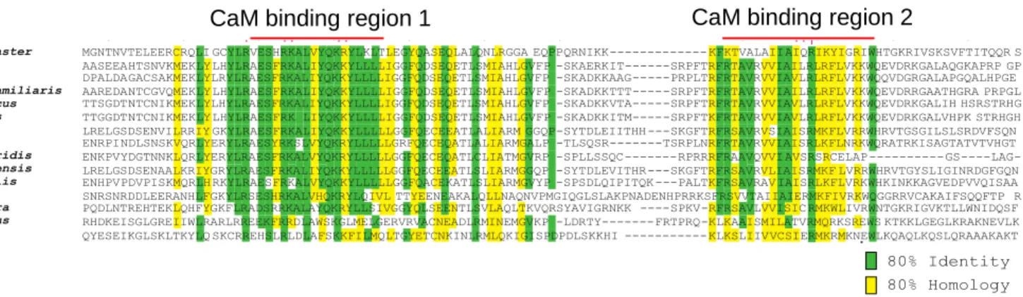
Figure 2. Multiple-sequence alignment of amino acids 2687-2796 in the PACT domain of PLP. Highly conserved residues align with two Calmodulin-binding regions shown with red lines. A minimal region of this fragment containing the second CaM binding region was synthesized as PACTP.
23
Figure 3. A) SDS-PAGE gel of purified Asl and CNN fragments. Note presence of multiple species in Asl F3 despite being run through a denaturing gel. B) SDS-PAGE gel of purified Asl 842-961 chosen in order to eliminate unstructured regions present within the full-length Asl F3.
Figure 4. SDS-PAGE gel of CaM purified alone and with PACTF before and after
digestion of the affinity tag with PreScission protease. Red boxes outline soluble CaM bands. Orange boxes outline soluble GST-PACTF in
“HIS-CAM+GST-PACT” lane and GST alone in “Digested CaM+PACTF” lane. Further analysis will determine if PACTF is remaining soluble after digestion of GST-tag.
24
25
Figure 6. CD spectra of Asl 842-961 as a function of pH at 20°C. pH 2.8, red; pH 4.85, green; pH 7.5, purple. Peaks at 208 nm and 222 nm marked with dotted lines indicate formation of helical structure within Asl 842-961 at pH 2.8.
26
27