The journal homepage www.jpacr.ub.ac.id ISSN : 2302 ‐ 4690
19
Characterization of Polyclonal Antibody Induced by
Autoantibody TPO (Thyroidperoxidase) From Autoimmune
Thyroid Disease (AITD) Serum with ELISA and Western
Blotting
Maulidya Aulia Fiqriyana,1 Aulani’am Aulanni’am,1* and Anna Roosdiana1
Department of Chemistry, Faculty of Mathematics and Natural Sciences, Brawijaya University, Jl. Veteran, Malang 6514, East Java,Indonesia
Email author: lidyaulia@gmail.com; Tel. : +62341575838; Mobile: +6281805080381 Received 7 February 2013; Revised 16 February 2013; Accepted 16 February 2013;
Published online for 1 April 2013 ABSTRACT
Autoantibody TPO is a potential marker for early detection of autoimmune thyroid disease (AITD). Autoantibody TPO has a specifity and a sensitivity ranging from 82% to100% in comparison to other AITD serology markers. Concentration of autoantibody TPO in sera had a positive correlation with activities of chronic AITD. This research have been conducted to investigate the characteristic of polyclonal antibody TPO induced by autoantibody TPO from serum of AITD patients. The autoantibody TPO was isolated from serum of AITD patients and its band proteins was confirmed using western blotting, acquiring molecular weight of 51.81 kDa. The protein was induced to a rabbit (Oryctolagus cuniculus) and recognized as an antigen. Serum of rabbit produced polyclonal antibody which was characterized using ELISA and Western blotting. The result showed that autoantibody TPO isolated from serum of AITD patients was immunogenic, which induced the polyclonal antibody anti-TPO (anti-anti-TPO). It was found that the highest polyclonal antibody was obtained on 3rd serum bleeding with the absorbance of 0.3225 and a molecular weight of 51.81 kDa, which is able to specifically recognize autoantibody TPO.
Key word: Autoimmune thyroiditis, Autoantibody TPO, TPO, polyclonal antibody, ELISA, Western blotting
INTRODUCTION
Autoimmune Thyroid Disease (AITD) is a disorder activity of thyroid gland. The
prevalence in the world occurs quite high in the range of 2% to 4% in women, and about 1% in men4. The disease mostly occurs in 30-50 years old women, and this prevalence increases according to appropriate longevity. The factor causing an increase in prevalence of autoimmune thyroiditis is the environmental condition13, which potentially initiates susceptible gene interaction. Susceptible genes in the pathogenesis are genes regulating immune responses as well as antibody and genes encoding the target autoantigen such as thyroglobulin (Tg), TSH-receptor (TSHR) and thyroid peroxidase (TPO)17.
Inflammation can cause disorder of the thyroid gland to synthesize its hormone. The primary hormogenesis enzyme responsible to the thyroid synthesis is TPO18. The enzyme catalyzes oxidation of iodine to iodide and incorporation of diiodothyrosin and
The journal homepage www.jpacr.ub.ac.id ISSN : 2302 ‐ 4690
20
monoiodothyrosin as well as diiodothyrosin and diiodothyrosin, producing hormones tri-iodo-threonin (T3) and tetra-iodo-thyroxine (T4)17, respectively.
In AITD, the presence of TPO is identified as non-self molecules. Non-self molecules cause the immune system produces autoantibodies that diminish the role of TPO1. The dominant specific autoantibodies in the patients are autoantibody TPO8, which can directly damage thyroid cells compared to other autoantibodies, i.e. anti-Tg and anti-TSHR9. It was also confirmed by Kohno et al.11 that the serum concentration containing autoantibodies TPO activity has a positive correlation to activity of chronic autoimmune thyroiditis. Therefore, autoantibody TPO are conceivably capable of an important marker for diagnosis of AITD. Thus, in this paper, polyclonal antibody induced by autoantibody TPO from patients sera is characterized using ELISA and Western blotting.
EXPERIMENTAL
Sera collection from AITD patients
Eleven samples (Sera of AITD Patients) were collected from SIMA clinical laboratory, Malang, Indonesia. Samples randomly selected from all ages of the patients.
Isolation and identification of autoantibody TPO
The autoantibody TPO was collected by electro elution technique. Western blot technique was applied to ensure the sample contains autoantibody TPO. Protein concentration was measured by the Nanodrop technique.
Protein isolation from sera of AITD patients
The mixture of 200 µL sera of the patients and homogenates was added to 2 mL PSMF solution and was transferred into a sterilized polypropylene tube. Then, this mixture was homogenized, sonicated for 10 min and centrifuged with 6000 rpm for 15 min. Furthermore, the supernatant was taken and added to cold absolute ethanol with a ratio of 1:1(v/v) and it finally left overnight forming a precipitate. After centrifuged (10000 rpm) for 15 minutes, the precipitate was taken and dried to remove ethanol. Then, the dried precipitate was homogenized to a solution of cold Tris-HCl (0.02 M, pH 6.5) with the ratio of 1:1 (w/v), then the crude was stored at -20°C.
Identification of autoantibody TPO by Western Blotting
The isolated protein was confirmed by Western blotting to determine its molecular weight. In the first step, separation of protein autoantibody TPO was performed by SDS-PAGE electrophoresis. Electrophoresis was performed using gel polyacrylamide 12% and run constantly at 20 mA and 250 volts for 1-1.5 hours. Gel was cut at the well (marker) and stained by Comassie Brilliant Blue R-250 for 30-60 minutes, followed by washing the gel in de-staining solution for 24 hours. The protein was identified based on the position of the stained marker. The next step is the identification of autoantibody TPO using Western Blotting.
Isolation of autoantibody TPO With Electro elution
Bands of autoantibody TPO confirmed by Western Blotting were isolated by the Electro-elution. The gel was cut and put in cellophane, also added to1.5 ml of 0.1 M phosphate buffer pH 7. Electro elution was operated at 20 mA and 250 volts, and also 4°C overnight. The results were precipitated using cold ethanol (1:1), incubated at 4°C for 10-15
The journal homepage www.jpacr.ub.ac.id ISSN : 2302 ‐ 4690
21
minutes, and centrifuged (700 rpm) at room temperature for 20 minutes. The precipitate was dissolved in 600 μl of 20 mM Tris-Cl pH 8. The solution wass stored at temperature of -20°C prior to be used.
Production of Polyclonal Antibodies Against autoantibody TPO Emulsion Preparation of Protein in Adjuvant
Autoantibody TPO was added to 150 mL of Complete Freund's Adjuvant (CFA). Similarly, autoantibody TPO protein was added to equal volume of Incomplete Freund's Adjuvant (IFA). The mixture was homogenized for approximately 60 minutes to form a stable emulsion. When it was stable, the protein was ready to be injected into rabbits.
Preparation of Experimental Animals
Two male New Zealand white rabbits (Oryctolagus cuniculus) (6 months old) were used in the study. First rabbit is used as controls and the other was immunized with autoantibody-TPO. All conditions and handling of animals in this study were conducted with protocols approved by the Brawijaya University Committee on Animal Use and Care (UB 109-UB-PEM).
Immunization of Experimental Animals
At first immunization, rabbit was injected subcutaneously in the dorsal region with CFA combining protein. In the same method, second (day 14) and third (day-60) immunization were performed using IFA combining protein. As the control, rabbit was immunized with PBS. Before immunization, the protein level of autoantibody TPO isolates was measured by nanodrop technique, confirming the content of 0.56 mg / mL (560 µL/ml). Thus, the amount of antigen autoantibody TPO in 200 µL solution of protein used by each immunization is 112 µL per rabbit.
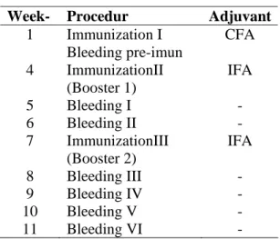
Table 1. Schedule of immunization and bleeding in rabbits
Week- Procedur Adjuvant
1 Immunization I Bleeding pre-imun
CFA
4 ImmunizationII (Booster 1)
IFA
5 Bleeding I - 6 Bleeding II - 7 ImmunizationIII
(Booster 2)
IFA
8 Bleeding III - 9 Bleeding IV - 10 Bleeding V - 11 Bleeding VI -
Sera Collection of Animals
Rabbit’s blood was collected from auricular vein. Collecting blood was approximately 2 to 3 mL and allowed to stand for 2-3 hours in sterile Eppendorf tubes. Bleeding process
The journal homepage www.jpacr.ub.ac.id ISSN : 2302 ‐ 4690
22
were performed on days of 29, 36, 43, 50, 57, 67, 74, 81, 88 and 95 after the first immunization (Table 1). They were centrifuged at 1500 rpm for 20 min, and then the supernatant was taken. Supernatant was re-centrifuged at 3000 rpm for 10 minutes. Supernatant containing anti-TPO antibodies was transferred in sterile Eppendorf tubes and stored at -20oC.
Characterization polyclonal antibody of autoantibody TPO (anti-anti-TPO) Specificity test of polyclonal antibody induced by autoantibody TPO (anti-anti-TPO) with Enzyme linked immunosorbent assay (ELISA)
Characterization of anti-anti TPO was performed by indirect ELISA. The first step was antigen coating buffer. Antigen performed by autoantibody TPO were optimally diluted in 0.05 M phosphate buffer (pH 7.5) and used as coating buffer (50µl/Well). The antigen was incubated at 40C overnight in a moist chamber. After washing, anti-TPO was coated with anti rabbit IgG AP conjugated as secondary antibody (1:2500) which were optimally diluted in PBS, and incubated for 2 hours at room temperature. The wells were washed 3 times for 3 minutes to remove unbound conjugate, and 50µl/Well of pNPP substrate in diethanolamine 10% was added, then 30 min incubation at room temperature was stopped by adding 50µl/Well of 3M NaOH. The optimal density (OD) values were recorded in a microplate ELISA reader at 405 nm.
Specificity test of polyclonal antibody induced by autoantibody TPO (anti-anti-TPO) with Western Blotting method
Western blotting was performed as immunological analysis. Protein transferring was done by wet transferred device produced by BIORAD. Protein transferring was processed from unstained gels to nitrocellulose membrane on 25 volt for 15 hours. The result was stained with Ponceau for 5-10 min, determining whether protein on the gel was transferred. Nitrocellulose membranes containing blotted antigens were cut into strips and blocked for 1 hour at room temperature in 5% skim milk containing PBS. The strips were washed in PBS-Tween 3 times for 5 minutes. To confirm the bands target, membrane nitrocellulose incubated with primary antibodies was optimally diluted in PBS-T skim milk 5% (1:200) overnight at 40C, and washed 3 times for 5 minutes with TBS. There after, membrane nitrocellulose was incubated with AP conjugated secondary antibody (1:2500 in TBS) for 1 hour room temperature, and washed PBS-T for 4 times for 5 minutes. Furthermore, protein bands were detected by adding a substrate Western Blue. The reaction was terminated by washing the membrane in distilled deionized water.
RESULTS AND DISCUSSIONS
Protein Isolated Autoantibody TPO from Serum of AITD Patients
Thyroidperoxidase (TPO) is the main enzyme involved in thyroid hormogenesis. TPO is expressed in thyroid with releasing iodine for addition to tyrosine residues in thyroglobulin (Tg) for the production of thyroxine (T4) or triiodothyronine (T3). In humans, TPO is a microsomal antigen that induces an autoimmune responses, leading role in autoimmune thyroid disease (AITD)6. It is consistent with studies Kohno et al,10 that the sera of thyroiditis containing autoantibody TPO and antigens in microsomes are major TPO itself. Epitopes TPO are recognized by autoantibody TPO. TPO can be a specific autoantigen, when cellular is damaged by hormonal and cellular immune mechanisms15. Cellular damage occurs when T lymphocytes is sensitized, and autoantibodies bind to the cell membrane causing cell lysis
The journal homepage www.jpacr.ub.ac.id ISSN : 2302 ‐ 4690
23
and inflammatory reactions. Meanwhile, changes in thyroid function occur because of stimulating autoantibody activity or blocking autoantibody in the membrane cell receptors 12.
Autoantibody TPO was used as antigen obtained by isolated autoantibody TPO. The results must be assessed of the molecular weight by SDS-PAGE using a discontinuous system with a 12.5% separating gel (Figure 1).
Figure 1. SDS-PAGE proteins isolated
from serum of AITD patients (above)
Figure 2. The Result of confirmation of
autoantibody TPO by western blotting (Right)
Crude protein containing few protein bands with various molecular weights. Relative molecular weight of each band was confirmed using the equation from a line marker protein bands. The results of protein bands profile with SDS-PAGE are 216.37 kDa, 169.10 kDa, 103.29 kDa, 62.64 kDa, 51.81 kDa, 27.29 kDa, 19.32 kDa, 16.67 kDa, and 13.69 kDa. Also, it was followed by western blotting (WB) to confirm the molecular weight of the autoantibody TPO (Figure 2).
In western blotting method, Autoantibody TPO as antigen react with autoantibody TPO monoclonal antibody, so that positive reaction of antigen-antibody is indicated by the appearance of purplish blue stain (Figure 2). The result confirms a positive reaction autoantibody TPO against monoclonal antibody TPO as visualized by a purplish blue band at 51.81 kDa region. This band is supposed as autoantibody TPO. According to Fauci et al,6
AITD patients is positive with high titers of autoantibody TPO. Therefore, autoantibody TPO acts as the best marker of AITD. Autoantibody TPO has a specificity and a sensitivity approximately 82%-100% compared to others4.
Polyclonal Antibody Production induced by autoantibody TPO (anti-anti TPO) in rabbits (Oryctolagus cuniculus)
The supposed autoantibody TPO band (51.81 kDa) was purified by the electro elution. Then, the obtained pure autoantibody TPO was induced into the rabbit, which is recognized as an antigen. As the result, the cellular system produce an immune responses to form antibody. Antibody can be found on the surface of B cells. The antibody stay at receptors or may be freely in the blood or lymph1. Specific binding of antigen causes B cells produce
The journal homepage www.jpacr.ub.ac.id ISSN : 2302 ‐ 4690
24
large amounts antigen-specific antibodies (polyclonal antibody). The polyclonal antibody specifically recognize the autoantibody TPO.
Characterization of polyclonal antibodies induced by autoantibody TPO (anti-anti-TPO) with indirect ELISA method
The highest titers of polyclonal antibodies obtained from induced autoantibody TPO were measured by ELISA. Using the method, it can detect the bleeding polyclonal antibody which requires the highest titers (Figure 3). It shows a graphic of anti-anti-TPO titer in the rabbits blood serum as much as six times bleeding after they were induced by autoantibody TPO. It was found that anti-anti-TPO concentrations increased gradually from the second week (first booster) and achieved the highest level in second bleeding with the absorbance value of 0.206. Highest titer showed an increasing number of antibodies as primary immune responses. Rabbit immune system recognizes the antigen, autoantibody TPO so that the immune responses constantly synthesize specific antibodies forming anti-anti-TPO to achieve an adequate antibodies titers. After second bleeding process, second booster was performed in order to increase anti-anti-TPO titers on third bleeding process which result in the absorbance of 0.3225. This increased absorbance is a secondary immune responses, in which B cells reactivate to produce anti-anti TPO after the rabbits was reimmunized with autoantibody TPO on second booster.
Figure 3. (Right), concentration values of
anti-TPO induced by autoantibody TPO (anti-anti-TPO). Note : 0 (preimun); 1 (serum of bleeding 1); 2 (serum of bleeding 2); 3 (serum of bleeding 3); 4 (serum of bleeding 4); 5 (serum of bleeding 5); 6 (serum of bleeding 6).
Figure 4. (Left), The autoantibody
TPO detection results in serum of AITD patients with polyclonal antibody anti-TPO.
Comparing to primary and secondary immune response absorbance value, the secondary is higher because the primary response activates native B cells which produce antibodies to response the first antigen entry16. Primary response produced the highest antibody concentrations on 14th day. The secondary immune response involved B cells memory which is faster memory proliferating than native B cells12. That is the reason why the secondary immune response was faster and produced more antibodies than the primary immune
The journal homepage www.jpacr.ub.ac.id ISSN : 2302 ‐ 4690
25
response. Titer secondary immune response was higher than the primary immune response, which can be seen on the absorbance value at the next bleeding. Compared to the second bleeding, the fourth value and next bleeding absorbance were higher, however the absorbance values of anti-anti-TPO was decreased compared to the third bleeding. Decreasing absorbance values was caused by the terminated of synthesis anti-anti-TPO so that the remain anti-anti-TPO was decomposed in the rabbit’s blood. It is caused of antigen (autoantibody TPO) which activates B cells not found in the blood.
Result from the ELISA indicated that autoantibody TPO has its immunogenicity. Immunogenicity is the ability of an antigen to induce a response in the body immune system, both humoral and cellular12. This immune response causes rabbit serum create specific polyclonal antibody to autoantibody TPO (anti-anti-TPO).
Specificity test of polyclonal antibody induced by autoantibody TPO (anti-anti-TPO) in Rabbit Blood Serum with Western Blotting Method
The aim of specifity test is to determine the ability of polyclonal antibody (anti-anti-TPO) to specifically recognize autoantibody TPO. For this purpose, Western blotting was employed to confirm the molecular weight of the anti-anti-TPO. In the Western blotting method, a protein antigen is separated according to its molecular weight, so that the antibody, which is reacted with proteins, only recognizes epitopes with specific molecular mass. The result of Western Blotting showed a positive reaction between anti-anti-TPO and autoantibody TPO (Figure 4). Positive reaction proved that the polyclonal antibody anti-anti-TPO bears a high specificity because it only recognized autoantibody anti-anti-TPO. The result was indicated by forming band 51.81 kDa on membrane nitrocelluse. Specificity antibody is determined by the amount of epitopes that can be bound to antibody, the lower epitop recognized, the better specificity of antibody. Generally, polyclonal antibody has a high affinity but a low specificity since this antibody can detect many types of injected protein epitopes. In this experiment, the result of specificity test indicates that autoantibody TPO has an appropriate epitopes to polyclonal antibody anti-anti-TPO, so that the anti-anti-TPO has a high specificity in which anti-anti-TPO only binds with autoantibody TPO epitopes. The higher level of specificity according to Goldsby et al7 will not cause any cross-reactivity. Antibody with a high specificity will not react to antigens that does not match with those antibody. Moreover, it will only react with the target (autoantibody TPO) which relates to AITD.
CONCLUSION
Autoantibody TPO isolated from serum of AITD patients has 51.81 kDa molecular weight. Autoantibody TPO is an immunogenic, so that it can induce polyclonal antibody anti-anti-TPO. The results of sensitivity and specificity of polyclonal antibody anti-anti-TPO is specified to autoantibody TPO according to ELISA and Western blotting method. Character of anti-anti-TPO has the highest titer on third bleeding with absorbance at 0.3225 and is able to recognize specific autoantibody TPO on 51.81 kDa region.
ACKNOWLEDGEMENT
The author would like to acknowledgement to rector of Brawijaya University and Directorate general of higher education (DIKTI) Indonesia for research grants 2012, and Dr.Sc Akhmad Sabarudin who helped reading manuscript. And Without whose participatioan this work would not be completed succesfully.
The journal homepage www.jpacr.ub.ac.id ISSN : 2302 ‐ 4690
26 REFERENCES
[1] Baratawidjaja, K.G., dan Rengganis. I., Imunologi Dasar, Edisi ke-8, 2009. Fakultas Kedokteran Universitas Indonesia. Jakarta.
[2] Barrett, E.J, The Thyroid gland. in Boron WF. Boulpaep EL. Medical physiology.A cellular and molecular approach.1st Edition, 2003, Saunders. Philadelphia. 1035-1048. [3] Baynes, J., and Dominiczak, M., Medical Biochemistry: Biochemical Endocrinology.
2003, Mosby, London.
[4] Chavan. A., M. Kumar., D. Prasad., S. Sundaresan., and T. Thangapannerselvem,
Inter. J. Res., 2010, 2 (1), 219-223.
[5] Darwish. A.I .Abdul-Rahman M Al-Obaid. and Hamoud A Al-Malaq, Chem. Central J.
2011, 5, 38.
[6] Fauci. S. A., D. L. Kasper., D. L. Longo., E. Braunwald., S. L. Hauser., J. L. Jameson., J. Loscalzo, Harrison’s Principles of Internal Medicine, 17th Edition, 2008, McGraw Hill Medical. New York.
[7] Goldsby. R.A., T. J. Kindt, and A. Osborne, Immunology, 2000, W. H. Freeman and Company. California.
[8] Ghoraishian, S.M., S. H.H. Moghaddam and M. A. Ardekani, Iran J. Immun,2006, 3, 3.
[9] Kindt. T.J., R.A.Goldsby., B.A. Osborne., and J. Kuby, Immunology, 2007, W.H. Freeman. New York.
[10] Kohno. Y. N. Naito. K. Saito. A Hoshioka.H.Niimi. H. Nakajima and T. Hosoya, J. Clin. Exp. Immun.,1988, 75, 217-221.
[11] Kohno. Y., F. Yamaguchi., K. Saito, H. Niimi ., T. Nishikawa., and T. Hosoya, J. Clin. Exp. Immun.,1991, 85, 459-463.
[12] Kresno, S. B., Imunologi : Diagnosis dan Prosedur Laboratorium, Edisi kelima, 2010, Badan Penerbit Fakultas Kedokteran Universitas Indonesia. Jakarta
[13] Kuby, J., The Anatomy of The Immune System. Infection and immunity Rev. 2004. [14] Masjhur, S. J., Penyakit Tiroid Autoimun. 2010, Fakultas Kedokteran Universitas
Padjadjaran. RS Dr. Hasan Sadikin. Bandung.
[15] Rifa’i. M., Imunologi dan Bioregulator. 2009, Laboratorium Faal, Fakultas Kedokteran Universitas Brawijaya. Malang.
[16] Rifa’i. M, Autoimun dan Bioregulator. 2010, Universitas Brawijaya Press. Malang. [17] Ridgway, E. C., Y. Tomer., and S. M. McLachlan. J. Clin. Endocrin. Metab.,2007, 92,
3755-3761.