57
Squamous cell carcinoma ex pleomorphic adenoma; a case report
Sara Amanpour1, Sorena Fardisi2, Reza Tabrizi3, Mohammad Reza Zarei4, Maryam Raoof5*,
Roya Khatami 6
1. Department of Oral and Maxillofacial Pathology, School of Dentistry, Kerman University of Medical Sciences, Kerman, Iran
2. Department of Oral and Maxillofacial Surgery, School of Dentistry, Shiraz University of Medical Sciences, Shiraz, Iran
3. Department of Oral and Maxillofacial Surgery, School of Dentistry, Shahid Beheshti University of Medical Sciences, Tehran, Iran
4. Department of Oral Medicine, Kerman Oral and Dental Diseases Research Center, Kerman, Iran 5. Laboratory of Molecular Neuroscience, Neuroscience Research Center, Institute of
Neuropharmacology, Kerman University of Medical Sciences, Kerman, Iran 6. Department of Oral Medicine, School of Dentistry, Shahed University, Tehran, Iran
Abstract
A rare case of squamous cell carcinoma ex pleomorphic adenoma in maxillary sinus is presented. The patient is a 50-year-old woman presenting with a slow-progressive swelling in left side of her face that she has noticed 2 years earlier. The lesion was not painful and caused asymmetry and mild exophthalmos. Microscopic examination revealed that the tumor was composed of two components; partly of a pleomorphic adenoma and partly of a squamous cell carcinoma. Immunohistochemical examination for Ki-67 and SMA and mucicarmin staining were also done and confirmed the diagnosis of carcinoma ex pleomorphic adenoma. There is no evidence of recurrence 12 months after operation.
Keywords: Pleomorphic adenoma, Malignant transformation, Squamous cell carcinoma, Case report
Introduction
Among different salivary gland tumors, carcinoma ex pleomorphic adenoma (Ca ex PA) accounts for approximately 3.6% (1). It constitutes 6.2% of all pleomorphic adenoma (PA) and 11.7% of all malignant
salivary gland neoplasms (1, 2).
Furthermore, the risk of malignant transformation in PAs in the first five years after evolution of the lesion is 1.6% (3). Car ex PA is defined as an epithelial malignant transformation within a primary or previous PA, and is often considered as a diagnostic challenge for the pathologists and clinicians, since it is difficult to
diagnose this entity pre-operatively due to unspecific clinical presentation [4, 5]. In fact, most of Ca ex PA tumors shows clinical presentation similar to PA. It means that they can be asymptomatic and
not invasive in gross examination.
However, early diagnosis and accurate surgical intervention is important to increase the patients' survival rates because of aggressiveness and destructive behavior of this tumor (1-6). WHO states that the malignant component of Ca ex PA is mostly adenocarcinoma, not otherwise specified (NOS), and sometimes salivary
Corresponding author: Tel: +98 9133416108 Fax: +98 343 2118073
Address: Neuroscience Research Center, Institute of Neuropharmacology, Kerman University of Medical Sciences, Kermam, Iran
E-mail: Maryam.raoof@gmail.com
Received; 2015/06/19 accepted; 2015/08/25
Received; 2015/04/5 revised; 2015/05/25 accepted; 2015/06/7
58
duct carcinoma (SDC), undifferentiated carcinoma, adenoid cystic carcinoma, or mucoepidermoid carcinoma (7).
We discuss the presentation,
histopathologic picture, and management of a rare case of squamous cell carcinoma
(SCC) ex pleomorphic adenoma
originating in the maxillary sinus.
Case report
A 50-year-old woman presented to our clinic with a chief complaint of asymmetry due to a long-standing swelling on the left side of her face which had first appeared 2 years earlier. There were no other symptoms such as pain and facial paralysis. The swelling was bony hard and non-tender in palpation. There was also a mild exophthalmos without pain and visual disturbance. Clinical examination of cervical lymph nodes showed no abnormal findings. Waters radiograph revealed a space-occupying destructive mass filling the left antrum completely. Computed tomography scans depicted a large ill- defined mass in left maxillary sinus causing destruction of medial, infraorbital and floor of the sinus with extention to the left orbital region (Figures 1, 2 and 3).
Figure 1. Waters view. Complete opacification of left maxillary sinus.
Figure 2. Computed tomography. Axial view; a destructive lesion in left antrum.
Figure 3. Computed tomography. Cronal view;
distruction of palate, lateral nasal wall and maxillary sinus.
Incisional biopsy of the lesion was
performed and histopathological
examination revealed a case of
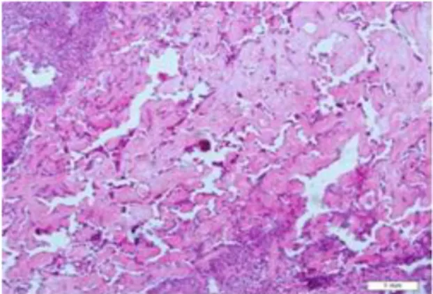
pleomorphic adenoma in a hyalinizing fibrotic and chondromyxoid stroma with the malignant areas of carcinomatous component consisting dysplastic squamous cells which exhibited large nuclei with pleomorphic shapes, prominent nucleoli and few abnormal mitotic figures. Focal areas of calcification were present. Moreover, clear cells arranged in small nests or as individual cells intermixed with squamous cells were also detected (Figures 4, 5 and 6).
59
Figure 4. Histopathologic features of PA
component of tumor (Hematoxylin-Eosin, original magnification ×100).
Figure 5. Squamous cell carcinoma cell component
of the tumor (Hematoxylin-Eosin, original
magnification ×100).
Figure 6. Extensive hyalinization and areas of
calcification close to the malignant cells
(Hematoxylin-Eosin, original magnification ×100). Mucicarmin staining was done and all clear cells were negative, therefore, the
diagnosis of mucoepdermoid carcinoma was ruled out for the malignant component
of the tumor. Immunohistochemical
staining was positive for ki-67 in more than 60% of squamous cells whereas pleomorphic adenoma component was negative for this marker. Additional immunohistochemical staining was also conducted. The clear cells in malignant component were negative for SMA, but clear cells of PA showed reactivity for SMA which proved the myoepithelial origin of these cells. These findings were consistent with the diagnosis of squamous cell carcinoma ex pleomorphic adenoma for this case.
Discussion
Based on World Health Organization classification in 2005, Ca ex PA is the most common malignant change in PA and
accounts for 11.7% of salivary
malignancies (3, 8). Malignant mixed tumors are divided into three categories: 1. carcinoma ex pleomorphic adenoma, 2. Carcinosarcoma and 3. metastasizing mixed tumor. (9)
Although PA mostly arise in the parotid gland or minor salivary glands, it is also
the most frequent tumor of the
seromucinous glands of other organs such as nasal cavity, sinuses and trachea (10). Any type of carcinoma can arise in a PA, but adenocarcinoma NOS and salivary duct carcinoma are the most common types. (4, 5, 11). The frequency of malignant components of Ca ex-PA is 43– 90% in adenocarcinoma, not otherwise specified (NOS), 10–34% in SDC, and rare in mucoepidermoid carcinoma and SCC (9, 11). In sinonasal tract, Ca ex PA is considered as the least common malignancy (12).
Generally, there is a history of a painless and slow-growing mass which start to enlarge rapidly over a short period of time (13). Patients usually present with signs
and symptoms indicating malignant
transformation (e.g., facial nerve palsy, pain, skin perforation and trismus).
60
Approximately 23% to 40% of cases shows facial nerve weakness or palsy. (14) Cellular pleomorphism, cellular anaplasia, atypical mitosis, destruction of normal
tissues, invasiveness and abnormal
architectural patterns are defined by Gnepp and Wenig as the criteria of malignancy in a PA. (15). Gerughty et al. suggested that microscopic evidence of an invasive
growth pattern, neural or vascular
invasion, focal areas of calcification and necrosis are related to a poor prognosis. (16) In agreement with Gnepp and Wenig and according to the criteria proposed by Gerughty, this patient is definitely a case of carcinomatous transformation in the PA.
Several cases of Ca ex PA have been described in previous reports, (17-21) but SCC ex pleomorphic adenoma is appear to be rare as mentioned by Peel and Gnepp (22). Seifert et al. recognized SCC components in 4 out of 38 (10%) Ca-ex-PA patients (23). In contrast, Lewis et al. (11) found no cases with SCC components among 73 patients, nor did Matsubayashi et al. among 1010 cases. (9). In addition, in 37 cases of Car ex PA reported by Tortoledo et al. (24), no malignant components were diagnosed as SCC. A case of Ca ex PA with the presence of SCC and salivary duct carcinoma was reported by Nakamori et al. (25).
Therefore, SCC as the malignant
component of Car ex PA seems to be rare (25-28).
In differential diagnosis, it is important for the pathologists to distinguish SCC from squamous cell metaplasia, as well as SCC from mucoepidermoid carcinoma (29, 30). Ki-67 staining reported by Zhu et al. as a useful diagnostic marker to discriminate between malignant and benign tumors of salivary gland (31). In the current patient, the immunostaining of Ki-67 helped us to differential SCC from squamous cell metaplasia. Mucicarmin staining was also used to detect mucin cells in order to distinguish SCC from mucoepidermoid carcinoma.
In the present case, partial maxillectomy as well as left eye exenteration was done. As achieving a complete resection with wide margins in the head and neck region is not always possible, in order to reduce the probability of recurrence, the primary site
received external irradiation
postoperatively. The patient was then referred to a prosthodontist to fabricate maxillary and midface prosthesis. There was no evidence of recurrence 12 months after operation.
Conclusion
This is a rare case of carcinoma ex pleomorphic adenoma of the antrum with an unusual malignant component of squamous cell carcinoma. The SCC component has originated from the epithelial part of PA.
References
1. Gnepp DR: Malignant mixed tumours
of the salivary glands: a review. Pathol Annu. 1993;28(2):279–328.
2. Antony J, Gopalan V, Smith RA, Lam
AK: Carcinoma ex pleomorphic
adenoma: a comprehensive review of clinical, pathological and molecular data. Head Neck Pathol. 2011; 6(1): 1-9.
3. Furukawa M, Suzuki H, Matsuura K,
Takahashi E, Suzuki H, Tezuka F.
Carcinoma ex pleomorphic adenoma of the palatal minor salivary gland with extension into the nasopharynx. Auris Nasus Larynx. 2001; 28(5):279-81.
4. Gnepp DR, Brandwein-Gensler MS,
El-Naggar AK, Nagao T: Carcinoma ex pleomorphic adenoma. In World Health Organization Classification of Tumours: Pathology and Genetics of Head and Neck Tumours. Edited by Barnes L, Eveson JW, Reichart P,
61
Sidransky D. Lyon, France: IARC Press; 2005:242–43.
5. Ellis GL, Auclair PL: Carcinoma ex
pleomorphic adenoma, In: Tumors of the Salivary Glands. Atlas of tumor pathology. Washington, D.C: Armed
Forces Institute of Pathology;
2008:259–69.
6. Altemani A, Martins MT, Freitas L,
Soares F, Araújo NS, Araújo VC: Carcinoma ex pleomorphic adenoma (CXPA): immunoprofile of the cells involved in carcinomatous progression. Histopathology. 2005;46(3):635–41.
7. Yamada S, Nabeshima A, Tabata T,
Guo X, Tasaki T, Wang KY, Shimajiri S and Sasaguri Y. Invasive salivary
duct carcinoma ex pleomorphic
adenoma of the parotid gland: a teaching case giving rise to the genuine diagnostic difficulty on an inadequate cytology specimen. Diagnos Pathol. 2012; 7(7):61-9.
8. Grossmann Sde M, Johann AC, Castro
WH, Friedman H, Gomez RS,
Mesquita RA. Anterior midline nodule of the hard palate. Oral Surg Oral Med Oral Pathol Oral Radiol Endod. 2009; 108(6):808-11.
9. Matsubayashi S, Yoshihara T.
Carcinoma ex pleomorphic adenoma
of the salivary gland: an
immunohistochemical study. Eur Arch Otorhinolaryngol. 2007; 264(8):789– 95.
10.Cimino-Mathews A, Lin B. M,Chang
S. S, Boahene K. D, Bishop J. A. Carcinoma ex Pleomorphic Adenoma of the Nasal Cavity. Head Neck Pathol. 2011; 5(6):405–9.
11.Lewis JE, Olsen KD, Sebo TJ.
Carcinoma ex pleomorphic adenoma: pathologic analysis of 73 cases. Hum Pathol. 2001; 32(5): 596–604.
12.Cho KJ, el-Naggar AK, Mahanupab P,
Luna MA, Batsakis JG. Carcinoma ex-pleomorphic adenoma of the nasal cavity: a report of two cases. J Laryngol Otol. 1995; 109(10):677–9.
13.Livolsi VA, Perzin KH. Malignant
mixed tumors arising in salivary glands. I. Carcinomas arising in benign mixed tumors: a clinicopathologic study. Cancer. 1977; 39(5):2209-30.
14.Luers JC, Wittekindt C, Streppel M,
Guntinas-Lichius O. Carcinoma ex pleomorphic adenoma of the parotid gland. Study and implications for diagnostics and therapy. Acta Oncol. 2009; 48(1):132-6.
15.Gnepp DR, Wenig BM. Malignant
mixed tumors. In: Ellis GL, Auclair PL, Gnepp DR, eds. Major problems in pathology, vol 25, Philadelphia: W. B. Sanders, 1991; 350-68.
16.Gerughty RM, Scofield HH, Brown
FM, HennigarGR. Malignant mixed tumors of salivary gland origin. Cancer. 1969; 24(7): 471-86.
17.Harada H. Histomorphological
investigation regarding to malignant
transformation of pleomorphic
adenoma (socalled malignant mixed tumor) of the salivary gland origin: special reference to carcinosarcoma. Kurume Med J. 2000; 47(7):307–23.
18.Olsen KD, Lewis JE. Carcinoma ex
pleomorphic adenoma: a
clinicopathologic review. Head Neck. 2001; 23(5):705–12.
19.Rosa JC, Fonseca I, Felix A, Soares J.
Immunohistochemical study of c-erbB–2 expression in carcinoma
ex-pleomorphic adenoma.
Histopathology. 1996; 28(8):247–52.
20.Zbaren P, Zbaren S, Caversaccio MD,
Stauffer E. Carcinoma ex pleomorphic adenoma: diagnostic difficulty and outcome. Otolaryngol Head Neck Surg. 2008; 138(11):601–5.
21.Spiro RH, Huvos AG, Strong EW.
Malignant mixed tumor of salivary origin. A clinicopathologic study of 146 cases. Cancer. 1977; 39:388–96.
22.Peel RT, Gnepp DR. Diseases of the
salivary glands. In: Barnet Leon, editor. Surgical pathology of the head and neck. New York: Marcel Dekker; 1985: 593–6.
62
23.Seifert G, Schultz J, Donath K. A
pathological sub-classification of
carcinoma of salivary gland
pleomorphic adenoma. An analysis of 38 cases. HNO 1977; 25:337–348.
24.Tortoledo ME, Luna MA, Batsakis JG:
Carcinomas ex pleomorphic adenoma
and malignant mixed tumors.
Histomorphologic indexes. Arch
Otolaryngol. 1984; 110(6): 172–6.
25.Nakamori K, Ohuchi T, Hasegawa T,
et al. Carcinoma ex pleomorphic adenoma of the buccal region is composed of salivary duct carcinoma
and squamous cell carcinoma
components. Int J Oral Maxillofac Surg. 2009; 38(9): 1116-8.
26.Kim KM, Lee A, Yoon SH, Kang JH,
Shim SI: Carcinoma ex pleomorphic adenoma of the palate: a case report. J Korean Med Sci. 1997; 12(7):63–6.
27.Garth RJ: Squamous liver metastases
from a carcinoma arising within a pleomorphic adenoma of the parotid
gland. J Laryngol Otol. 1990;
104(6):152–3.
28.Iino M, Yamada H, Ishikawa H,
Suzuki M, Shomura E, Ide F, Saito I, Mori Y: Carcinoma ex pleomorphic adenoma of the submandibular gland: report of a case with an unusual malignant component of clear cell squamous cell carcinoma. Oral Surg Oral Med Oral Pathol Oral Radiol Endod. 2008; 106(3):30–4.
29.Aker H, Ozturk M, Ozec I, Ozer H. An
usual buccal adenoma with extensive
squamous metaplasia and cyst
formation. J Chin Med Assoc. 2003; 66(6):184–8.
30.Donath K, Seifert G.
Tumour-stimulating squamous cell Metaplasia (SCM) in necrotic aria of salivary gland tumours. Pathol Res Pract .1997; 193(7):689–93.
31.Zhu Q, Tipoe GL, White FH.
Proliferative activity as detected by
immunostaining with Ki-67 and
proliferating cell nuclear antigen in benign and malignant epithelial lesions of the human parotid gland. Anal Quant Cytol Histol. 1999; 21(2):336– 42.