Exploring
bEtapapillomavirus
infEctions and thEir
Colophon
Elsemieke Iebeliene Plasmeijer
Exploring betapapillomavirus infections and their association with cutaneous squamous-cell carcinoma development
Thesis, Leiden University, Leiden, The Netherlands
Cover image: Uluru 2008
Layout and printing: Optima Grafische Communicatie, Rotterdam, The Netherlands ISBN: 978-90-8559-066-8
©Elsemieke Plasmeijer, 2010
Exploring betapapillomavir us infections and
their association with cutaneous squamous-cell
carcinoma development
Proefschrift
ter verkrijging van
de graad van Doctor aan de Universiteit Leiden, op gezag van de Rector Magnificus prof.mr. P.F. van der Heijden,
volgens besluit van het College voor Promoties te verdedigen op dinsdag 26 oktober 2010
klokke 15.00 uur
door
Elsemieke Iebeliene Plasmeijer
promotiecommissie
Promotores: Prof.Dr. A.C.M. Kroes
Prof.Dr. A.C. Green (University of Queensland, Brisbane, Australia) Copromotor: Dr. M.C.W. Feltkamp
Overige leden: Prof.Dr. J.A. Bruijn Prof.Dr. J. Vandenbroucke Prof.Dr. R. Willemze
Als je goed om je heen kijkt zie je dat alles gekleurd is -K. Schippers
contEnts
Chapter 1 General introduction 9
Chapter 2 Transmission of betapapillomaviruses between domestic partners in an Australian community
27
Chapter 3 Betapapillomavirus infection profiles in tissue sets from cutaneous squamous cell-carcinoma patients
37
Chapter 4 Lack of association between the presence and persistence of betapapillomavirus DNA in eyebrow hairs and betapapillomavirus L1 antibodies in serum
53
Chapter 5 Persistence of betapapillomavirus infections as a risk factor for actinic keratoses, precur-sor to cutaneous squamous-cell carcinoma
71
Chapter 6 The association between cutaneous squamous cell carcinoma and betapapil-lomavirus seropositivity: a cohort study
89
Chapter 7 General discussion 105
Samenvatting 121
Publications 127
Authors and affiliations 131
Curriculum vitae 135
chaptEr 1
chapter 1 11
papillomaviridae
Papillomaviruses are small epitheliotropic DNA-viruses that can induce a wide variety of hyperproliferative lesions (papillomas, warts, carcinomas) in the skin and mucosa of mammals (rabbit, horse, dog, sheep, deer, elk, cattle, primates and humans) and birds. Papillomaviruses are subdivided into different genera (Figure): the human papillomavi-ruses (HPV) belong to the genera alpha, beta, gamma, mu and nu and include mucosal and cutaneous types.
In 1933, the etiologic agent of cutaneous warts in cottontail rabbits was identified by Richard Shope (1) as a transmittable virus later called the cottontail rabbit papillomavirus (CRPV). In 1949 Strauss and colleagues (2) were the first to detect viral particles in human warts by electron microscopy. Subsequently, at least 100 different full length HPV genomes have been described and new types are detected regularly (3-6). A new papillomavirus (PV) isolate is recognized as such if the complete genome has been cloned and the DNA sequence of the L1 open reading frame (ORF) differs by more than 10% from the closest known PV type (4).
HPV are known to be associated with benign anogenital and cutaneous warts (7), as well as to be involved in cancer development, in particular with anogenital cancer (8). Most nota-bly the carcinogenic role of high-risk mucosal HPV in cervical cancer is well established and was first proposed in 1976 by Zur Hausen (9), who was recently awarded the Nobel Prize for his pioneering work in this area.
A role for HPV in human skin carcinogenesis was suggested even earlier by Jablonska and co-workers (10) while working with patients suffering from a rare genodermatosis called epidemodysplasia verruciformis (EV) who are at increased risk of cutaneous squamous cell carcinoma (SCC). Several HPV types have been found in EV-associated SCC and subsequently they have been associated with non-EV SCC in epidemiological as well as laboratory studies (11-14). Types from the betagenus (betaPV) appear to be the most likely candidates involved in skin carcinogenesis, especially SCC.
betapapillomavir uses
12 chapter 1
of known betaPV types (3;4;15) (Figure). BetaPV DNA can be found in plucked eyebrow hairs, skin swabs and skin biopsies, as well as betaPV antibodies being detected in serum.
detection methods
betapv dna
The presence of betaPV in plucked eyebrow hairs has been used as a measure of betaPV infection in several epidemiologic studies. Detection of betaPV DNA in DNA extracted from plucked hairs, skin swabs or biopsies is usually performed with polymerase chain
Chapter
1
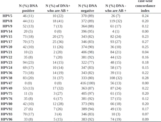
Fig
ur
e
Figure. P hylo genetic tr ee in fe rre d f ro m th e L 1 nu cleotide sequen ces of th e c ur re nt ly k now n 1 89 pa pil lo m av iru se s. Figure fr om a nd le ge nd a da pt ed fr om (6 ).Figure. Phylogenetic tree inferred from the L1 nucleotide sequences of the currently known 189
chapter 1 13
reaction (PCR) whereby preferential areas of the genome can be amplified. Next to type-specific PCRs for betaPV genotypes, several broad-spectrum PCR methods have been developed to detect cutaneous HPV-types, species or genera: CPI/IIs (16), FAP59/64 (17), F/G (18), modified F/G (MaHa) (19), HPV-type specific PCR (20), degenerate nested PCR (21) and PM-PCR (22). Broad spectrum PCR methods can be combined with either cloning and sequencing or direct sequencing of the amplimer, but these methods are too laborious for large epidemiological studies. On the other hand the development of a reverse hybrid-ization assay (RHA) in combination with the PM-PCR enables quick and simultaneous identification of 25 betaPV types (22). Other detection methods are the APEX (23) and the reverse line blotting (RLB) methods (24).
BetaPV antibodies
Antibodies against betaPV proteins can be detected to determine a person’s betaPV sero-logical status. These antibodies can be detected against the major capsid protein L1 and the non-structural protein E6 using HPV-virus like particle (VLP) or GST-HPV fusion proteins as antigen in ELISA (12;25) or multiplex (26). The latter method (Luminex®) is a new method based on fluorescent bead technology that allows simultaneous detection of anti-bodies against up to 100 different in situ affinity-purified recombinant HPV proteins (27).
natural histor y
acquisition and transmission
14 chapter 1
BetaPV can be found on different parts of the skin, as demonstrated by skin swabs of the forehead (17), arms and legs; and by plucked hairs from eyebrows, arms and legs (30;32). It is likely that betaPV infection is acquired early in life by close skin contact since children appear to be infected with the same cutaneous HPV types as their parents within months after birth (33). The exact transmission route of betaPV is unknown but it is hypothesized to be transmitted through skin and hair derivates (34). Recent studies have given contradictory results however, with one study suggesting that transmission between parents and children also occurs at later ages and in adulthood as well (35), while another has suggested that transmission occurs rarely between family members (36). The issue of betaPV transmission is the topic of Chapter 2, which suggests that close skin contact is the primary means of transmission.
prevalence and per sistence
dna prevalence
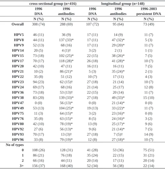
The overall prevalence of betaPV DNA is high, but varies depending on the population, anatomic site assessed - whether eyebrow hairs, skin swabs or biopsies of normal skin are being sampled - and the method used for detection. Various studies showed a prevalence of betaPV, measured by either (multiple) skin swabs or plucked eyebrow hairs from multiple sites, to be between 45% and 80% (30;33;34;37). The largest study so far comprised 1405 persons without skin cancer (845 immunocompetent, 560 immunosuppressed) in 6 coun-tries (38). The overall betaPV prevalence ranged from 84-91% between immunocompetent and immunosuppressed respectively, with HPV23 the most prevalent type. Multiple betaPV types per person were often found and there was no predominant type. Only age, and for immunosuppressed participants time of immunosuppression, was associated with betaPV (38). Sun exposure and skin type were not associated.
In biopsies from normal skin the prevalence varies between 50% and 80% (39;40). In Chapter 3 the intraperson distribution of betaPV DNA in normal skin, perilesional skin and SCC biopsies as well as plucked eyebrow hairs is addressed in detail.
seroprevalence
chapter 1 15
age 13-20 years and higher betaPV positivity (44). A higher lifetime sun exposure, how-ever, was associated with decreased HPV infection. On the other hand, a US case-control study showed no significant relations between HPV seropositivity and age, skin sensitivity and number of sunburns (26). In Chapter 4 it is also shown that sunburn does not initiate the betaPV antibody response.
persistence
Persistence of viral DNA is considered an important aspect of mucosal HPV infections in relation to cervical carcinogenesis (45). Recent studies indicate that also betaPV DNA infections persist. In a small cohort of 23 healthy adults it was demonstrated that the major-ity of detected betaPV infections persisted for up to 2 years (46). Eyebrow hairs were plucked at 8 time-points over 2 years and showed that 74% of the participants had at least one persisting infection. Another recent study showed persistent betaPV DNA positivity in 48% of the 42 healthy individuals after 7 years (37). It is unknown whether persistence plays a role in the betaPV related carcinogenesis and this issue is the topic of Chapter 5.
No previous studies have involved the persistence of betaPV L1 antibodies, while in Chap-ter 5 we saw that antibodies are stable over 8 years, with 89% of people remaining antibody positive or negative.
disease associations
Epidermodysplasia verruciformis
EV was first described in 1922 by Lewandowsky and Lutz (47) as a disease where patients develop pityriasis versicolor-like lesions and flat warts as well as numerous SCC, but not basal cell carcinomas (BCC), on sun-exposed sites at a young age. In the SCC of EV patients mainly betaPV types 5 and 8 are found (48). Recently it was shown that EV-patients harbor multiple betaPV types in both eyebrow hairs and skin biopsies (49) with viral loads ranging from less than 1 betaPV copy per 100 cells up to 400 copies per cell.
16 chapter 1 Ta bl e 1. Ep id em io lo gi ca l s tu di es a bo ut th e as so ci at io n be tw ee n be ta PV D N A p re va le nc e an d SC C d ev el op m en t Author (r ef)
Study type/ population Infection marker
Method HPV -types Cases Contr ols
Adjusted odds ratio (95% CI)
Comments
Boxman (19)
Nested case-control/ Australia
DNA
in
eyebrow hairs
Nested PCR
betaPV
64 NMSC* 51 BCC 25 SCC 64 51 25 0.8 (0.3-1.8) 0.6 (0.2-1.5) 2.0 (0.5-8.0) *BCC/SCC/intra-epithial carcinoma/NMSC undefined
Boxman (56)
Cross- sectional/ Australia
DNA in eyebrow hairs Nested PCR betaPV 276 AK 231
3.4 (1.8-6.5) (M) 1.0 (0.6-1.8) (F) Significant association between betaPV
and
AK only in men
Struijk (57)
Case-control/ Netherlands
DNA
in
eyebrow hairs
Type-specific PCR 2, 5, 8, 15, 16, 20, 24, 38
155 SCC
371
1.7 (1.1-2.7)
Association between betaPV and SCC with increasing age and male sex
Harwood (53)
Case-control/ UK
DNA
in normal
skin biopsies
Degenerate/ Nested PCR
betaPV 10 NMSC* 29 6.4 (1.8-22.9) *BCC/SCC Struijk (12)
Case-control/ Australia
DNA
in
eyebrow hairs
Type-specific PCR 5, 8, 15, 20, 24, 38
126
AK
64 SCC
57
1.6 (0.8-3.0) 0.9 (0.4-2.0)
McBride (55)
Prospective/ Australia
DNA in eyebrow hairs Nested PCR betaPV 71 1-10 AK
41 > 10
AK
179
1.8 (0.7-4.4)
Association with having more than 10
AK. Significant
associations with age over 60 years, fair skin color
, high sun
chapter 1 17
suggests that EVER-defects in zinc-metabolism may also play a role in the susceptibility of EV-patients to HPV-infections (50;51).
Keratinocyte skin cancer
Keratinocyte skin cancer is a common malignancy in mainly Caucasian populations, consisting of BCC and SCC and several epidemiological studies have investigated the association between markers of HPV infection, in particular betaPV infection, and kera-tinocyte skin cancer. Although basal cell carcinomas (BCC) are the most common kerati-nocyte skin cancer, no clear associations with betaPV DNA or antibodies have been found (16;25;26;41;52-54).
Studies that have investigated the role of betaPV DNA, detected in eyebrow hairs and skin biopsies, in the development of AK and SCC are summarized in Table 1. Associa-tions have been found between the presence of betaPV DNA and AK (12;55;56) and SCC (12;19;53;57), but no specific high risk types were identified.
Studies investigating the association between antibodies against betaPV and AK or SCC are summarized in Table 2. Seroreactivity to betaPV L1 was associated with AK and SCC in a number of studies (12;26;53;54;58-61) and the presence of AK was inversely associ-ated with seroreactivity to betaPV E6 (12). E6 and L1 antibodies were hardly ever found concomitantly, suggesting that antibody responses to the early (non-structural, intracel-lular) and late (structural, also extracelintracel-lular) betaPV proteins take place at different times and phases during betaPV infection or betaPV-associated tumor development (12). It was also shown that HPV DNA positivity and L1 seropositivity were correlated, and E6 sero-positivity was inversely correlated with HPV DNA sero-positivity, somewhat in line with the hypotheses either that E6 antibodies partly protect against SCC or that SCC patients have difficulties inducing immune responses to cutaneous HPV E6 proteins (12;57).
18 chapter 1 Ta bl e 2. Ep id em io lo gi ca l s tu di es a bo ut th e as so ci at io n be tw ee n be ta PV s er op re va le nc e an d SC C d ev el op m en t Author (r ef)
Study type/ population Infection marker
Method HPV -types Cases Contr ols
Adjusted odds ratio (95% CI)
Comments
Steger (59)
Case-control/ Germany
L1 serology W estern blot 8 11 445 10.7 (2.5-63.2) Stark (61)
Case-control/ Germany
L1 serology ELISA 8 14 SCC 210 30.3 (7.4-142.5)
Bouwes Bavinck (60) Case-control/ Netherlands
L1 serology ELISA 8 13 SCC 82 3.1 (0.7-13.3) Feltkamp (25)
Case-control/ Netherlands
L1 serology
ELISA
5, 8, 15, 20, 24, 38
540 SCC
333
1.4 (0.8-2.5)
Masini (58)
Case-control/ Italy
L1 serology
ELISA
8, 15, 23, 36
46 SCC
84
3.2 (1.3-7.9) (HPV8) 0.4 (0.2-0.9) (HPV15) 1.0 (0.3-3.3) HPV23) 2.8 (0.8-10.0) (HPV36)
Karagas (26)
Case-control/ USA
L1 serology multiplex betaPV 252 SCC 461 1.5 (1.1-2.1) Struijk (12)
Case-control/ Australia L1/E6 serology
ELISA
5, 8, 15, 16, 20, 24, 38
126
AK
64 SCC
57
2.3 (0.9-4.9)(L1) 0.6 (0.3-1.3) (E6) 3.9 (1.4-10.7) (L1) 0.5 (0.2-1.1) (E6)
Associations between betaPV
L1
and E6 serology and
AK/SCC
Casabonne (26)
Nested case- control/ UK
L1 serology
multiplex
betaPV
39 SCC
80
0.5 (0.1-1.7)* 1.0 (0.4-2.5) ** Association between 1* or 2+** betaPV
type(s) and SCC
Karagas (54) Case-control/ USA L1 serology multiplex betaPV 663 SCC 805
chapter 1 19
Australia, that started in 1986 with the enrollment of 2095 participants (66) who were then followed up until 2007. All studies described in this thesis have been performed in Australian participants, Chapters 2, 4, 5 and 6 as part of the Nambour Skin Cancer Study and Chapter 3 in a small cohort of SCC-patients in Northern Queensland.
scope of this thesis
Chapter 2 describes the transmission of betaPV between opposite-sex partners as com-pared with age and sex matched controls.
Chapter 3 describes the distribution of betaPV as measured in eyebrow hairs and biopsies of normal skin, SCC tumor tissue and perilesional skin of 21 SCC-patients.
Chapter 4 describes the relation between two frequently used markers for betaPV research: betaPV DNA in eyebrow hairs and betaPV antibodies from serum, both cross-sectionally and longitudinally.
Chapter 5 describes the association between persistent betaPV infection as indicated by viral DNA in eyebrow hairs and the risk of AK on the whole body and on the face. Chapter 6 describes the association between betaPV antibodies in serum and the develop-ment of SCC in a longitudinal study.
20 chapter 1
references
1 Shope R, Weston Hurst E. Infectious papillomatosis of rabbits. J Exp Med 1933;58(november):607-24. 2 Strauss MJ, Shaw EW. Crystalline virus-like particles from skin papillomas characterized by intranuclear
inclusion bodies. Proc Soc Exp Biol Med 1949 Oct;72(1):46-50.
3 De Villiers EM, Gunst K. Characterization of seven novel human papillomavirus types isolated from cutane-ous tissue, but also present in mucosal lesions. J Gen Virol 2009 Aug;90(Pt 8):1999-2004.
4 De Villiers EM, Fauquet C, Broker TR, Bernard HU, zur Hausen H. Classification of papillomaviruses. Virology 2004 Jun 20;324(1):17-27.
5 zur Hausen H. Papillomaviruses causing cancer: evasion from host-cell control in early events in carcinogen-esis. J Natl Cancer Inst 2000 May 3;92(9):690-8.
6 Bernard HU, Burk RD, Chen Z, van DK, zur Hausen H, De Villiers EM. Classification of papillomaviruses (PVs) based on 189 PV types and proposal of taxonomic amendments. Virology 2010 May 25;401(1):70-9. 7 Jablonska S, Orth G. Cutaneous warts: clinical, histological and virological correlations. Arch Dermatol Res
1995;287(6):616-8.
8 Schiffman M, Castle PE, Jeronimo J, Rodriguez AC, Wacholder S. Human papillomavirus and cervical cancer. Lancet 2007 Sep 8;370(9590):890-907.
9 zur Hausen H. Condylomata acuminata and human genital cancer. Cancer Res 1976 Feb;36(2 pt 2):794. 10 Jablonska S, Dabrowski J, Jakubowicz K. Epidermodysplasia verruciformis as a model in studies on the role
of papovaviruses in oncogenesis. Cancer Res 1972 Mar;32(3):583-9.
11 Weissenborn SJ, Nindl I, Purdie K, Harwood C, Proby C, Breuer J, et al. Human papillomavirus-DNA loads in actinic keratoses exceed those in non-melanoma skin cancers. J Invest Dermatol 2005 Jul;125(1):93-7. 12 Struijk L, Hall L, Van der Meijden E, Wanningen P, Bouwes Bavinck JN, Neale R, et al. Markers of
cutane-ous human papillomavirus infection in individuals with tumor-free skin, actinic keratoses, and squamcutane-ous cell carcinoma. Cancer Epidemiol Biomarkers Prev 2006 Mar;15(3):529-35.
13 Schaper ID, Marcuzzi GP, Weissenborn SJ, Kasper HU, Dries V, Smyth N, et al. Development of skin tumors in mice transgenic for early genes of human papillomavirus type 8. Cancer Research 2005 Feb 15;65(4):1394-400.
14 Caldeira S, Zehbe I, Accardi R, Malanchi I, Dong W, Giarre M, et al. The E6 and E7 proteins of the cutaneous human papillomavirus type 38 display transforming properties. J Virol 2003 Feb;77(3):2195-206.
15 Pfister H. Chapter 8: Human papillomavirus and skin cancer. J Natl Cancer Inst Monogr 2003;(31):52-6. 16 Tieben LM, Ter Schegget J, Minnaar RP, Bouwes Bavinck JN, Berkhout RJ, Vermeer BJ, et al. Detection of
cutaneous and genital HPV types in clinical samples by PCR using consensus primers. J Virol Methods 1993 May;42(2-3):265-79.
17 Forslund O, Antonsson A, Nordin P, Stenquist B, Hansson BG. A broad range of human papillomavirus types detected with a general PCR method suitable for analysis of cutaneous tumours and normal skin. J Gen Virol 1999 Sep;80 ( Pt 9):2437-43.
chapter 1 21
19 Boxman ILA, Russell A, Mulder LHC, Bouwes Bavinck JN, Ter Schegget J, Green A. Case-control study in a subtropical Australian population to assess the relation between non-melanoma skin cancer and epider-modysplasia verruciformis human papillomavirus DNA in plucked eyebrow hairs. International Journal of Cancer 2000 Apr 1;86(1):118-21.
20 Shamanin V, Delius H, De Villiers EM. Development of a broad spectrum PCR assay for papillomaviruses and its application in screening lung cancer biopsies. J Gen Virol 1994 May;75 ( Pt 5):1149-56.
21 Harwood CA, Spink PJ, Surentheran T, Leigh IM, De Villiers EM, McGregor JM, et al. Degenerate and nested PCR: a highly sensitive and specific method for detection of human papillomavirus infection in cutaneous warts. J Clin Microbiol 1999 Nov;37(11):3545-55.
22 de Koning M, Quint W, Struijk L, Kleter B, Wanningen P, van Doorn LJ, et al. Evaluation of a novel highly sensitive, broad-spectrum PCR-reverse hybridization assay for detection and identification of beta-papillomavirus DNA. J Clin Microbiol 2006 May;44(5):1792-800.
23 Gheit T, Billoud G, de Koning MN, Gemignani F, Forslund O, Sylla BS, et al. Development of a sensitive and specific multiplex PCR method combined with DNA microarray primer extension to detect Betapapil-lomavirus types. J Clin Microbiol 2007 Aug;45(8):2537-44.
24 Brink AA, Lloveras B, Nindl I, Heideman DA, Kramer D, Pol R, et al. Development of a general-primer-PCR-reverse-line-blotting system for detection of beta and gamma cutaneous human papillomaviruses. J Clin Microbiol 2005 Nov;43(11):5581-7.
25 Feltkamp MCW, Broer R, di Summa FM, Struijk L, Van der Meijden E, Verlaan BPJ, et al. Seroreactivity to epidermodysplasia verruciformis-related human papillomavirus types is associated with nonmelanoma skin cancer. Cancer Research 2003 May 15;63(10):2695-700.
26 Karagas MR, Nelson HH, Sehr P, Waterboer T, Stukel TA, Andrew A, et al. Human papillomavirus infec-tion and incidence of squamous cell and basal cell carcinomas of the skin. J Natl Cancer Inst 2006 Mar 15;98(6):389-95.
27 Waterboer T, Sehr P, Michael KM, Franceschi S, Nieland JD, Joos TO, et al. Multiplex human papilloma-virus serology based on in situ-purified glutathione S-transferase fusion proteins. Clinical Chemistry 2005 Oct;51(10):1845-53.
28 Orth G. Genetics of epidermodysplasia verruciformis: Insights into host defense against papillomaviruses. Semin Immunol 2006 Dec;18(6):362-74.
29 Munoz N, Castellsague X, de Gonzalez AB, Gissmann L. Chapter 1: HPV in the etiology of human cancer. Vaccine 2006 Aug 21;24S3:S1-S10.
30 Boxman IL, Berkhout RJ, Mulder LH, Wolkers MC, Bouwes Bavinck JN, Vermeer BJ, et al. Detection of human papillomavirus DNA in plucked hairs from renal transplant recipients and healthy volunteers. J Invest Dermatol 1997 May;108(5):712-5.
31 Schmitt A, Rochat A, Zeltner R, Borenstein L, Barrandon Y, Wettstein FO, et al. The primary target cells of the high-risk cottontail rabbit papillomavirus colocalize with hair follicle stem cells. J Virol 1996 Mar;70(3):1912-22.
22 chapter 1
33 Antonsson A, Karanfilovska S, Lindqvist PG, Hansson BG. General acquisition of human papillomavirus infections of skin occurs in early infancy. Journal of Clinical Microbiology 2003 Jun;41(6):2509-14. 34 Antonsson A, Forslund O, Ekberg H, Sterner G, Hansson BG. The ubiquity and impressive genomic diversity
of human skin papillomaviruses suggest a commensalic nature of these viruses. Journal of Virology 2000 Dec;74(24):11636-41.
35 Weissenborn SJ, de Koning MN, Wieland U, Quint WG, Pfister HJ. Intrafamilial transmission and family-specific spectra of cutaneous betapapillomaviruses. J Virol 2009 Jan;83(2):811-6.
36 Gottschling M, Goker M, Kohler A, Lehmann MD, Stockfleth E, Nindl I. Cutaneotropic human beta-/gamma-papillomaviruses are rarely shared between family members. J Invest Dermatol 2009 Oct;129(10):2427-34. 37 Hazard K, Karlsson A, Andersson K, Ekberg H, Dillner J, Forslund O. Cutaneous Human Papillomaviruses
Persist on Healthy Skin. J Invest Dermatol 2006 Oct 5.
38 de Koning MN, Weissenborn SJ, Abeni D, Bouwes Bavinck JN, Euvrard S, Green AC, et al. Prevalence and associated factors of betapapillomavirus infections in individuals without cutaneous squamous cell carcinoma. J Gen Virol 2009 Mar 25.
39 Asgari MM, Kiviat NB, Critchlow CW, Stern JE, Argenyi ZB, Raugi GJ, et al. Detection of human papil-lomavirus DNA in cutaneous squamous cell carcinoma among immunocompetent individuals. J Invest Dermatol 2008 Jun;128(6):1409-17.
40 Rollison DE, Pawlita M, Giuliano AR, Iannacone MR, Sondak VK, Messina JL, et al. Measures of cutaneous human papillomavirus infection in normal tissues as biomarkers of HPV in corresponding nonmelanoma skin cancers. Int J Cancer 2008 Nov 15;123(10):2337-42.
41 Waterboer T, Neale R, Michael KM, Sehr P, de Koning MN, Weissenborn SJ, et al. Antibody responses to 26 skin human papillomavirus types in the Netherlands, Italy and Australia. J Gen Virol 2009 Aug;90(Pt 8):1986-98.
42 Michael KM, Waterboer T, Sehr P, Rother A, Reidel U, Boeing H, et al. Seroprevalence of 34 human papil-lomavirus types in the German general population. PLoS Pathog 2008 Jun;4(6):e1000091.
43 Casabonne D, Waterboer T, Michael KM, Pawlita M, Mitchell L, Newton R, et al. The seroprevalence of human papillomavirus by immune status and by ethnicity in London. Infect Agent Cancer 2009;4:14. 44 Termorshuizen F, Feltkamp MC, Struijk L, de Gruijl FR, Bouwes Bavinck JN, van Loveren H. Sunlight
exposure and (sero)prevalence of epidermodysplasia verruciformis-associated human papillomavirus. J Invest Dermatol 2004 Jun;122(6):1456-62.
45 Schlecht NF, Kulaga S, Robitaille J, Ferreira S, Santos M, Miyamura RA, et al. Persistent human papil-lomavirus infection as a predictor of cervical intraepithelial neoplasia. JAMA 2001 Dec 26;286(24):3106-14. 46 de Koning MN, Struijk L, Bouwes Bavinck JN, Kleter B, Ter Schegget J, Quint WG, et al.
Betapapillomavi-ruses frequently persist in the skin of healthy individuals. J Gen Virol 2007 May;88(Pt 5):1489-95. 47 Lewandowsky F., Lutz W. A case of a previously undescribed skin disease (epidemodysplasia verruciformis).
Arch Dermatol Syphilol 1922;141: 193-203.
48 Majewski S, Jablonska S, Orth G. Epidermodysplasia verruciformis. Immunological and nonimmunological surveillance mechanisms: role in tumor progression. Clin Dermatol 1997 May;15(3):321-34.
chapter 1 23
50 Lazarczyk M, Cassonnet P, Pons C, Jacob Y, Favre M. The EVER proteins as a natural barrier against papillomaviruses: a new insight into the pathogenesis of human papillomavirus infections. Microbiol Mol Biol Rev 2009 Jun;73(2):348-70.
51 Lazarczyk M, Pons C, Mendoza JA, Cassonnet P, Jacob Y, Favre M. Regulation of cellular zinc balance as a potential mechanism of EVER-mediated protection against pathogenesis by cutaneous oncogenic human papillomaviruses. J Exp Med 2008 Jan 21;205(1):35-42.
52 Wieland U, Ritzkowsky A, Stoltidis M, Weissenborn S, Stark S, Ploner M, et al. Communication: papil-lomavirus DNA in basal cell carcinomas of immunocompetent patients: an accidental association?TITLE. J Invest Dermatol 2000 Jul;115(1):124-8.
53 Harwood CA, Surentheran T, Sasieni P, Proby CM, Bordea C, Leigh IM, et al. Increased risk of skin cancer associated with the presence of epidermodysplasia verruciformis human papillomavirus types in normal skin. Br J Dermatol 2004 May;150(5):949-57.
54 Karagas MR, Waterboer T, Li Z, Nelson HH, Michael KM, Bavinck JN, et al. Genus beta human papil-lomaviruses and incidence of basal cell and squamous cell carcinomas of skin: population based case-control study. BMJ 2010;341:c2986.
55 McBride P, Neale R, Pandeya N, Green A. Sun-related factors, betapapillomavirus, and actinic keratoses: a prospective study. Arch Dermatol 2007 Jul;143(7):862-8.
56 Boxman ILA, Russell A, Mulder LHC, Bouwes Bavinck JN, Ter Schegget J, Green A. Association between epidermodysplasia verruciformis- associated human papillomavirus DNA in plucked eyebrow hair and solar keratoses. Journal of Investigative Dermatology 2001 Nov;117(5):1108-12.
57 Struijk L, Bouwes Bavinck JN, Wanningen P, Van der Meijden E, Westendorp RGJ, Ter Schegget J, et al. Presence of human papillomavirus DNA in plucked eyebrow hairs is associated with a history of cutaneous squamous cell carcinoma. Journal of Investigative Dermatology 2003 Dec;121(6):1531-5.
58 Masini C, Fuchs PG, Gabrielli F, Stark S, Sera F, Ploner M, et al. Evidence for the association of human papillomavirus infection and cutaneous squamous cell carcinoma in immunocompetent individuals. Arch Dermatol 2003 Jul;139(7):890-4.
59 Steger G, Olszewsky M, Stockfleth E, Pfister H. Prevalence of antibodies to human papillomavirus type 8 in human sera. J Virol 1990 Sep;64(9):4399-406.
60 Bouwes Bavinck JN, Stark S, Petridis AK, Marugg ME, Ter Schegget J, Westendorp RG, et al. The presence of antibodies against virus-like particles of epidermodysplasia verruciformis-associated humanpapillomavi-rus type 8 in patients with actinic keratoses. Br J Dermatol 2000 Jan;142(1):103-9.
61 Stark S, Petridis AK, Ghim SJ, Jenson AB, Bouwes Bavinck JN, Gross G, et al. Prevalence of antibodies against virus-like particles of Epidermodysplasia verruciformis-associated HPV8 in patients at risk of skin cancer. J Invest Dermatol 1998 Oct;111(4):696-701.
62 Bouwes Bavinck JN, De Boer A, Vermeer BJ, Hartevelt MM, van der Woude FJ, Claas FH, et al. Sunlight, keratotic skin lesions and skin cancer in renal transplant recipients. Br J Dermatol 1993 Sep;129(3):242-9. 63 Brash DE, Rudolph JA, Simon JA, Lin A, McKenna GJ, Baden HP, et al. A role for sunlight in skin cancer:
24 chapter 1
65 Green A, Battistutta D, Hart V, Leslie D, Weedon D. Skin cancer in a subtropical Australian population: incidence and lack of association with occupation. The Nambour Study Group. Am J Epidemiol 1996 Dec 1;144(11):1034-40.
chaptEr 2
transmission of
bEtapapillomavirusEs
bEtwEEn domEstic
partnErs in an australian
community
Elsemieke I. Plasmeijer, Adele C. Green,Maurits
N.C. de Koning, Peter O’Rourke, Wim G.V. Quint, Mariet C.W. Feltkamp and Rachel E. Neale
28 chapter 2
abstract
Betapapillomaviruses may be associated with the development of cutaneous squamous cell carcinoma but little is known about their transmission. One suggestion is that they are transmitted through close skin contact.
To test this hypothesis we assessed whether co-habiting opposite-sex couples were more or less likely to share betaPV types than each member of the couple and an age-matched, opposite-sex control.Betapapillomavirus was measured in eyebrow hairs of 57 couples and 114 age- and sex-matched controls. We compared the proportion of partners who shared at least one betaPV type with the proportion of control partnerships sharing a betaPV type. We further subdivided those who shared at least one type into those who shared only one and those who shared more than one. We tested the significance of differences in these proportions using Chi-squared tests. A case-wise concordance index was used to calculate the overall concordance of the partners and the control pairings.
At least one betaPV type was shared by 39% of the co-habiting couples and 26% of the control pairs (p=0.10). When restricted to all people with at least one virus infection (26 couples) 74% of the partners and 46% of the control pairs shared at least one type (p=0.02). The case-wise concordance index for partners was 0.28 (95% CI 0.21-0.35) and for the matched control pairs 0.16 (95% CI 0.12-0.20) (p<0.001).
chapter 2 29
introduction
Human papillomaviruses of the beta-genus (betaPV) are cutanotropic viruses that are associated with cutaneous squamous cell carcinoma (SCC) (1). So far 31 different betaPV types have been fully sequenced and more than 100 types partially sequenced (2;3). Epi-demiological studies have shown that all currently identified betaPV types are frequently found in hair bulbs of eyebrows and body hairs, normal skin swabs and biopsies from healthy controls and transplant recipients, as well as in tumour tissue from patients with SCC (4-6). Usually multiple infections are detected within a sample (7).
Little is known about the transmission of betaPV. In healthy people no clinical signs of ini-tial infection are observed. We have found only 5 previous studies addressing transmission of betaPV, several of which are very small (8-12). The data about transmission between parents and children is ambiguous: one study involving 38 infants showed parents and babies as young as 4 weeks of age to share betaPV types (8) and another study showed that transmission between parents and children occurs frequently (13;14). However in a third study transmission between parents and children was observed rarely (15). In this cohort transmission between couples was also infrequently seen (15). A cohort of 23 participants showed that the 5 students sharing a household were not likely to obtain each other’s betaPV, but instead kept their own infection profile (10). Despite different outcomes, all of these studies concluded that betaPV transmission probably takes place during close (skin-to-skin) contact.
To test this hypothesis we assessed whether co-habiting married or de facto opposite-sex couples (herein called ‘partners’) were more or less likely to share betaPV types than each member of the couple and an age-matched, opposite-sex control.
material and methods
study population and sample collection
30 chapter 2
cohort participated in a sub-study aiming to understand the association between HPV and skin cancer (18), and 10 eyebrow hairs were plucked from each participant and processed as described below. Participants’ relationships with one another in 1996 were recorded. For the analysis described here we selected all 57 male-female co-habiting couples. For each of these 114 people, we randomly selected an opposite-sex control from the remaining 393 participants, matched to the age of his/her partner. For example, a 60-year-old man and his 50-year-old wife were matched to a 50-year-old woman and a 60-year-old man respectively.
dna isolation, pcr and hybridization
DNA from eyebrow hairs was isolated according to a method described previously (19). BetaPV detection and genotyping were performed using a reversed hybridization assay as described previously (20). All amplimers generated with the broad spectrum PCR were analysed with a reverse hybridization assay (RHA) that permitted specific detection and identification of 25 established betaPV genotypes (i.e., 5, 8, 9, 12, 14, 15, 17, 19-25, 36-38, 47, 49, 75, 76, 80, 92, 93 and 96). The RHA was performed according to the manufacturer’s instructions (skin (beta) HPV prototype research assay; Diassay BV, Rijswijk, The Neth-erlands).
statistical analyses
chapter 2 31
results
The mean age of the men was 55 years (SD 11) and of the women 51 (SD 11). Seventy-four percent of the male partners were betaPV-positive (median number of types: 2, range 1-12), compared with 86% of the male controls (p=0.07) (median number of types: 2, range 1-15), 70% of the female partners (median number of types: 2, range 1-11) and 74% of the female controls (p=0.65) (median number of types: 1, range 1-11).
At least one betaPV type was shared by 39% of the co-habiting couples (Table). For the control pairs this was 26% (p=0.10). Fourteen percent of partners versus 11% of control pairs shared more than one type (p=0.25). When we repeated the analyses for all people with at least one virus infection (26 couples) 74% of the partners and 46% of the control pairs shared at least one type (p=0.02), and 32% versus 19% shared more than 1 type (p=0.08) (Table). The case-wise concordance index for partners was 0.28 (95% CI 0.21-0.35) and for the matched control pairs 0.16 (95% CI 0.12-0.20) (p<0.001).
Table: Number of betaPV types shared by partners and by partners and their controls.
No. of shared types Partners, n=57
N (%) Control-pairs, n=114N (%)
Including all participants
0 35 (61) 84 (74)
1+ 22 (39) 30 (26)
Chi-square 2.71 (p=0.10)
0 35 (61) 84 (74)
1 14 (25) 18 (16)
>1 8 (14) 12 (11)
Chi-square 2.79 (p=0.25)
No. of shared types Partners, n=26
N (%) Control-pairs, n=52N (%)
Including only betaPV positive participants
0 7 (26) 28 (54)
1+ 19 (74) 24 (46)
Chi-square 5.08 (p=0.02)
0 7 (26) 28 (54)
1 11 (42) 14 (27)
>1 8 (32) 10 (19)
32 chapter 2
discussion
In this study we found that participants more often shared at least one betaPV type with their opposite-sex domestic partner than with random controls of the same age and sex as their partner. This difference was significant when the analysis was restricted to people who had at least one betaPV infection. Partners also were likely to share more than one type than control pairs, although due to small numbers significant differences could not be observed. We found a highly significant difference in the concordance index. We assessed skin type, sun exposure and skin cancer rate as possible confounders and found no differ-ences between the male and female partners and male and female controls. The borderline significant difference in betaPV prevalence between the male partners and male controls is most likely to be due to random sampling error and is not likely to have caused differences. The higher number of viruses seen in male controls than in male partners suggests that they would have an increased chance of sharing types with the female partner, so if anything, this variability may have led to an underestimation of the difference in shared types found.
chapter 2 33
Our data are cross-sectional and we therefore cannot address the issue of whether or not persistent betaPV types are shared between couples.
In conclusion, these cross-sectional data demonstrate that co-habiting partners of the opposite-sex share a greater number of betaPV types than with randomly selected matched members of the population. This finding supports the hypothesis that close contact is the primary means of betaPV transmission, probably through skin-to-skin contact. Larger, longitudinal studies are needed to confirm this finding and to give more insight into the sustainability of the shared infections.
acknowledgements
34 chapter 2
references
1 zur Hausen H. Papillomaviruses in human cancers. Proceedings of the Association of American Physicians 1999 Nov;111(6):581-7.
2 De Villiers EM, Gunst K. Characterization of seven novel human papillomavirus types isolated from cutane-ous tissue, but also present in mucosal lesions. J Gen Virol 2009 Aug;90(Pt 8):1999-2004.
3 Pfister H. Chapter 8: Human papillomavirus and skin cancer. J Natl Cancer Inst Monogr 2003;(31):52-6. 4 Bouwes Bavinck JN, Feltkamp M, Struijk L, Ter Schegget J. Human papillomavirus infection and skin
cancer risk in organ transplant recipients. J Investig Dermatol Symp Proc 2001 Dec;6(3):207-11.
5 Boxman IL, Berkhout RJ, Mulder LH, Wolkers MC, Bouwes Bavinck JN, Vermeer BJ, et al. Detection of human papillomavirus DNA in plucked hairs from renal transplant recipients and healthy volunteers. J Invest Dermatol 1997 May;108(5):712-5.
6 Struijk L, Hall L, Van der Meijden E, Wanningen P, Bavinck JN, Neale R, et al. Markers of cutaneous human papillomavirus infection in individuals with tumor-free skin, actinic keratoses, and squamous cell carcinoma. Cancer Epidemiol Biomarkers Prev 2006 Mar;15(3):529-35.
7 de Koning MN, Weissenborn SJ, Abeni D, Bouwes Bavinck JN, Euvrard S, Green AC, et al. Prevalence and associated factors of betapapillomavirus infections in individuals without cutaneous squamous cell carcinoma. J Gen Virol 2009 Mar 25.
8 Antonsson A, Karanfilovska S, Lindqvist PG, Hansson BG. General acquisition of human papillomavirus infections of skin occurs in early infancy. Journal of Clinical Microbiology 2003 Jun;41(6):2509-14. 9 Hsu JY, Chen AC, Keleher A, McMillan NA, Antonsson A. Shared and persistent asymptomatic cutaneous
human papillomavirus infections in healthy skin. J Med Virol 2009 Aug;81(8):1444-9.
10 de Koning MN, Struijk L, Bouwes Bavinck JN, Kleter B, Ter Schegget J, Quint WG, et al. Betapapillomavi-ruses frequently persist in the skin of healthy individuals. J Gen Virol 2007 May;88(Pt 5):1489-95. 11 Weissenborn SJ, de Koning MN, Wieland U, Quint WG, Pfister HJ. Intrafamilial transmission and
family-specific spectra of cutaneous betapapillomaviruses. J Virol 2009 Jan;83(2):811-6.
12 Gottschling M, Goker M, Kohler A, Lehmann MD, Stockfleth E, Nindl I. Cutaneotropic human beta-/gamma-papillomaviruses are rarely shared between family members. J Invest Dermatol 2009 Oct;129(10):2427-34. 13 Hsu JY, Chen AC, Keleher A, McMillan NA, Antonsson A. Shared and persistent asymptomatic cutaneous
human papillomavirus infections in healthy skin. J Med Virol 2009 Aug;81(8):1444-9.
14 Weissenborn SJ, de Koning MN, Wieland U, Quint WG, Pfister HJ. Intrafamilial transmission and family-specific spectra of cutaneous betapapillomaviruses. J Virol 2009 Jan;83(2):811-6.
15 Gottschling M, Goker M, Kohler A, Lehmann MD, Stockfleth E, Nindl I. Cutaneotropic human beta-/gamma-papillomaviruses are rarely shared between family members. J Invest Dermatol 2009 Oct;129(10):2427-34. 16 Green A, Battistutta D, Hart V, Leslie D, Weedon D. Skin cancer in a subtropical Australian population:
incidence and lack of association with occupation. The Nambour Study Group. Am J Epidemiol 1996 Dec 1;144(11):1034-40.
chapter 2 35
18 Boxman ILA, Russell A, Mulder LHC, Bavinck JNB, Ter Schegget J, Green A. Association between epi-dermodysplasia verruciformis- associated human papillomavirus DNA in plucked eyebrow hair and solar keratoses. Journal of Investigative Dermatology 2001 Nov;117(5):1108-12.
19 Boom R, Sol CJ, Salimans MM, Jansen CL, Wertheim-van Dillen PM, van der Noordaa J. Rapid and simple method for purification of nucleic acids. J Clin Microbiol 1990 Mar;28(3):495-503.
20 de Koning M, Quint W, Struijk L, Kleter B, Wanningen P, van Doorn LJ, et al. Evaluation of a novel highly sensitive, broad-spectrum PCR-reverse hybridization assay for detection and identification of beta-papillomavirus DNA. J Clin Microbiol 2006 May;44(5):1792-800.
21 Huang JK, Tai JJ. Twin concordances test for ascertained trichotomous traits data. Stat Med 2007 Feb 20;26(4):869-94.
chaptEr 3
bEtapapillomavirus
infEction profilEs
in tissuE sEts from
cutanEous squamous
cEll-carcinoma patiEnts
Elsemieke I. Plasmeijer, Rachel E. Neale, Petra G. Buettner, Maurits N.C. de Koning, Jan ter Schegget,
Wim G.V. Quint, Adele C. Greenand
Mariet C.W. Feltkamp
38 chapter 3
abstract
chapter 3 39
introduction
Infection with human papillomaviruses (HPV) from the beta-genus (betaPV) is associated with the development of actinic keratoses (AK) and squamous cell-carcinoma (SCC) in immune-competent persons as well as in organ transplant patients (1-7) The majority of people are infected with multiple betaPV (8),and a substantial proportion of these infec-tions remains detectable over time, indicative of persistent infection (9;10).
Different mechanisms by which betaPV play a role in carcinogenesis have been proposed, for example the “hit-and-run” hypothesis, whereby betaPV act early in carcinogenesis and is not necessary for maintenance of the malignant phenotype (11;12). BetaPV may act within or contribute to field cancerisation where a discrete area of tissue is at increased risk of developing cancer (13),as seen for SCC of the oesophagus (14) and cutaneous actinic keratoses (15-18). A postulated mechanism of transformation is betaPV-mediated impairment of host cell defenses against excessive sun light exposure, such as inhibition of DNA repair and apoptosis (19-21).
In epidemiological studies, the presence of betaPV DNA in eyebrow hairs, skin swabs, and normal skin biopsies have all been used as markers of betaPV-infection (22-26). Which of these is the most appropriate indicator of the betaPV types found in the tumour and/or the surrounding area is currently unknown. It has been proposed that the hair follicle is the natural reservoir of cutaneous HPV (22;27) with support from studies showing that HPV is present in hair follicles obtained from different body sites such as scalp, eyebrow, arm, trunk, leg and pubic region (22;28).
Here we have investigated within a series of SCC patients the prevalence and distribution of 25 different betaPV types in sets comprising four sample types (SCC, perilesional skin, normal skin on the mirror image site of the SCC, and plucked eyebrow hairs) to gain possible insights into viral pathogenesis of SCC and assess if plucked eyebrow hairs are indeed sentinel for betaPV present in the tumour.
material and methods
study population and sample collection
40 chapter 3
between April 2002 and April 2003 were recruited from the Townsville Hospital by local specialist doctors and general practitioners. Ten eyebrow hairs were plucked from each participant using sterile tweezers and gloves, and biopsies were collected from the SCC, perilesional skin immediately adjacent to the SCC and normal skin from the mirror image site of the SCC. Because of ethical, cosmetically reasons, for the patients with a facial SCC (n=3) a biopsy of the forearm was used as normal skin. All samples were snap frozen and stored at -70°C. Age, sex and information about sun exposure were recorded for all participants. The study was approved by the ethics committee of James Cook University and by the Townsville Health Service District Institutional Ethics Committee.
dna isolation, pcr and hybridization
DNA from eyebrow hairs and biopsies were isolated using a QIAamp DNA mini kit (Qiagen). Briefly, hairs and biopsies were pre-treated overnight with proteinase K solu-tion according to the manufacturer’s instrucsolu-tions. After lysis with 200 ul AL buffer, half of the volume was stored at -70oC, whilst the other half was processed according to the manufacturer’s instructions.
BetaPV detection and genotyping were performed using a reversed hybridization assay as described before (23). Briefly, PM-PCR was performed in a final reaction volume of 50 ul, containing 10 ul of the isolated DNA, 2.5 mM MgCl2, 1x GeneAmp PCR buffer II, 0.2 deoxynucleotide triphosphates, 1.5 U AmpliTaq Gold DNA polymerase and 10 ul of the PM primer mix. The PCR was performed by a 9 min pre-heating step at 94oC, followed by 35 cycles of amplification comprising 30 s at 94oC, 45 s at 52oC, and 45 s at 72 oC. The PCR was ended by a final elongation step at 5 min at 72 oC. All amplimers were subsequently analyzed with a reverse hybridization assay (RHA) that permitted specific detection and identification of the 25 established betaPV genotypes (i.e. 5, 8, 9, 12, 14, 15, 17, 19-25, 36-38, 47, 49, 75, 76, 80, 92, 93 and 96). The RHA was performed according to the manufacturer’s instructions (skin (beta) HPV prototype research assay; Diassay BV, Rijswijk, The Netherlands).
statistical analyses
dif-chapter 3 41
ferences in the median number of betaPV types between the four sample types. Wilcoxon tests were used to estimate the significance of differences between any two sample types.
Overall betaPV agreement was defined as the proportion of cases where both samples being compared were either betaPV-positive or -negative. To compare the number of types in common between the SCC tissue and other samples obtained from each patient, we derived the proportion of the total number of infections found in SCCs (type-specific per patient and summed across all participants) that were also found in normal skin, perilesional skin and hairs, respectively.
We estimated the sensitivity of testing hair follicles for betaPV DNA, using SCC tissue as the reference. We first calculated sensitivity assuming that for the test to be classified as ‘positive’, 100% of the types found in the SCC also had to be detected in the hair follicles. We then recalculated sensitivity with test concordance defined as 50% of the types in com-mon between SCC and hair follicles. Finally we repeated the sensitivity analyses taking as the reference the perilesional field of skinadjacent to the SCC rather than the SCC itself.
Analyses were performed in SPSS 14.0 and SAS 9.1.
results
Patients participating in this study were an unselected sample of 37 patients with SCC who had other tissue samples, next to the SCC, collected and available for analysis. We included only those 21 participants for whom complete sample sets were available. The mean age of these patients was 71 years (range 35-87) and 80% were males.
betapv presence and prevalence
An overview of all betaPV types found in the four different samples of all 21 SCC cases is shown in Table 1. Only one SCC patient (# 1) was entirely betaPV negative, with no betaPV DNA detected in any of his samples.
42 chapter 3
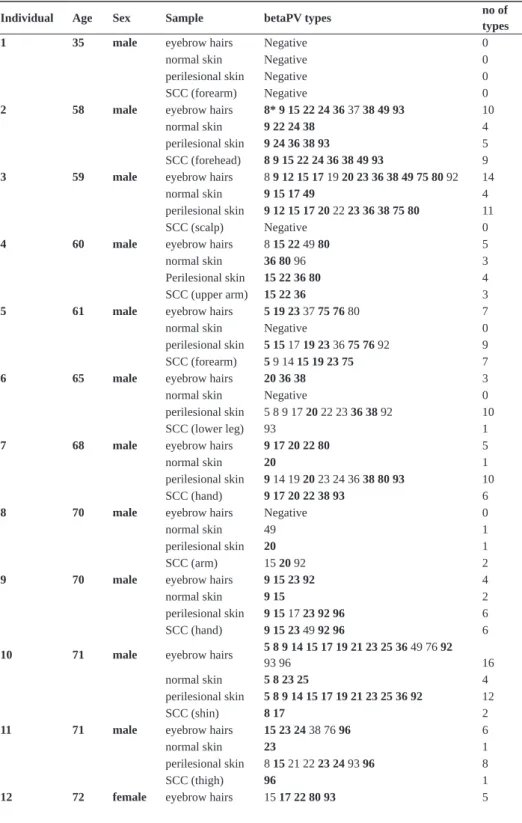
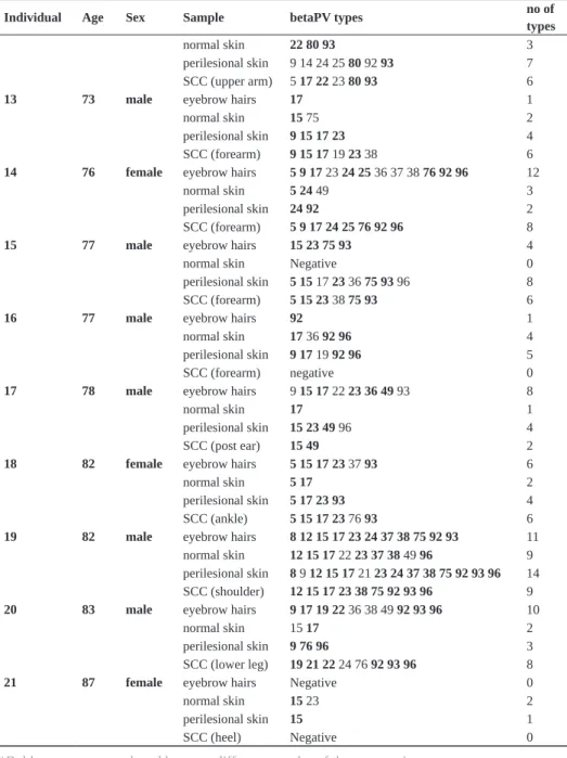
Table 1. Individual sample listed with all betaPV types detected.
Individual Age Sex Sample betaPV types no of types
1 35 male eyebrow hairs Negative 0
normal skin Negative 0
perilesional skin Negative 0
SCC (forearm) Negative 0
2 58 male eyebrow hairs 8* 9 15 22 24 36 37 38 49 93 10
normal skin 9 22 24 38 4
perilesional skin 9 24 36 38 93 5
SCC (forehead) 8 9 15 22 24 36 38 49 93 9 3 59 male eyebrow hairs 8 9 12 15 17 19 20 23 36 38 49 75 80 92 14
normal skin 9 15 17 49 4
perilesional skin 9 12 15 17 20 22 23 36 38 75 80 11
SCC (scalp) Negative 0
4 60 male eyebrow hairs 8 15 22 49 80 5
normal skin 36 80 96 3
Perilesional skin 15 22 36 80 4
SCC (upper arm) 15 22 36 3
5 61 male eyebrow hairs 5 19 23 37 75 76 80 7
normal skin Negative 0
perilesional skin 5 15 17 19 23 36 75 76 92 9
SCC (forearm) 5 9 14 15 19 23 75 7
6 65 male eyebrow hairs 20 36 38 3
normal skin Negative 0
perilesional skin 5 8 9 17 20 22 23 36 38 92 10
SCC (lower leg) 93 1
7 68 male eyebrow hairs 9 17 20 22 80 5
normal skin 20 1
perilesional skin 9 14 19 20 23 24 36 38 80 93 10
SCC (hand) 9 17 20 22 38 93 6
8 70 male eyebrow hairs Negative 0
normal skin 49 1
perilesional skin 20 1
SCC (arm) 15 20 92 2
9 70 male eyebrow hairs 9 15 23 92 4
normal skin 9 15 2
perilesional skin 9 15 17 23 92 96 6
SCC (hand) 9 15 23 49 92 96 6
10 71 male eyebrow hairs 5 8 9 14 15 17 19 21 23 25 3693 96 49 76 92 16
normal skin 5 8 23 25 4
perilesional skin 5 8 9 14 15 17 19 21 23 25 36 92 12
SCC (shin) 8 17 2
11 71 male eyebrow hairs 15 23 24 38 76 96 6
normal skin 23 1
perilesional skin 8 15 21 22 23 24 93 96 8
SCC (thigh) 96 1
chapter 3 43
betaPV prevalence (95%) with a median number of types of 5, which was also significantly higher than in normal skin (p<0.001) (Table 2).
Individual Age Sex Sample betaPV types no of types
normal skin 22 80 93 3
perilesional skin 9 14 24 25 80 92 93 7 SCC (upper arm) 5 17 22 23 80 93 6
13 73 male eyebrow hairs 17 1
normal skin 15 75 2
perilesional skin 9 15 17 23 4
SCC (forearm) 9 15 17 19 23 38 6
14 76 female eyebrow hairs 5 9 17 23 24 25 36 37 38 76 92 96 12
normal skin 5 24 49 3
perilesional skin 24 92 2
SCC (forearm) 5 9 17 24 25 76 92 96 8
15 77 male eyebrow hairs 15 23 75 93 4
normal skin Negative 0
perilesional skin 5 15 17 23 36 75 93 96 8
SCC (forearm) 5 15 23 38 75 93 6
16 77 male eyebrow hairs 92 1
normal skin 17 36 92 96 4
perilesional skin 9 17 19 92 96 5
SCC (forearm) negative 0
17 78 male eyebrow hairs 9 15 17 22 2336 49 93 8
normal skin 17 1
perilesional skin 15 23 49 96 4
SCC (post ear) 15 49 2
18 82 female eyebrow hairs 5 15 17 23 37 93 6
normal skin 5 17 2
perilesional skin 5 17 23 93 4
SCC (ankle) 5 15 17 23 76 93 6
19 82 male eyebrow hairs 8 12 15 17 23 24 37 38 75 92 93 11 normal skin 12 15 17 22 23 37 38 49 96 9 perilesional skin 8 9 12 15 17 21 23 24 37 38 75 92 93 96 14 SCC (shoulder) 12 15 17 23 38 75 92 93 96 9 20 83 male eyebrow hairs 9 17 1922 36 38 49 92 93 96 10
normal skin 15 17 2
perilesional skin 9 76 96 3
SCC (lower leg) 19 21 22 24 76 92 93 96 8
21 87 female eyebrow hairs Negative 0
normal skin 15 23 2
perilesional skin 15 1
SCC (heel) Negative 0
*Bold types are types shared between different samples of the same patient.
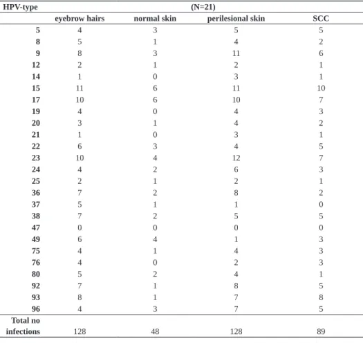
44 chapter 3
Overall, the prevalence of most individual betaPV-types was lowest in normal skin, except for HPV37 and 80 where the prevalence was lowest in the SCC biopsies and for HPV 49 that was lowest in perilesional skin. The most prevalent types across all tissue samples were HPV15, HPV17 and HPV23 (Table 3).
comparisons between scc, perilesional and normal skin biopsies
We observed a high degree of overlap between the betaPV types in the different samples from each patient (Table 1). No type was found exclusively in any of the samples, including SCC, when betaPV type distribution was compared.
Overall betaPV agreement (having both samples concordant for the presence or absence of betaPV, irrespective of type) between tumour tissue and perilesional skin was 86% (18/21; 95%CI 64-97), and between tumour tissue and normal skin was 71% (15/21; 95%CI 48-89).
A total of 128 betaPV infections were found in the eyebrow hairs of the 21 participants, 48 in normal skin biopsies, 128 in perilesional biopsies and 89 in tumour tissue biopsies (Table 3). Of the 89 betaPV infections found in tumour tissue, 56 infections with the same betaPV type were also found in the perilesional skin of the same patient (56/89, 63%; 95%CI 52-73). In comparison, 23 overlapping infections were observed between SCC and normal skin, a proportion of 26% (23/89; 95% CI 17-36). There were 33 overlapping infections between perilesional skin and normal skin (26%, 33/128; 95% CI 19-34).
betapv in eyebrow hairs as marker of betapv infection in scc
Overall betaPV agreement was 86% between eyebrow hairs and tumour tissue and 90% between hairs and perilesional skin. Of the 89 betaPV infections found in SCC tissue, 63 type-specific infections were also found in the eyebrow hair follicles of the same patient 63/89, 71%; 95% CI 60-80) and 79 overlapping infections were detected between perile-sional skin and eyebrow hairs (79/128, 62%; 95% CI 53-70).
Table 2. Overall betaPV detection in samples of SCC-cases.
(N=21)
eyebrow hairs normal skin perilesional skin SCC
Detection of betaPV, n (%)
positive 18 (86) 17 (81) 20 (95) 17 (81)
median no betaPV types (IQR*) 5 (2-10) 2 (1-4) 5 (4-10) 6 (1-7)
range 0-16 0-9 0-14 0-9
chapter 3 45
In three individuals SCC was present on the face (# 2, 3, 17 in table 1). In the two betaPV positive SCC (# 2 and 17) all types present in the SCC were also found in the eyebrow hairs of the corresponding individuals.
When we defined hair samples as being concordant if they contained all of the types found in the SCC biopsy, sensitivity was 29% (95%-CI 10-56). Using a less stringent definition of concordance whereby hair samples were classified as concordant if they contained 50% of the betaPV types found in the SCC, sensitivity was 82% (95% CI 57-96). When the perilesional skin was taken as the reference category (instead of SCC tissue), sensitivities for these comparisons were 25% (95% CI 9-50) and 65% (95% CI 41-85) respectively.
Table 3. BetaPV prevalence in 21 SCC patients shown per sample.
HPV-type (N=21)
eyebrow hairs normal skin perilesional skin SCC
5 4 3 5 5
8 5 1 4 2
9 8 3 11 6
12 2 1 2 1
14 1 0 3 1
15 11 6 11 10
17 10 6 10 7
19 4 0 4 3
20 3 1 4 2
21 1 0 3 1
22 6 3 4 5
23 10 4 12 7
24 4 2 6 3
25 2 1 2 1
36 7 2 8 2
37 5 1 1 0
38 7 2 5 5
47 0 0 0 0
49 6 4 1 3
75 4 1 4 3
76 4 0 2 3
80 5 2 4 1
92 7 1 8 5
93 8 1 7 8
96 4 3 7 5
Total no
46 chapter 3
discussion
In this study we systematically explored and compared type-specific betaPV prevalence and distribution in sets of four different tissue samples taken from 21 incident SCC patients.
The overall betaPV DNA positivity was high, ranging from 81% in normal skin and SCC tissue to 86% in the eyebrow hairs and 95% in perilesional skin. These percentages underscore the ubiquity of cutaneous betaPV infections that has been previously reported (8;9;29). The multiplicity of infections was also found to be high, with a median number of infecting types of 5 in eyebrow hairs and perilesional skin and 6 in the tumour, and up to 14 and 16 different betaPV types present in single samples from perilesional skin and eyebrow hair, respectively. The number of betaPV types detected in normal skin was considerably less than in the other tissues, in support of previous data showing that normal skin has fewer betaPV types than SCC tissue (30;31).
The lower number of betaPV types found in normal skin than in the tissue near the SCC supports the hypothesis that perilesional skin represents an area of field cancerisation from which the tumour arose (15-18).The localised presence of betaPV may have contributed to the field change, in conjunction with other factors such as sunburn or chronic sun exposure. Alternatively focal damage may have enhanced betaPV infection, increasing the viral load above the detection limit of the test, or less likely, may have rendered the affected skin more susceptible to infection with betaPV. We obtained the normal skin biopsies from the mirror image site of the SCC so that betaPV detection would be unconfounded by local photo immune suppression or to stimulation of viral replication by UV irradiation.
chapter 3 47
The detection of betaPV in eyebrow hairs has been used in epidemiological studies as a marker of infection, not only because the bulb is regarded as a reservoir of infection but also because of the ease of obtaining eyebrow hairs. The greater diversity of types found in hair follicles compared with normal skin lends support to the notion that hair follicle is a reservoir for betaPV (22), with the epidermal stem cells residing in the bulge as the probable main site of persistent infection.
We compared betaPV DNA in eyebrow hairs with biopsies of the SCC and perilesional skin to obtain information about the comparability of these samples. The type-specific agree-ment was slightly higher for SCC than for perilesional skin. To calculate the sensitivity of eyebrow hair follicle testing as a measure of relevant betaPV infection, we first took SCC-tissue as the reference tissue. The sensitivity ranged from 25-82% depending on the definition of concordance and whether the SCC or the perilesional tissue is used as the reference. The relevance of the high agreement in the two individuals with a betaPV posi-tive SCC present on the face needs to be explored further in larger datasets given the small number of participants. Although eyebrow hairs are frequently used as markers of infection in betaPV studies, it is notable that substantial differences can exist between type-specific detection rates in SCC tumour and eyebrow hairs of individuals.
48 chapter 3
A possible limitation of this study is the small patient group. However, based on the comprehensiveness of our sample sets, the high number of included HPV types and the unique study design, we believe the generated data provide valuable new information that is generalisable. Our study population was representative of SCC patients generally seen in the Townville area in terms of average age (70 versus 67 years) as the key risk factor for SCC in a high-risk population like Townsville, though it contained a higher proportion of males (80%) than seen overall (61%) (34).
In summary, this series of samples of SCC-patients show that perilesional skin is clearly different from normal skin with respect to betaPV infection, supporting the field change hypothesis for cutaneous SCC development. The contribution of betaPV to field cancerisa-tion is unknown but might be related to its property to impair cellular defenses against UV-induced DNA damage (35;36). The clinical relevance, if any, of the difference in betaPV types is unknown. Since no specific types were identified in any of the particular biopsies that were not present in other samples, no obvious high-risk types emerged in this series. Possibly, the number of types or a combination of certain types increases the risk of developing a SCC (25;32). It is also possible that detection of more betaPV types represents higher viral loads accompanied by greater viral gene expression and an increased risk of SCC.
Similar analyses in larger datasets, ideally including measures of viral load, may help to elucidate the role of betaPV in cutaneous carcinogenesis, and to determine whether the betaPV status of eyebrow hairs is a sufficiently good marker of infection of the tumour field to warrant its continued use in epidemiological studies.
acknowledgements
chapter 3 49
references
1 Berkhout RJ, Tieben LM, Smits HL, Bavinck JN, Vermeer BJ, Ter Schegget J. Nested PCR approach for detection and typing of epidermodysplasia verruciformis-associated human papillomavirus types in cutane-ous cancers from renal transplant recipients. J Clin Microbiol 1995 Mar;33(3):690-5.
2 Bouwes Bavinck JN, Stark S, Petridis AK, Marugg ME, Ter Schegget J, Westendorp RG, et al. The presence of antibodies against virus-like particles of epidermodysplasia verruciformis-associated humanpapillomavi-rus type 8 in patients with actinic keratoses. Br J Dermatol 2000 Jan;142(1):103-9.
3 Boxman ILA, Russell A, Mulder LHC, Bavinck JNB, Ter Schegget J, Green A. Association between epi-dermodysplasia verruciformis- associated human papillomavirus DNA in plucked eyebrow hair and solar keratoses. Journal of Investigative Dermatology 2001 Nov;117(5):1108-12.
4 Jong-Tieben LM, Berkhout RJ, Smits HL, Bouwes Bavinck JN, Vermeer BJ, van der Woude FJ, et al. High frequency of detection of epidermodysplasia verruciformis-associated human papillomavirus DNA in biop-sies from malignant and premalignant skin lesions from renal transplant recipients. J Invest Dermatol 1995 Sep;105(3):367-71.
5 Karagas MR, Nelson HH, Sehr P, Waterboer T, Stukel TA, Andrew A, et al. Human papillomavirus infec-tion and incidence of squamous cell and basal cell carcinomas of the skin. J Natl Cancer Inst 2006 Mar 15;98(6):389-95.
6 Schaper ID, Marcuzzi GP, Weissenborn SJ, Kasper HU, Dries V, Smyth N, et al. Development of skin tumors in mice transgenic for early genes of human papillomavirus type 8. Cancer Research 2005 Feb 15;65(4):1394-400.
7 Stockfleth E, Nindl I, Sterry W, Ulrich C, Schmook T, Meyer T. Human papillomaviruses in transplant-associated skin cancers. Dermatol Surg 2004 Apr;30(4 Pt 2):604-9.
8 de Koning MN, Weissenborn SJ, Abeni D, Bouwes Bavinck JN, Euvrard S, Green AC, et al. Prevalence and associated factors of betapapillomavirus infections in individuals without cutaneous squamous cell carcinoma. J Gen Virol 2009 Mar 25.
9 de Koning MN, Struijk L, Bavinck JN, Kleter B, Ter Schegget J, Quint WG, et al. Betapapillomaviruses frequently persist in the skin of healthy individuals. J Gen Virol 2007 May;88(Pt 5):1489-95.
10 Berkhout RJM, Bavinck JNB, Ter Schegget J. Persistence of human papillomavirus DNA in benign and (pre)malignant skin lesions from renal transplant recipients. Journal of Clinical Microbiology 2000 Jun;38(6):2087-96.
11 Bavinck JNB, Feltkamp MCW. Milk of human kindness? HAMLET, human papillomavirus, and warts. New England Journal of Medicine 2004 Jun 24;350(26):2639-42.
12 Pfister H, Fuchs PG, Majewski S, Jablonska S, Pniewska I, Malejczyk M. High prevalence of epidermodys-plasia verruciformis-associated human papillomavirus DNA in actinic keratoses of the immunocompetent population. Archives of Dermatological Research 2003 Dec;295(7):273-9.
13 Slaughter DP, Southwick HW, Smejkal W. Field cancerization in oral stratified squamous epithelium; clinical implications of multicentric origin. Cancer 1953 Sep;6(5):963-8.
50 chapter 3
15 Ulrich M, Maltusch A, Rowert-Huber J, Gonzalez S, Sterry W, Stockfleth E, et al. Actinic keratoses: non-invasive diagnosis for field cancerisation. Br J Dermatol 2007 May;156 Suppl 3:13-7.
16 Vatve M, Ortonne JP, Birch-Machin MA, Gupta G. Management of field change in actinic keratosis. Br J Dermatol 2007 Dec;157 Suppl 2:21-4.
17 Carlson JA, Scott D, Wharton J, Sell S. Incidental histopathologic patterns: possible evidence of ‘field cancerization’ surrounding skin tumors. Am J Dermatopathol 2001 Oct;23(5):494-6.
18 Rohwedder A, Foong H, Tyring SK, Rady P, Carlson JA. Incidental epidermodysplasia verruciformis human papillomavirus infection (EV acanthoma): evidence for ‘field cancerization’ and a putative cofactor in sebor-rheic keratosis. J Cutan Pathol 2008 Dec;35(12):1151-5.
19 Giampieri S, Storey A. Repair of UV-induced thymine dimers is compromised in cells expressing the E6 protein from human papillomaviruses types 5 and 18. Br J Cancer 2004 Jun 1;90(11):2203-9.
20 Struijk L, Van der Meijden E, Kazem S, Ter Schegget J, de Gruijl FR, Steenbergen RD, et al. Specific betapapillomaviruses associated with squamous cell carcinoma of the skin inhibit UVB-induced apoptosis of primary human keratinocytes. J Gen Virol 2008 Sep;89(Pt 9):2303-14.
21 Underbrink MP, Howie HL, Bedard KM, Koop JI, Galloway DA. E6 proteins from multiple human betapap-illomavirus types degrade Bak and protect keratinocytes from apoptosis after UVB irradiation. J Virol 2008 Nov;82(21):10408-17.
22 Boxman IL, Berkhout RJ, Mulder LH, Wolkers MC, Bouwes Bavinck JN, Vermeer BJ, et al. Detection of human papillomavirus DNA in plucked hairs from renal transplant recipients and healthy volunteers. J Invest Dermatol 1997 May;108(5):712-5.
23 de Koning M, Quint W, Struijk L, Kleter B, Wanningen P, van Doorn LJ, et al. Evaluation of a novel highly sensitive, broad-spectrum PCR-reverse hybridization assay for detection and identification of beta-papillomavirus DNA. J Clin Microbiol 2006 May;44(5):1792-800.
24 Forslund O, Antonsson A, Nordin P, Stenquist B, Hansson BG. A broad range of human papillomavirus types detected with a general PCR method suitable for analysis of cutaneous tumours and normal skin. J Gen Virol 1999 Sep;80 ( Pt 9):2437-43.
25 Struijk L, Hall L, Van der Meijden E, Wanningen P, Bouwes Bavinck JN, Neale R, et al. Markers of cutane-ous human papillomavirus infection in individuals with tumor-free skin, actinic keratoses, and squamcutane-ous cell carcinoma. Cancer Epidemiol Biomarkers Prev 2006 Mar;15(3):529-35.
26 Waterboer T, Sehr P, Michael KM, Franceschi S, Nieland JD, Joos TO, et al. Multiplex human papilloma-virus serology based on in situ-purified glutathione S-transferase fusion proteins. Clinical Chemistry 2005 Oct;51(10):1845-53.
27 Bouwes Bavinck JN, Feltkamp M, Struijk L, Ter Schegget J. Human papillomavirus infection and skin cancer risk in organ transplant recipients. J Investig Dermatol Symp Proc 2001 Dec;6(3):207-11.
28 Kohler A, Forschner T, Meyer T, Ulrich C, Gottschling M, Stockfleth E, et al. Multifocal distribution of cuta-neous human papillomavirus types in hairs from different skin areas. Br J Dermatol 2007 May;156(5):1078-80.