Dr Brajamohan Mishra et al JMSCR Volume 08 Issue 02 February 2020 Page 590
Role of CT scan in Management of Blunt Abdominal Injury
Authors
Dr Brajamohan Mishra, Dr Swasti Rekha Nayak, Dr Abinasha Mohapatra*
Dr Pramit Ballav Panigrahi
Department of Surgery, VIMSAR, Birla, Odisha, India 768017 *Corresponding Author
Dr Abinasha Mohapatra
Assistant Professor, Department of Surgery, VIMSAR, Burla, Odisha, India 768017
Abstract
Background and objectives: Evaluating patients who have sustained blunt abdominal injuries remains one of the most challenging and resource –intensives aspects of acute trauma care. Missed intra-abdominal injuries continue to cause preventable deaths. Objective is to asses efficacy of CT Scan (computed tomography as accurate diagnostic tool for blunt abdominal injuries patients.
Methods: 96 cases of blunt abdominal injury admitted in VIMSAR, Medical College, Burla, Sambalpur during the period of October 2017 to October 2019 were included in my study after taking informed consent. All these patients were thoroughly investigated.
CT Scan was done for all heamodynamically stable patients. Recorded data included age, sex, types of organ injuries and scan results. Organ injuries were grading using the OIS (Organ Injury Scale) guidelines.
Results: The study comprised of 96 patients having blunt abdominal injuries. Majority of patients were in age group of 20-39 years male .Most common injury were splenic (40%), liver(23%)and hemoperitoneum (55%).95% (92 patients) were positive for abdominal injury and 5% (4 patients) were negative. The CT findings of hemoperitoneum and/or solid organ injury were confirmed in the17 cases taken up for surgery rest conservatively managed.
Conclusions: In this study CT scan was 100% sensitive in diagnosis of blunt abdominal injuries. Negative CT scan discourage unnecessary urgent abdominal exploration.
Introduction
The lack of historical data and the presence of distracting injuries or altered mental status, from head injuries or intoxication, can make blunt abdominal injuries difficult to diagnose and manage. Patients are frequently kept for observation following BAI, despite initially negative evaluations.
Victims of BAI often have both intra- abdominal and extra- abdominal injuries further complicating
care. The majority of cases related to RTA (75%), blows to abdomen (15%) and (6-9%) due to fall. Trauma is the leading cause of death in persons under 45 years of age, with 10% of these fatalities attributable to abdominal injury.
The most commonly injured organ are spleen, liver, retroperitoneum, small bowel, kidneys, bladder, colon, diaphragm, and pancreas. Computed tomography (CT) scan of the abdomen can reveal others associated injuries, notably
http://jmscr.igmpublication.org/home/ ISSN (e)-2347-176x ISSN (p) 2455-0450
Dr Brajamohan Mishra et al JMSCR Volume 08 Issue 02 February 2020 Page 591 vertebral and pelvic fractures and injuries in the
thoracic cavity. CT scans, unlike direct peritoneal lavage (DPL) or Focused Assessment with Sonography for Trauma (FAST) examinations, have the capability to determine the source of haemorrhage. Many retroperitoneal injuries go unnoticed with DPL and FAST examinations. CT scans provide excellent imaging of the pancreas, duodenum, and genitourinary systems. The images can help quantitate the amount of blood in the abdomen and can reveal individual organs with precision. Imaging plays a critical role in the evaluation of patients with blunt abdominal injuries. CT as the sole modality, enables evaluation of others associated injuries in addition to global evaluation of abdomen.
Trauma has been defined as damage to the body caused by exchange with environmental energy that is beyond the body’s resilence[1]. It is the leading cause of death. Indian statistics reveal a disproportionate involvement of younger age groups (15-25 years). The Indian fatality rates for abdomen trauma are 20 times that for developed countries[2]. About 30% of such death can be preventable. Swift recognition of injury with prompt and appropriate treatment to reduce morbidity and mortality is the goal of modern trauma care and hence accurate diagnosis is essential.
The challenge in the imaging of abdominal trauma is to accurately identify the injuries that require early exploration and at the same time avoid unnecessary operative intervention in cases can be managed conservatively. Laboratory tests are nonspecific, plain X-ray abdomen are usually not helpful in early post injury period. For all these reasons, several diagnostic modalities in practice have evolved till date and still they are evolving. The modalities in practice are, Abdominal Paracentesis, DPL (Diagnostic Peritoneal Lavage), X-Ray Abdomen, Ultrasound of Abdomen, Computed Tomography (CT) Scan of Abdomen, Laparoscopic Exploration of abdomen. To ascertain degree of trauma, a rapid, cost effective, safe and reproducible investigation used
is ultrasonography. FAST (Focussed assessment for the sonographic examination of trauma patients) is needed in most cases nowadays to quantify the degree of trauma[3,4]. The inability of USG detect many parenchymal injuries and assess the retroperitoneum, active bleeding which limits its value[5,6].
Over the last decade, CT Scan has gained widespread clinical acceptance in evaluation of haemodynamically stable patients with BAT. CT not only allows comprehensive evaluation of presence and extent of injuries to solid organ, retroperitoneum, bowel, mesentery and associated haemorrhage but also allows surgeons to reach vital decisions regarding the need of surgery. Routine use CT has substantially reduced the number of additional radiographic studies as well as need of DPL[7].
Aims & Objectives
The present work is undertaken with the following aims
General Objective
1. To assess the role of CT Scan in evaluation of patients of Blunt abdominal injuries
Specific Objective
1. To assess CT is the choice of investigation in solid organ injuries in hemodynamically stable patients.
2. To assess the role of CT in management of BAT patients i.e either conservative or laparotomy.
Secondary Objective
1. To compare FAST and CT Scan in diagnosis of BAI injuries in emergency patients.
2. To assess its limitations in management of BAI patients in our tertiary hospital.
Material
Dr Brajamohan Mishra et al JMSCR Volume 08 Issue 02 February 2020 Page 592 Burla, ODISHA among patients with history of
blunt abdominal trauma admitted in surgery Department.
Period of Study- Nov. 2017 to Oct. 2019
Calculated Sample Size – n = Total cases of blunt trauma of abdomen = 96 [Male=83(87%), Female=13(13%) i.e. 1.11 % 0f total admission] (Out of total surgical admission = 8012)
Inclusion Criteria: All patients with suspected abdominal organ injury by blunt trauma were included. All age groups of both sexes were included in this study.
Exclusion Criteria: Patients with other associated injuries e.g. Chest injury, Head injury, Pelvic injury, Spine injury, Bone injury etc.
Methods
On admission, all the patients were evaluated after necessary resuscitative measure. A quick detailed history and thorough clinical examination was carried out to reach at a provisional diagnosis regarding nature of injury. Histories were taken which consists of Allergic medication (patients was on), Previous illness, Last mealtime, Events preceding the injury. Primary Survey was done and the patients were examined in the following manner:
General physical examination ( pulse rate, blood pressure at 15 minutes interval for 1 hr then hourly interval for 6hours and then 2 hourly, respiratory rate, pallor, cyanosis and capillary refill at lip of mucosa ) Abdominal examination
Per rectal examination was done to exclude bleeding per rectum or any injury to distal part of colon.
All extended injuries were managed accordingly. All patients were given tetanus toxoid, human anti-tetanus immunoglobin and antibiotic in the ward. All routine investigations [CBC, Blood
group, Serum electrolytes, LFT, Serum Amylase and lipase, Urine for routine and microscopic, X-ray abdomen and chest, USG abdomen, pelvis and FAST – After
initial resuscitation, it was done in all cases
CECT Scan abdomen and pelvis – Done to grade solid organ injury those who were hemodynamically stable or were managed conservatively after USG
Abdominal paracentesis, and Diagnostic peritoneal laparotomy (DPL)]
Management – surgical and non surgical (conservatively)
Observation and Results
Table 1: Age & Sex Distribution of Patients (n=96)
Age group in year Male Female No of cases Percentage
0-10 2 2 5 5%
11-20 15 2 18 18%
21-30 25 5 30 30%
31-40 20 2 22 22%
41-50 17 1 20 20%
51-60 2 1 3 3%
60 & above 2 0 2 2%
Total 83 13 96 100
The above table reveals that the majority of cases (70%) were in the age group of 11-40 years and only 25% were in the age group above 40 years. Peak incidence was in the third decade (30%). Male and female cases were 87% and 13% respectively.
Table 2- Cause & Incidence (n=96)
Cause No. of cases Percentage
Road traffic accident 74 74%
Fall from height 14 14%
Blunt weapons blow 7 7%
Bullock cart 3 3%
Dr Brajamohan Mishra et al JMSCR Volume 08 Issue 02 February 2020 Page 593 In this cases road traffic accident was the
commonest cause and accounted for about 74% of cases and the cause next in the order was injury by fall (14%). Injuries arising from blunt weapons blow, bullock cart and animal horn thrust were
almost minor in proportion. The cases sustaining injury by fall from height hailed from construction sites and belonged mainly to labourer class. Whereas victims of local traffic accidents were mainly from affluent class.
Table -3 Clinical manifestation (n=96)
Clinical manifestation No. of cases Percentage
Abdominal pain 92 92%
Chest pain 14 14%
Vomiting 16 16%
Absolute constipation 18 18%
Hematuria 3 3%
Pallor 14 14%
Abdominal tenderness 82 82%
Abdominal rigidity 61 61%
Abdominal distension 46 46%
Absent bowel sound 40 40%
Hematuria 3 3%
Shifting dullness 15 15%
Obliteration of liver dullness 24 24%
The commonest presentation was of abdominal pain and tenderness which were present in 92% and 82% of cases respectively, either with or without an external bursts, scratch mark or skin erythema over the site of impact. It was followed
by rigidity (61%), abdominal distension (46%), absence of bowel sounds (40%), absolute constipation (18%), vomiting (16%), and hematuria (3%).
Table-4 Abdominal and Chest roentgenogram findings (n=96)
Finding Number Percentage
Gas under diaphragm 26 26%
Inter loop collections 13 13%
Distended loops with fluid & gas 10 10%
Fracture ribs 6 6%
Ground glass appearance 4 4%
Hemothorax 2 2%
No. abnormality 37 37%
In 37 (37%) cases, patient’s X-ray showed no signs of injury.
In patients with bowel injury (26%), X-rays showed gas under diaphragm in all the cases. It was virtually diagnostic. Around 10% of the cases showed distended bowel loops.
Some patients with hemoperitoneum showed ground glass appearance which was not so helpful diagnosis.
Some cases of splenic injury and chest injury showed fractured ribs. So this findings was an indirect evidence of splenic injury.
Dr Brajamohan Mishra et al JMSCR Volume 08 Issue 02 February 2020 Page 594
Table-5: Mode of Management
Type of management No. of cases percentage
Conservative 62 65%
Operative 34 35%
Total 96 100
Out of 62 patients who were managed by conservative means, majority were associated with solid viscera injuries-3 patients dies of shock due to multiple injuries within 6hrs of admission while they were being resuscitated. 10 patients showed no evidence of visceral injury and were discharged after improvement of symptoms. Out of 38 cases that were managed surgically, which also included those who, at first, were being
managed conservatively but later on operated, majority were injuries to the hollow viscus.
Out of the 62 patients managed conservatively, 10 showed no signs of visceral-3 patients dies of shock due to multiple injuries within 6 hours of admission,. So CECT was done in 49 patients injuries were graded according to the AAST grading (American Association of Surgery for Trauma).
Table 6: CECT finding (n=49)
Patients findings No. of cases Percentage
Splenic injury 29 Grade I 15 59.18%
Grade II 12
Grade III 2
Liver injury 17 Grade I 11 34.69%
Grade II 06
Mesenteric hematoma 3 6.1%
Table 7: Pattern of visceral involvement (n=96)
Organs No. of cases
Spleen 36
Liver 20
Small intestine 16
Colon and Rectum 2
Urinary bladder 2
Mesentery 3
Spleen and liver 2
Spleen and small intestine 1
Liver and small intestine 1
Small intestine and mesentery 2
No injury 10
Total 96
Table 8 - Spectrum of Intra Peritoneal Organ Involvement (N-96)
ORGAN INJURED NO. OF CASES PERCENTAGE
SPLEEN 40 40%
LIVER 23 23%
SMALL INTESTINE 22 22%
COLON AND RECTUM 3 3%
URINARY BLADDER 2 2%
MESENTERY 6 6%
Spleen was the most commonly injured organ and involved 40% of cases. Liver injury was present in 23% of cases whereas small intestinal injury was present in 22% of cases. Mesenteric injury was
Dr Brajamohan Mishra et al JMSCR Volume 08 Issue 02 February 2020 Page 595 Grade I injuries (11 patients) were managed
conservatively.
Grade II injuries were present in 8 patients. At first all of them were kept under conservative management. But 2 patients required hepatorrhaphy later on as they didn’t respond to treatment because of the associated splenic injury.
With grade III injuries there were 2 patients. Both were operated! Case was associated with ileal perforation liver repair and closure of the perforation was done but the patient succumbed in postoperative period due to septicemia.
1 case, each of Grade IV and V died while resuscitation due to associated head chest and pelvic injury.
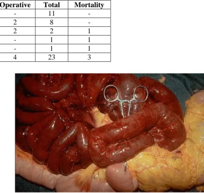
Table 11 (A): Management – Mortality chart
Grade Conservative Operative Total Mortality
Grade I 11 - 11 -
Grade II 6 2 8 -
Grade III - 2 2 1
Grade IV 1 - 1 1
Grade V 1 - 1 1
Total 19 4 23 3
Small Intestine Injury
Out of the 22 cases of small gut injury, 12 cases were having ileal perforation and 10 cases had jejuna perforation. In 10 cases, perforation was of <1 cm diameter. 1 out of these cases was associated with Grade V splenic injury where splenectomy was done. In all these cases simple closure with single layer of interrupted lambert suture was done. One case of ileal perforation was around 3 cm in diameter and associated with Grade III liver injury and pyoperitoneum. Resection and end-to end anastomosis with liver repair and toileting was done. Subsequently the patient dies due to septicemia 3 cases of ileal perforation had associated mesenteric injury with injury to superior mesenteric vessels. In all these cases resection and anastomosis of the devitalized segment with repair of the mesentery and ligation of the mesenteric vessels were done1 case developed entero-cutaneous fistula postoperatively and subsequently died of its complications. Rest of the cases were having multiple perforation with devitalized tissues around. In these cases resection with either end-to-end anastomosis or ileo-transverse anastomosis was done. In all cases peritoneal toileting was done. Care was taken to avoid luminal narrowing by repair.
Mesentric Injury
Out of 6 patients with mesenteric injury, 3 cases were associated with small intestine injury. They were all operated 1 case was complicated with entero-cutaneous fistula and subsequently dies. There were managed conservatively as their CT scan showed mesenteric hematoma.
Morbidity
Dr Brajamohan Mishra et al JMSCR Volume 08 Issue 02 February 2020 Page 596
Mortality
Table – 11 (B): Management – Mortality Chart (N=96)
Total No.
of cases
Conservative Management
Operative Management
No. of Deaths
Percentage
96 62 34 6 6
In case study the overall mortality rate due to blunt trauma abdomen was 6%.
3 of these cases died within 6hrs of admission before any surgical intervention, as he patients were in deep shock with severe associated injuries.
1 case of splenic injury with associated head injury dies postoperatively as that patient couldn’t
recover from the anesthesia and went in cardiac arrest.
1 case of liver injury with intestinal injury died post operatively due to septicemia and shock. 1 case of small intestinal injury with mesenteric injury developed entero-cutaneous fistula post operatively and died of its complications.
Table 12: Haemoperitoneum C.T. Quantification
Location of Hemorrhage CT Quantification Approximate Quantity
Fluid in only one space Mild 100-200ml
Fluid in two or more space Moderate 250-500ml
Fluid in all spaces, pelvis anterior/superior to urinary bladder
Gross >500ml
Table – 13: Correlation between injury grading and management in patients (N=96)
Injury Grade Total no. of patients No.of conservatively managed cases No.of operated cases Chi-Square Test (p-value) Liver injury
Grade I 1 1 NIL
0.091
Grade II 3 3 NIL
Grade III 6 6 NIL
Grade IV 5 3 2
Grade V 1 NIL 1
TOTAL 16 13 3
Splenic injury
0.643
Grade I NIL NIL NIL
Grade II 4 2 2
Grade III 1 1 NIL
Grade IV NIL NIL NIL
TOTAL 5 3 2
Renal injury
0.286
Grade I NIL NIL NIL
Grade II NIL NIL NIL
Grade III 3 3 NIL
Grade IV 4 2 2
Grade V NIL NIL NIL
Total 7 5 2
Pancreatic injury
0.667
Grade I 1 1 NIL
Grade II 2 1 1
Grade III NIL NIL NIL
Grade IV NIL NIL NIL
Grade V NIL NIL NIL
Total 3 2 1
Solid organ
0.659
Grade I 4 4 0
Grade II 10 7 3
Grade III 10 10 0
Grade IV 9 5 4
Grade V 1 0 1
Dr Brajamohan Mishra et al JMSCR Volume 08 Issue 02 February 2020 Page 597
CT showing Haemoperitoneum
CT showing Abdomen Injury
Large Intestine Injury
Discussion
No age is bar for blunt trauma injuries. The maximum blunt abdominal injuries occurs in age group 20-45.This was because patient in this age group lead more active life and have more outdoor
activities. Patient in age group >50 years , lead a less active life, have less incidence of injuries. In this study, nearly 70% of patients were more age group 10-40 years .This age group represent working population.
Poor results of USG may be due to overlying bowel shadow, surgical emphysema, empty bladder and lack of skilled radiologist at emergency hours.
Mallik k et al. (8) study demonstrates the superiority of CT over USG as diagnostic tool in blunt trauma abdomen. CT Scan altered the diagnosis and choice of managements.
Table – 16
Organ Present
study (n=96)
Cox EF 1984 In n=870
Davis et al 1976 n=437
Spleen 40 42.6 20
Liver 23 35.6 29
stomach 22 4.7 15
Large intestine 3 <0.1 -
Mesentery 6 - 7
Urinary bladder
2 3.2 29
P Value=0.0044(<0.05)
With the above comparison it is clear that the pattern of visceral injuries is not common and it varies from series to series.
Morbidity & Mortality
Most common complication in our study of blunt trauma was wound infection (5%)
The overall mortality rate was 6% in our series of blunt trauma. It is less than the reported mortality of 13.3% (davis et al, 1976) and of 17% by cox et at (1984).
Poor prognostic factors in blunt trauma are delay transportation and treatment, multiple visceral injuries, associated other organ system injuries and presence of sepsis and shock.
Conclusion
Dr Brajamohan Mishra et al JMSCR Volume 08 Issue 02 February 2020 Page 598 majority of our patients. Result of this study
shows that CT scan is a superior diagnostic modality in the diagnosis and management of blunt trauma abdominal trauma. Spleen is the most commonly injured organ in blunt abdominal injury. Negative CT scan discourage unnecessary urgent exploratory laparotomy.
CT Quantification of Hemoperitoneumtable
Road traffic accidents is the commonest cause according for 74% of all admissions.
Most commonly associated injury was chest injury (24%) followed diagnosis and planning of management of the patients.
CECT abdomen is the most important tool in grading the solid organ injuries and deciding further management to tackle emergencies.
Spleen (40%) was found to be the most common intra-peritoneal organ injured followed by liver (23) and small intestine (22%). Few subset of patients had multiple organ injury too, which need either single setting or multiple setting surgical intervention.
Wound infection rate was high in the post-operative cased of blunt trauma because of inadequate preparation preoperatively.
Mortality in the series was 6% mostly due other associated injuries leading to shock at the time of presentation (3%). Postoperative sequence and multiorgan failure accounted for the rest 3% of deaths.
The essence of management of these blunt abdominal trauma thus lies on early resuscitation, prompt first aid and accurate diagnosis with smart surgical interventions or deemed proper conservation management.
Conclusion
The incidence of blunt trauma abdomen was 1.11% of all cases admitted to the surgery department.cct
Age: In this study it was seen that the age of the patient varied between 2 ½ years to 72 years. The majority of cases (70%) were in the age group of 11-40 years. 45% were in the age group of 31-60
years. Bag well (1980)64 observed 56% cases in the 35-61 age group. The incidence observed in this series was comparable to the above series.
Sex: In our series, the male and female cases were 87% and 13% respectively. In the study conducted by Canty TG (1999)7 and Davis (1976)16 there was male preponderance (80% & 82% respectively). In all these studies males predominated females because they were more exposed to different outdoor activities including accidents in contrast to female. The finding was well marked among Indian females who usually confine themselves to the indoor.
Spectrum of Blunt Trauma Abdomen
Majority of our patients (74%) sustained motor vehicle accident either as an occupant of vehicle or as pedestrian, 14% were due to fall from height, 7% due to blunt blow, 3 due to bullock cart injury and 2 cases were due to animal horn thrust.
In the study of Ciftic et al (1998)4 and Davis et al (1976)16 accidents were the cause in 60% & 70% respectively which is comparable with our results.
Clinical Manifestations
In our series, the clinical manifestations were abdominal pain (92%), tenderness (82%), abdominal rigidity (61%), abdominal distension (46%) and absence of bowel sounds 40%). The incidence of clinical manifestations in the series of Nwabrinke T et al65 was tenderness 69% pain 52%, rigidity 25%, abdominal distension 48%, pallor 37%.
The above comparison depicts that incidence of clinical manifestations varied from series to series.
Associated Injuries
Dr Brajamohan Mishra et al JMSCR Volume 08 Issue 02 February 2020 Page 599
X-ray Abdomen and Chest
Main diagnostic value of X-ray was in diagnosing bowel injury where it showed gas under diaphragm. Rest of the findings were not diagnostic.
Ultrasonography and Fast
After initial resuscitation, USG abdomen/Fast done in all the patients. The commonest finding was free peritoneal fluid seen in 55 (55%) patients followed by splenic injury in 40 (40%) patients and liver injury in 23 (23%). Grading of solid organ injury was further done by C.T. scanning (those who were managed conservatively) and laparotomy findings. Patients with bowel injuries conservatively and laparotomy findings. Patients with bowel injuries usually showed distended bowel loops. But it was an indirect evidence and not diagnostic. 10 cases (10%) who were stable and showed no evidence of abdominal injury in X-ray or U.S.G. were managed conservatively without any further investigations.
CECT Abdomen
CT was done in patients who were hemodynamically stable and were managed conservatively after USG abdomen showed hemoperitoneum or organ injury. The main role of CT was to grade injuries in hemodynamically stable patients so that the treatment options i.e. conservative operative could be decided.
CT was done in 49 patients of whom most common organ injured was spleen (29 cases/59%). Out of these 15 cases, 12 cases and 2 cases showed Grade, I, II, III injuries respectively. CT also diagnosed 11 cases of grade 1 and 6 cases of Grade II liver injuries and 3 cases of mesenteric hematomas all of which were managed conservatively.
Intra Peritoneal Visceral Involvement
Spleen was the most commonly involved organ and accounted for 40% of cases, followed by liver
(23%) small intestine (22%) mesentery (6%), large bowel (3%) & bladder (2%).
Summary
Although expensive and potentially time consuming, CT scan provides the most detailed images of traumatic pathology and assist in determination of operative intervention. It also a standard technique for detection of solid organ injury, vertebral and pelvic fracture and injuries in thoracic cavity.
References
1. Gackowski W, Najninigier B et al, Treatment of liver injuries in personal clinical material from 1990-1006, 1997; Su Ipt 2; 223-7
2. Wilson RF, Walt AJ, Et al, Injury to stomach and small bowel-EDS-Management of trauma, Pitfalls & Practice. 1996; 497-509 (Motz 1890) 3. Ruddock et al 1937, Rao, Ivatury, Louis F.
Zantat, Surg. C.N.A.M. 1291, 796, 1999 4. CifticAo, Tanyel FC, Salman AB et al, GI
perforation due to blunt abdominal trauma, Pedia, Surg. INt. 1998, Apr. 13(4); 259-64. 5. Timothy G, Canty St., Timothy G, Canty Jr., Carlos Brown J., Injuries of GI tract from Blunt trauma in children-A twelve year experience at a designated pediatric centre, J Trauma 1999, 46(5):847
6. Champion HR, Sacco WJ, Garner SP et al, Emerging technology for vehicular safety and emergency response to road way crashes, Tr care in new millennium, 79 (6) 1999, 1229.
7. Canty TG Sr., Canty TG Jr., Brown C et al, Injuries of GI tract from blunt abdominal trauma in children, A 12 year experience at a designated trauma centre, J Trauma 1999 Feb 46 (2) 234-40.