Cyclic
compression
increases
F508
Del
CFTR
expression
in
ciliated
human
airway
epithelium
XNadzeya Marozkina,1Jürgen Bosch,1Calvin Cotton,1Laura Smith,1James Seckler,1
XKhalequz Zaman,1Shagufta Rehman,2Ammasi Periasamy,2Herbert Gaston,3Ghaith Altawallbeh,1 Michael Davis,7David R. Jones,4Robert Schilz,6Scott H. Randell,5and Benjamin Gaston1,8
1Pediatric Pulmonology Division, Department of Pediatrics, Case Western Reserve University School of Medicine, Cleveland, Ohio;2W. M. Keck Center for Cellular Imaging, Department of Biology, University of Virginia, Charlottesville, Virginia; 3Lake Effect Pharma, LLC, Gates Mills, Ohio;4Thoracic Surgery Service, Memorial Sloan Kettering Cancer Center, New York, New York;5Department of Cell Biology and Physiology, University of North Carolina, Chapel Hill, North Carolina; 6Pulmonology and Critical Care Medicine University Hospitals, Cleveland, Ohio;7Department of Pediatrics, Division of Pulmonary Medicine, Children’s Hospital of Richmond at Virginia Commonwealth University, Richmond, Virginia; and 8Pediatric Pulmonology Division, Rainbow Babies and Children’s Hospital, Cleveland, Ohio
Submitted 22 January 2019; accepted in final form 15 May 2019
Marozkina N, Bosch J, Cotton C, Smith L, Seckler J, Zaman K, Rehman S, Periasamy A, Gaston H, Altawallbeh G, Davis M, Jones DR, Schilz R, Randell SH, Gaston B. Cyclic compression increases F508 Del CFTR expression in ciliated human airway epi-thelium.Am J Physiol Lung Cell Mol Physiol317: L247–L258, 2019. First published May 22, 2019; doi:10.1152/ajplung.00020.2019.— The mechanisms by which transepithelial pressure changes observed during exercise and airway clearance can benefit lung health are challenging to study. Here, we have studied 117 mature, fully ciliated airway epithelial cell filters grown at air-liquid interface grown from 10 cystic fibrosis (CF) and 19 control subjects. These were exposed to cyclic increases in apical air pressure of 15 cmH2O for varying times. We measured the effect on proteins relevant to lung health, with a focus on the CF transmembrane regulator (CFTR). Immunofloures-cence and immunoblot data were concordant in demonstrating that air pressure increased F508Del CFTR expression and maturation. This effect was in part dependent on the presence of cilia, on Ca2⫹influx,
and on formation of nitrogen oxides. These data provide a mechano-sensory mechanism by which changes in luminal air pressure, like those observed during exercise and airway clearance, can affect epithelial protein expression and benefit patients with diseases of the airways.
airway cilia; airway clearance; airway pressure; cystic fibrosis; S-nitrosocysteine
INTRODUCTION
Airway epithelial cilia clear secretions and inhaled particles from the lungs (7, 47). Additionally, airway cilia have sensory functions, as do cilia in the kidney, ear, and elsewhere (13, 14, 30, 36, 39, 54). Specifically, they sense chemical stimuli (such as bitter taste agonists) (30, 36) and mechanical stimuli (such as air movement) (6, 60). Exercise and a variety of airway clearance procedures increase air pressure gradients across the ciliated airway epithelial surface (27, 48, 62). Clinically,
meth-ods that change transepithelial pressure may benefit certain patients with airways diseases, such as asthma and cystic fibrosis (CF) (4, 45). In asthma, increased airway pressure may dilate airways because of the intrinsic mechanical properties of airway smooth muscle (35). In CF and other lung diseases, ATP release associated with ciliary motion may benefit pa-tients by augmenting airway hydration through purinergic receptors (8). However, these potential mechanisms underlying the benefits of airflow and pressure are challenging to study in detail. This is, in part, because each pseudostratified epithelial culture takes 5– 6 wk per experiment to grow and because standardized and commercial systems are not yet available for air pressure and flow experiments in vitro. Here, we have studied the effect of changes in air pressure to signal changes in airway epithelial protein expression, signaling mediated, in part, by nitrogen oxides.
Whereas inducible nitric oxide synthase (iNOS) is expressed constitutively in the normal human airway epithelium (3, 34), high levels of expression are not observed for any NOS isoform in CF airway epithelial cells (33). Low-level expres-sion of endothelial NOS (eNOS) has been reported in ciliated cell basal bodies (64, 67), and a functional role for airway eNOS is suggested by the fact that eNOS⫺/⫺mice have greater
airway methacholine responsiveness following antigen sensi-tization than do inducible (iNOS)⫺/⫺ or neuronal (nNOS)⫺/⫺
mice (11). Of note, eNOS is activated by calcium flux that leads to calmodulin binding (25). Calcium flux through the transient receptor potential vanilloid 4 (TRPV4) channel is established to occur during airway epithelial ciliary motion (1, 39). Therefore, we hypothesized that mechanical stimulation of airway cilia could activate ciliated cell eNOS by increasing apical calcium flux.
Activation of eNOS can signal bioactivities by producing nitric oxide (NO) orS-nitrosothiols; alternatively, it can form inert end products such as nitrate (5, 23, 25, 40, 41, 58). The distribution and bioactivities of these chemical NOS products are regulated by colocalized proteins (5, 23, 41, 58).S -nitro-sothiols can have a number of beneficial airway effects (2, 10, 22, 23, 26, 28, 29, 37, 40, 42, 61, 65, 66). These include airway
smooth muscle relaxation and increased cilia beat frequency. S-nitrosoglutathione (GSNO) and other S-nitrosothiols also increase expression (28, 29, 42, 65) and function (10) of the CFTR protein. However, the absence of iNOS (33), as well as impaired eNOS activity, in the CF airway appears to result in impaired GSNO production (26). Indeed, these low levels suggest that inhibiting GSNO catabolism may be futile as a therapy. We hypothesized that airway ciliary mechanotrans-duction could increase nitrogen oxide promechanotrans-duction and, in turn, CFTR expression.
MATERIALS AND METHODS
Reagents
All reagents were purchased from Sigma Aldrich (St. Louis, MO) unless otherwise noted.
Cell Cultures
Primary human airway pseudostratified epithelial cell cultures were grown at air-liquid interface (ALI) from bronchial (lung transplant or lobectomy) or nasal epithelial cells obtained from patients homozy-gous for F508Del CFTR or without CF [wild type (WT)] as previ-ously described (9, 18, 42). Primary cells purchased from Charles River (company near our institution in Cleveland) from donors at the University of North Carolina (UNC) were also grown at ALI with minor modifications (19). These cultures were derived from subjects at UNC under the UNC Institutional Review Board (see below). These cells are annotated as in Table 1 (ALI 1–9). Additionally, CF bron-chial epithelial cells (CFBE41o⫺), F508del or WT-transfected, were a gift from Dr. Eric Sorcher and grown as previously described (42, 66).
Ex Vivo Human Airway Epithelial Preparations
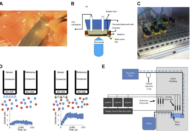
Tumor-free human bronchi obtained at operative lobectomy (24) were transported in lactated Ringer’s solution on wet ice. A subepi-thelial blister was raised with sterile saline, dissected sharply (see Fig. 1A), plated on human fibronectin cellware culture slides (BD Biosci-ences), and incubated at least 72 h (21% oxygen-5% CO2, 37°C) in UNC medium (18) with daily medium changes before permeabiliza-tion, staining, and imaging.
Cyclic Compressive Stress Procedures
In situ cyclic compressive stress (CCS) for microscopy is described in the immunofluorescence section. For the remainder of the studies, the ciliated, lumenal side of primary human airway pseudostratified epithelial cells in six-well transwell plates was ventilated through a sealed manifold (Fig. 1C) using a humidified rodent ventilator circuit
(New England Medical Instruments, Medway, MA) at pressures 20/5 cm water and a frequency of 14 breaths per minute (in a 37°C, 21% oxygen-5% CO2). The pressures represent “middle range” transepi-thelial (mucosal-to-submucosal) pressure gradient of 15 cm water (27, 48, 62); not as high as with dramatic transluminal pressures observed in asthma, but above pressures predicted during quiet breathing. Control cells in the same incubator were not attached to the ventilator. In the initial biochemical experiments, CCS was delivered similarly but as described by Button and coworkers (9). In selected experi-ments, CCS was also performed after a 2-h preincubation with the TRPV4 inhibitor HC067047 (500 nM) or the NOS inhibitor nitro-L
-arginine methyl ester (10 mM L-NAME; Cayman Chemical, Ann
Arbor, MI).
Immunofluorescence
Primary human airway pseudostratified epithelial cells and ex vivo human airway epithelia were fixed in 4% paraformaldehyde for 20 min, permeabalized in 0.05% Triton X-100 for 5 min, rinsed in phosphate buffered saline (PBS) three times (5 min), incubated overnight with primary antibodies, rinsed again in PBS three times (5 min), incubated 30 min with secondary antibodies, and then mounted with mount media and visualized by confocal microscopy (custom-ized Zeiss equipment) (42, 53). The primary antibodies were mouse anti-eNOS mAB (BD Transduction Laboratories; 1:200 dilution), rabbit anti-eNOS pAB (Cell Signaling; 1:200 dilution), and mouse anti-Dynein MAB (Thermo Scientific; 1:200). DAPI was from Ther-moScientific. The isotype controls were Purified Rabbit IgG Isotype Standard (no. 550875; BD Biosciences) and Purified Mouse IgG1 Isotype Control (no. 557273; BD Biosciences). The secondary anti-bodies were Alexa Fluor 488 chicken anti-mouse (Invitrogen; 1:500 dilution) and Alexa Fluor 568 goat anti-rabbit (Invitrogen; 1:250 dilution). In separate experiments at UNC, an Olympus FV10000 confocal microscope was used to image eNOS (Ab PA3031A; ThermoFisher; 1:200) in whole mount cells using donkey anti-rabbit AlexaFluor 488 (1:1,000) secondary (Ab 715-545-151; Jackson Im-munoResearch). Normal rabbit IgG was used as a control (5g/ml; Ab 550875; BD Biosciences). Cells were also stained with phalloidin for filamentous actin (Phalloidin Alexa 647; Invitrogen; 1:50) as well as for cilia (rat anti-tubulin MAB1864; 3g/ml; Millipore) (donkey anti-rat; Ab 712-295-153; Jackson ImmunoResearch; 1:1,000). DNA was imaged with Hoescht 33342 (0.01 mg/ml; H1399; Invitrogen).
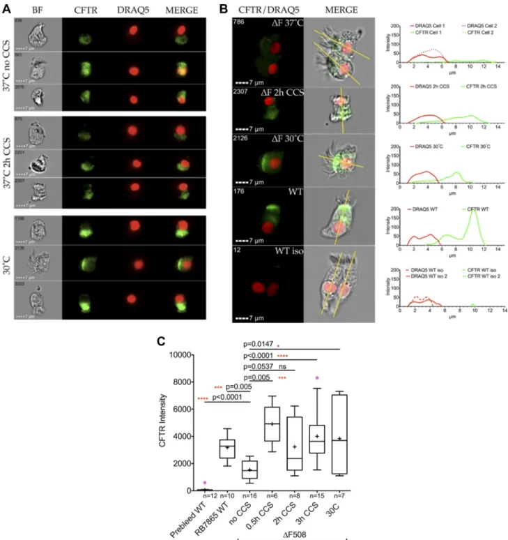
Imagestream
Psuedostratified WT and F508/F508 ALI cultures grown at 30°C for 72 h or at 37°C and treated with CCS as above for 0, 0.5, 2, or 3 h. Cells were removed from filters with Accutase and calcium/ magnesium-free PBS (1 mM EDTA) treatment for 10 min at 37°C. Released cells were then fixed in 4% paraformaldehyde in PBS (final concentration) in solution for 15 min at 4°C using mild agitation on an Eppendorf rotator. The paraformaldehyde-treated cells were incubated with primary and secondary antibodies as follows: CFTR (RB7865; 1:5,000; Ref. 67); eNOS (BD Transduc-tion Laboratories; 1:500); AF488 anti-rabbit (Molecular Probes; 1:15,000) AF546 anti-mouse (Molecular Probes; 1:15,000); and DRAQ5 (Cell Signaling; 1:10,000). Only cells treated with anti-eNOS were permeabilized with 0.2% Triton X-100 for 15 min (room temperature). After incubation with the secondary antibod-ies, cells were visualized on ImageStream X Mark II (lasers set to the following powers: 488 nm⫽50 mW, 658 nm⫽60 mW, and 768 nm⫽3.5 mW). Compensation matrices were collected for DRAQ5 and unstained cells to account for their autofluorescence and applied throughout the analysis. About 50% of the cells were in focus based on gradient root mean square analysis of the bright-field for further evaluation. The focused cells were then separated by aspect ratio and area observed in the DRAQ5 channel, allowing the identification of single versus multiple cells based on
Table 1. Sources and cystic fibrosis genotypes and growth for the human pseudostratified bronchial epithelial cell cultures grown at air-fluid interface
Annotation Genotype/No. Source
ALI 1 F508/F508-1 Charles River
ALI 2 F508/F508-2 UNC
ALI 3 F508/F508-3 Charles River
ALI 4 WT-1 Charles River
ALI 5 WT-2 UNC
ALI 6 WT-3 CWRU
ALI 7 WT-4-6 UVA
ALI 8 F508/F508-4 CWRU
ALI 9 WT-7 CWRU
the nucleus. Using this feature as opposed to the standard bright-field option allowed for a much tighter separation of clustered cells. All further analysis was then carried out on single cells. To identify representative intact cells, the size of the nucleus was tested for overlapping colocalization with CFTR. Cells that showed low colocalization of CFTR with DRAQ5 were then evaluated by the total area of the nucleus, allowing separation of viable intact cells from dead or apoptotic cells. Using this separa-tion method yielded typically about ~10% of cells useful for analysis per sample.
ImageJ Area Under the Curve Calculations
Images of cells were exported from IDEAS and analyzed using the Plot Profile option of ImageJ. For each cell, the area under the curve for the CFTR signal was calculated using GraphPad Prism. Statistical significance analysis was carried out in GraphPad Prism (Tukey method to flag outliers, nonparametric two-tailed Mann-Whitney).
Calcium Flux Experiments
The bottom of a microdish chamber (Ibidi, Munich, Germany) was replaced with a glass coverslip. A rubber stopper in the transwell was attached to a manually operated piston for CCS with air injections at a rate of ~1/min, each injection lasting ~20 s (see Fig. 1B). Cells were incubated with 50 l/well of 12 mM fluo 3-acetoxymethyl ester (FAM; Life Technologies; in air with 5% CO2; 37°C; 30 min) and imaged with an inverted microscope DMI6000 integrated to a Leica TCS-SPX confocal workstation with a⫻20 N.A. 0.7 objective lens. Images were collected with⫻3 digital zoom. To minimize focal drift, the stage and imaging chamber were equilibrated for 60 min before the experiment, the laser dwell time per pixel was increased to ~1,000 Hz with bidirectional mode scanning, and the pinhole was set at 1.5 Airy units. An argon laser of 488-nm line at 15% power was used to excite the cells loaded with fluo 3-AM, and fluorescence emission was recorded in the range of 493–582 nm with ~40% quantum efficiency Fig. 1. Materials and methods.A: isolation of cells ex vivo from a human bronchus using a submucosal normal saline blister, followed by culture and growth on human fibronectin cellware slides.B: cultures loaded with fluo-3 AM underwent cyclic compressive stress (CCS) on the microscope stage using a syringe attached to the sealed chamber. Emission (493–582 nm; 15% power) was imaged on a Leica TCS-SP5 confocal microscope.C: CCS on intact wells of cultured, ciliated epithelial cells at air-liquid interface (ALI) was generated using a custom manifold sealed to each transwell (culture medium on the basolateral side) attached to a heated and humidified rodent ventilator in a 37°C, in 5% CO2incubator. Control cells (right) were treated the same
PMT (hybrid detector) set at 75 V. Imaging was done in “xyt” mode using time series, and images were recorded at every 5 s up to 7 min and 40 s. Data from 33 different regions of interest were monitored to study the Ca2⫹flux.
Human Fasting Exhaled NO Measurements: In Vivo CCS
The fraction of exhaled NO (FENO) was measured using the Aerocrine MINO (Stockholm, Sweden) according to the manufac-turer’s instructions immediately before and immediately after treat-ment in the airway clearance vest (Hill Rom, Chicago, IL; frequency: 10 Hz; pressure: 10 cm water; 10 min). Subjects were nonsmoking volunteers, 20 – 60 yr old, with no pulmonary disease. Assays were performed in the morning after an overnight fast to prevent both1) gastric nitrite conversion to NO (40, 59), possibly compressed from the stomach into the exhaled air by the vest; and 2) an airway-reducing environment that would deplete NOx (40).
Immunoblots
Cell lysates were run on a 4 –15% SDS-page gel, and the membrane was incubated overnight with primary antibody. For human CFTR blots, we used anti-CFTR MAB25031 (2060; R&D Systems SC; 1:500), with secondary (SC-2061; Santa Cruz Biotechnology; 1:3,000). For murine CFTR blots, we used anti-CFTR NB300-511 (Novus; 1:1,000) and secondary SC-2973 (1:3,000). For anti--actin we used 13E5 (Cell Signaling; 1:2,000) and secondary was SC-2054 (Santa Cruz Biotechnology; 1:3,000). For nNOS, the primary was ab76067 (Abcam; 1:500) and secondary was SC-2054 (Santa Cruz Biotechnology; 1:3,000). For iNOS, the primary was ab3523 (Abcam; 1:500) and secondary was SC-2054 (Santa Cruz Biotechnology; 1:3,000). For phospho-eNOS, the primary AB (pS 1177) was from BD Transduction Laboratories (1:1,000) and secondary was SC-2060 (Santa Cruz Biotechnology; 1:3,000). For GSNO reductase (GSNOR; ADH5), the primary was 11051-1-AP (Proteintech; 1:1,000) and secondary was SC2054 (Santa Cruz Biotechnology; 1:3,000). Blots were visualized using the Biorad Chemidoc System.
S-Nitrosothiol Assays
S-nitrosothiols were measured by three complementary methods. Anaerobic reduction/chemiluminescence was performed on whole cell lysates as previously described (15). A novel method was used for measuringS-nitrosothiols in small, apical volumes using a polydop-amine-coated carbon fiber electrode (Fig. 1D; CFE-2; ALA Scientific) reacted with either monoclonal anti-S-nitrosocysteine (Creative Dy-namics; sensing electrode) or buffer (reference electrode) as previ-ously described using other antibodies (49, 50). Baseline measure-ments (sensing and reference electrodes in 10 mM Tris, pH 7.5, running buffer) used a 0.2-s current injection by means of a⫾50-mV step potential (ITC-1600; HEKA) with a Ag/AgCl2ground pellet. The baseline measurement was repeated (twice, adding fresh running buffer), and the current responses of the sensing and reference electrodes were recorded in fresh buffer to ensure a stable baseline. StandardS-nitrosothiol solutions (24) or airway cell apical medium samples were then added to the running buffer. The maximum difference in current response by the sensing and reference electrode after baseline subtraction was recorded (49, 50). This method was adapted as a chemical detection system for low concentrations of S-nitrosothiols [see Supplemental Data: https:/doi.org/10.17605/OSF.IO/ S6G24) (23, 31)].
Murine Studies
Immunoblot studies of eNOS⫺/⫺mice.eNOS⫺/⫺and background
(C57Bl6) lungs were isolated as described previously (59). Tissue was homogenized in CFTR lysis buffer [100 mM NaCl, 50 mM NaF, 50 mM Tris·HCl, pH 7.5, 1% NP-40, 0.25% sodium deoxycholate, 1 mM
EDTA, 1 mM EGTA, and protease inhibitor cocktail set III (Calbio-chem)] and immunoblotted for CFTR as described above.
Study approvals. Animal use protocols were approved by the Institutional Animal Care and Use Committee at Case Western Re-serve University (CWRU). Each of the clinical elements of the study [primary cell culture (PCC), ex vivo epithelial preparation (EVEP), and FENO/airway clearance vest (FAV) studies] was reviewed and approved by the Rainbow Babies and Children’s Hospital (PCC, FAV), University of Virginia (PCC and EVEP), and/or UNC, Chapel Hill (PCC) Institutional Review Boards, and subjects signed informed consent before participating.
Statistical Analysis
Pairwise comparisons were analyzed byt-test if normally distrib-uted with equal variance and rank-sum testing if not. Pairedt-test was used for pair-wise comparisons. Multiple comparisons were similarly analyzed either by ANOVA or ANOVA on ranks following Dunn’s test. Data are expressed as means ⫾ SE or median⫾interquartile range (IQR), as appropriate.P⬍0.05 was considered significant.
RESULTS
Endothelial NOS Is Present in Human Airway Epithelial Cells In Vitro and Ex Vivo
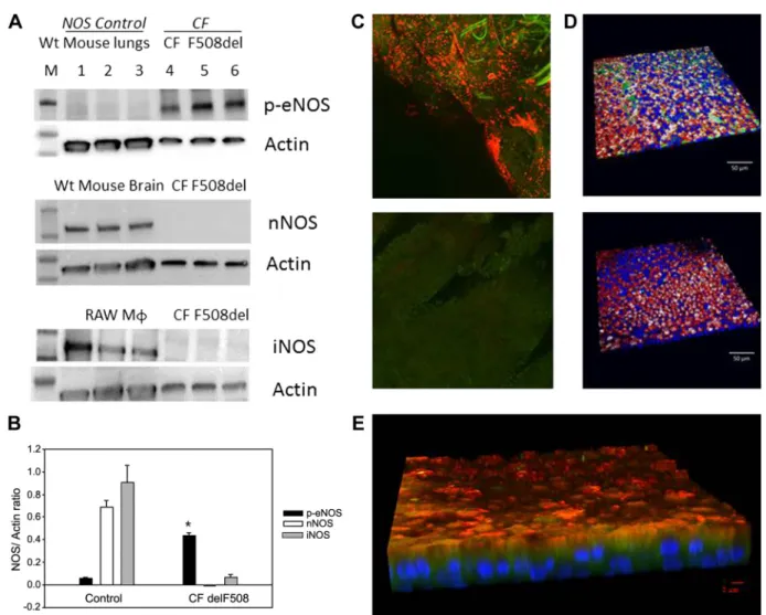
We used human ALI cultured cells from five normal subjects and four F508Del homozygous cells listed in Table 1 and annotated throughout as “ALIx.” We confirmed previous work (33) showing that iNOS is minimally expressed in F508Del homozygous cultures (n ⫽ 3 each; Fig. 2A; human primary ALI cultures, F508/F508, from Charles River and UNC; Table 1, ALI 1 and 2) and showed that nNOS was also essentially not detectable (Fig. 2A). However, eNOS was present and was phosphorylated at serine 1177, thus capable of activation by calcium flux and calmodulin binding (38) (n⫽3; Fig. 2,Aand B; human primary ALI cultures, F508/F508, from Charles River and UNC; Table1: ALI 1 and 2). Expression of phospho-eNOS was greater than that of nNOS and iNOS (P ⬍0.001; Fig. 2B). Controls were WT murine lung homogenate (eNOS), WT murine brain homogenate (nNOS), and the macrophage cell line RAW 264.7 (iNOS) as shown in Fig. 2A. Consistent with previous reports (64, 67), eNOS expression was apical in situ in airway epithelium of operative lobectomy specimens from humans with lung cancer (41) (Fig. 2C;n⫽3 subjects). It was also apical in WT human pseudostratified epithelium grown at ALI (Fig. 2D; human primary ALI cultures, WT, from UNC; Table1: ALI 5). In F508Del homozygous prepara-tions, it was apical and partially colocalized with dynein (Fig. 2E; n ⫽ 4, human primary ALI cultures, F508/F508, from UNC; Table1: ALI 2).
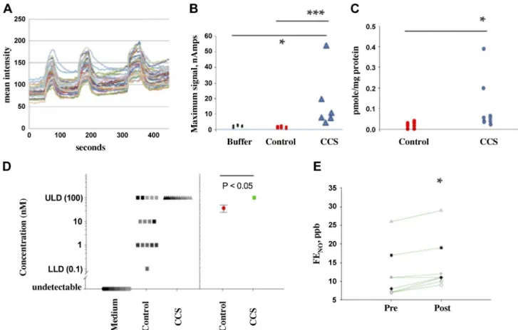
Cyclic Compression Leads to Cyclic Fluxes of Calcium into Human Airway Epithelial Cells
Because eNOS is activated by Ca2⫹-calmodulin binding
(25), we next confirmed that pulses of CCS resulted in cyclic Ca2⫹ flux into pseudostratified epithelial cells (1, 39). Each
(47.2⫾18 M pre; 67.1 ⫾9.8 M post; n⫽ 3 each; P ⫽ NS), suggesting that eNOS was not activated by calcium flux in the absence of cilia, likely because there is very little present in unciliated airway epithelial cells in culture (3).
Cyclic Compression Leads to Nitrogen Oxide Production by Human Airway Epithelial Cells
To determine whether CCS-induced calcium flux could activate eNOS, we measured epithelial NOS-derived S -nitro-sothiols, chosen because of their potential roles in CFTR maturation (28, 42, 65, 66), before and after CCS (Fig. 3,B–E; human primary ALI cultures, F508/F508, from Charles River; Table1: ALI 1 and 2). The CCS⌬P was 15 cm of water, not as extreme as predicted during an asthma exacerbation, but sub-stantially higher than in quiet breathing (48). S-nitrosothiol levels in the airways are near the limit of detection using traditional assays (23, 24, 65), particularly in CF (26). We
therefore employed a recently developed class of capacitance-based S-nitrosothiol assay with nM sensitivity (31, 53, 55) (Fig. 1E) in addition to traditional reduction/chemiluminesce. All assays were performed by investigators blind to the CCS treatment status. In all, we studied 61 filters from three CF subjects. All assays showed that CCS increased epithelial S-nitrosothiol levels in CF pseudostratified airway epithelial cells harvested immediately after CCS. First, by reduction/ chemiluminescence, whole cellS-nitrosothiol levels increased from 0.015 (IQR 0.0025– 0.028) pmol/g protein (control;n⫽ 8) to 0.06 (IQR 0.04 – 0.145) pmol/g protein (CCS;n⫽ 9; P ⫽ 0.002; Fig. 3C; Table 1: ALI 2). Second, we used the capacitance-based immunosensor (31, 55) coupled with an anti-S-nitrosocysteine (CSNO) antibody (59) to measure the apical S-nitrosothiol signal (sensor placed in fluid from the apex, adjacent to the eNOS localization; Fig. 3B). This signal increased after CCS from 1 (IQR 0 –2) nAmp (control;n⫽3)
to 10.5 (IQR 8 –20) nAmp (P⬍0.05;n⫽6; Fig. 3B; ALI 3). Finally, we used this capacitance-based sensor system with formaldehyde pretreatment as recently described and standard-ized (53) (Fig. 1E) to measure whole cell lysateS-nitrosothiols. Each assay in Fig. 3D was performed on a fresh set of cells using a sensor (53) that acts as acts as an on/off toggle for the presence or absence of CSNO. We therefore performed serial dilutions of the cell lysate over four logs on WT primary airway epithelial cells) with cyclic compressive stress (labeled as “CCS”) and without CCS (labeled as “control”) and medium alone (collected from these cells). The difference between the Fig. 3D, left, and Fig. 3D, right, is that Fig. 3D, left, dots represent the individual signals from each set of cells (using the log scale with which the measurements were made), while Fig. 3D,right, is the geometric mean of the Fig. 3D,left, results (red: control with standard error; green: cells that received CCS). There is a significant difference in these geometric mean CSNO levels between WT cells at baseline and those and receiving CCS (Pⱕ0.05). This method confirmed that all cells had S-nitrosothiols [36⫾12 nM; greater than medium alone
(0⫾0 nM),n⫽5 samples, 3 replicates each;P⬍0.001]. CCS increasedS-nitrosothiol levels to the point that the sensor was saturated (ⱖ100 nM in all samples,n⫽5 samples, 3 replicates each,P⬍0.001 compared with untreated; human primary ALI cultures, F508/F508, from CWRU; Table 1: ALI 8; Fig. 3D). Finally, to confirm that the increase of S-nitrosothiol level associated with CCS was caused by increased production, rather than decreased catabolism (41), we measured the effect of CCS on GSNO reductase expression in human primary ALI cultures (F508/F508 from Charles River; Table 1: ALI 3). We have found no difference in GSNOR expression and activity following CCS treatment (densitometry⫽0.80 ⫾ 0.43, vs. control 0.55⫾0.53; n⫽ 3 each;P⫽ NS).
Measurement of airway S-nitrosothiols in vivo before and after CCS would require pre- and post-CCS bronchoscopy. To avoid this invasive procedure, we studied fraction of exhaled NO (FENO), a marker for airway GSNO (57): airway GSNO can be labile but increasing GSNO airway increases FENO because of homolytic GSNO cleavage to form NO (40, 57). Thus a small change in FENO can be a marker for GSNO
Fig. 3. Cyclic compressive stress (CCS) increases apical Ca2⫹flux and nitrogen oxide formation in human airway epithelial cells at air-liquid interface (ALI). A: Ca2⫹influx into the apex of these cells (n⫽16; ALI 5) with each pulse of CCS (blue arrows). This experiment was repeated 3 times.B: sensor response from the system shown in Fig. 1Din apical medium measured after CCS from cystic fibrosis (CF), ciliated cells at ALI untreated (n⫽3) or treated with CCS as shown in 1C (n⫽6; ALI 3). TheS-nitrosothiol signal was higher after CCS. *P⬍0.05, CCS vs. buffer; ***P⬍0.05, CCS-treated vs. control cells.C: after CCS, F508Del homozygous cells were snap frozen in liquid nitrogen. Blinded reduction-chemiluminescence assay showedS-nitrosothiol content/protein after CCS (n⫽9) was greater than control (n⫽8; *P⫽0.002; ALI 1 and 2).D: assays were also performed on a fresh set of cells using a modified sensor as described in Ref. 53.Leftdots represent the individual signals from each set of cells (using the log scale with which the measurements were made), while therightdots represent the geometric mean of the results atleft(red: control with standard error; green: cells received CCS). Because this sensor acts as an on/off toggle for the presence or absence ofS-nitrosocysteine, we performed serial dilutions of the cell lysate and medium over 4 logs (0.01–100 nM). All cells hadS-nitrosothiols [36⫾12 nM; greater than medium alone (0⫾0 nM),n⫽5 samples, 3 replicates each;P⬍0.001]. CCS increasedS-nitrosothiol levels to the point that the sensor was saturated (ⱖ100 nM in all samples,n⫽5 samples, 3 replicates each,P⬍0.001 compared with untreated; ALI 8).E: normal subjects were studied after an overnight fast for fraction of exhaled NO (FENO) analysis before and after airway clearance vest treatment: 10 Hz, 10 cm water, 10 min;n⫽8). FENO
formation downstream of eNOS activation. However, the de-terminants of FENOare complex (12, 21, 40, 51, 57) and eNOS can activate counterregulatory GSNO reductase (5), which, in an NADH-dependent fashion, gradually decreases GSNO (38). Feeding increases cellular NADH (63), which increases GSNOR activity (38, 61). Therefore, we studied FENO in human subjects after an overnight fast. Measurements were made before and after in vivo airway clearance vest treatment, which cyclically increases pressure and shear stress across the ALI in vivo. The airway clearance system consistently in-creased fasting FENOin humans in vivo from 12.7 to 14.8 ppb (n⫽8;P⫽0.006; Fig. 3E). Since it does not do so in the fed state (56), these in vivo data are consistent with the observation that CCS leads to S-nitrosothiol formation in vitro.
Cyclic Compression Leads to Increased F508Del CFTR Maturation in Human Airway Epithelial Cells in a Cilia-Dependent Fashion
Single cell flow-based Imagestream analysis of cells grown in ALI cultures using area under the curve (AUC) calculation of total CFTR density (Fig. 4; Supplemental Figures S1, S2, and S3; see Supplemental Data: https:/doi.org/10.17605/ OSF.IO/S6G24) showed very little CFTR staining in the iso-type control and weak intracellular staining in F508del ho-mozygous cells. However, there was strong, apical expression in healthy, control cells and in F508Del CFTR homozygous cells treated with either CCS or 30°C. Specifically, filters of ALI cultures were placed in our custom CCS manifold (as described above) at 37°C, 5% CO2as above for 30 min, 2 h, or 3 h (Fig. 4C). Control F508 Del homozygous ALI cultures were placed into a 30°C/5% CO2incubator overnight (positive control) or in the CCS incubator (37°C; negative control). Non-CF ALI cultures (37°C) were used as an additional con-trol. Assessment and quantification of CFTR were performed using imaging flow cytometry with RB7865, a polyclonal antibody directed toward the ECL-1 loop of CFTR (43), for eNOS and DRAQ5 for nuclear DNA. Without CCS treatment the F508Del CFTR localization was minimal, and diffuse in the cell. For an in-depth analysis cells were selected and cross sectionally assessed as indicated in the Supplemental Fig. S3 (see Supplemental Data: https:/doi.org/10.17605/OSF.IO/ S6G24). An example of cells of each population is given with the corresponding profile next to each cell. The green line shows the distribution of CFTR along the longitudinal cut of the cells. Importantly, WT cells stained with the prebleed serum of rabbit RB7865 showed very little CFTR stain (isotype control), while the immune serum showed a very clear signal on the same batch of cells. In all cases we observed a signif-icant difference when treating cells with CCS. The CFTR intensity was proportional to the area under the curve. By contrast, CCS did not change eNOS expression (see Supple-mental Fig. S4; https:/doi.org/10.17605/OSF.IO/S6G24).
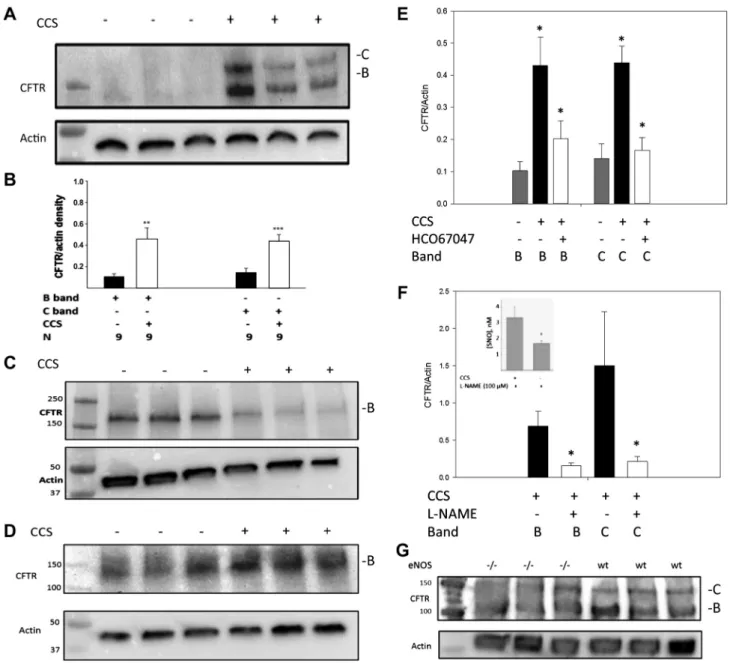
Immunoblot assays confirmed that expression of F508Del CFTR was increased by CCS in ciliated pseudostratified cul-tures (Fig. 5A; human primary ALI cultures, F508/F508, from Charles River; Table1: ALI 1). Specifically, partially mature CFTR (B band) increased relative to-actin (from 0.10⫾0.02 to 0.46⫾0.1;n⫽9 lanes, 2 wells/lane;P⬍0.001), and the fully mature CFTR (C band) increased from 0.12 ⫾ 0.03 relative to-actin to 0.42⫾0.02;n⫽9 lanes;P⬍0.001; (Fig.
5B; human primary ALI cultures, F508/F508, from Charles River and UNC (Table1: ALI 1 and 2). On the other hand, CCS did not result in any change F508Del CFTR expression in nonciliated, monolayer CF epithelial cell lines (CFBE41o⫺; n⫽3 lanes; 6 wells;P⫽NS; Fig. 5C), consistent with the lack of eNOS activation using calcium ionophore in these cells (above). If anything, there was a tendency for the B band to decrease with CCS in the absence of cilia. CCS also did not significantly affect WT CFTR maturation in WT CFBE41o⫺ cells (n⫽ 3; Fig. 5D), again suggesting an essential role for cilia.
Increased F508Del CFTR Maturation in Human Airway Epithelial Cells Exposed to Cyclic Stress Is Dependent on TRPV4 and eNOS
We next studied the role of TRPV4-dependent Ca2⫹flux (1,
39) and of NOS on the CCS effect to increase F508Del CFTR maturation (Fig. 5, E and F). Note that these cells express TRPV4 mRNA from early passage (S. H. Randell, unpublished observations) consistent with previous publications regarding TRPV4 protein expression (1, 39). CCS-induced CFTR matu-ration was inhibited by the TRPV4 inhibitor HC067047 (500 nM, 2 h, (n⫽4 lanes, 8 wells each;P⬍0.01; Fig. 5E; human primary ALI cultures, F508/F508, from UNC; Table1: ALI 2). Furthermore, CCS-induced WT CFTR maturation was inhib-ited by the NOS inhibitorL-NAME (10 mM, 2 h;n⫽9 lanes each; P ⬍ 0.01; Fig. 5F; human primary ALI cultures, WT, from CWRU and UNC; Table1: ALI 5 and 6), which inhibited S-nitrosothiol production from 3.3⫾0.7 to 1.8⫾0.1 nM;n⫽ 3 each;P⬍0.05; Fig. 5F,inset; human primary ALI cultures, WT, from UNC; Table1: ALI 5). Both B- and C-band CFTR, while present, were attenuated in eNOS⫺/⫺ mouse lung
ho-mogenates (n ⫽ 3 each; Fig. 5G). CCS in vitro decreased transepithelial resistance in Ussing chambers, likely because of disrupted cell junctions, precluding using this measure of CFTR function.
DISCUSSION
It is increasingly appreciated that cyclic changes in air pressure and flow affect the biology of ciliated airway epithe-lial cells in culture (6, 60), just as fluid flow alters endotheepithe-lial cell biology (17). Here, we have studied the effect of changes in apical air pressure on human airway epithelial CFTR mat-uration. We report that cyclic compression, at least in part through TRPV4 and NOS, leads to increased CFTR expression in ciliated airway epithelial cultures.
Airway epithelial cilia have motor and sensory functions (14, 30, 36, 54). Sensory functions are both chemosensory and mechanosensory. Recent evidence suggests that ciliary chemo-sensory functions involve NOx (32). Here, we provide evi-dence that airway mechanosensory functions also involve NOx. This observation is consistent with previous data regard-ing olfactory cilia, in which eNOS is activated by calcium flux to signal olfaction (13). These data suggest a novel role for eNOS in the sensory functions of airway cilia.
protein (Hop) for degradation, preventing Hop-CFTR interac-tion and, thereby, preventing CFTR degradainterac-tion (42). Other mechanisms are relevant as well (41, 65), but epithelial siRNA experiments suggest that the Hop-dependent mechanisms is foremost.
However, GSNOR inhibition only improves weight gain, not lung function, in CF patients in vivo, consistent with evidence
that GSNO production is decreased in the CF airway epithe-lium (33): inhibiting a catabolic enzyme is ineffective if there is little substrate. GSNO and other S-nitrosothiols are classi-cally produced by NOS isoforms (16, 22, 25, 52, 58, 61). Neither nNOS nor iNOS are expressed significantly in CF airway epithelial cells (26) (Fig. 2). Our current data suggest that CCS-driven eNOS activation may help augment GSNO
airway levels, and that increasing GSNO production, deficient in CF (26), may be more valuable for increasing CFTR mat-uration than inhibiting GSNO catabolism. Indeed, there may be little value to inhibiting GSNO catabolism if its substrate is deficient.
Genetic deletion of eNOS is associated with more metha-choline responsiveness in a murine asthma model than is deletion of other NOS isoforms (11). However, eNOS pro-duces low quantities of NO (ie, NO radical) relative to iNOS and is not frequently discussed in the context of airway NO
production (51). This paradox is consistent with evidence that NO itself may be less important than is commonly supposed in human airway biology downstream of NOS activation (22, 40). For example, human airway smooth muscle is less responsive to endogenous levels NO than endogenous levels of GSNO (20); levels of NO in the airway are normally too low to be of any physiological relevance (23, 57); and eNOS activation causes location-dependent formation ofS-nitrosothiols to sig-nal local effects independent of the quantity of NO radical produced (25, 41). In this regard, eNOS activation to produce
GSNO in the airway epithelium could have many beneficial effects. S-nitrosothiols increase ciliary motility (37), relax airway smooth muscle (20, 22), and alter Na⫹and Cl⫺currents (10, 29). Thus the pathway we describe here could help to explain certain benefits of airway pressure, like that in exercise. For example, positive pressure causes smooth muscle relax-ation in precision-cut human lung slices (35). This involves an effect on the biophysical properties of airway smooth muscle but could also involve epithelial eNOS activation (22, 25). Because iNOS is minimally present in the CF epithelium and is not upregulated posttranslationally by calcium, our data suggest that it is eNOS, activated by CCS, that increases F508Del CFTR maturation. eNOS activity may be underap-preciated in static cultures of airway epithelium.
AirwayS-nitrosothiols were not measured directly in vivo. We used change in fasting FENOas a surrogate to avoid pre-and post-CCS bronchoscopy. In CF patients, FENO increases with increased airway GSNO in vivo (57). Note, however, that determinants of FENOare complex (46) and affected by airway pH, the airway microbiome, airway ADMA and arginine me-tabolism, and airway GSNOR activity (12, 21, 40, 51, 57). GSNOR activity is regulated by NADH availability, which is decreased in fasting (26, 38): CCS increases FENO in fasted subjects, whereas it does not in fed subjects (56). Thus these in vivo observations are supportive of the in vitro evidence for an increase in airway epithelial GSNO formation resulting from CCS. GSNO formation could be enhanced by use of GSNOR inhibitor, because theS-nitrosothiol compounds are labile (41), but we have focused here purely on formation. A range of airway clearance techniques and devices provide in vivo CCS for CF patients. Our findings could ultimately help provide markers to determine which clinical techniques would be most beneficial and which would not (44).
In summary, human airway epithelial cells respond to cyclic increases in airway pressure with an increase in CFTR expres-sion. Consistent with previous publications (1, 39, 64, 67), this effect on CFTR likely involves Ca2⫹ flux into the cells to activate apical eNOS. CCS increasesS-nitrosothiol formation and increases F508Del maturation in a TRPV4- and NOS-dependent fashion. Moreover, nonciliated airway epithelial cells fail to increase F508Del CFTR maturation in response to CCS. Taken together, these data suggest the possibility that approaches to increase cyclic airway compression, whether through airway clearance techniques or vigorous exercise it-self, could benefit CF patients not only by mobilizing secre-tions, but also by correcting CFTR expression.
ACKNOWLEDGMENTS
We thank Dr. Douglas Hess for the gift of the GSNOR⫺/⫺, eNOS⫺/⫺, and background mice; Dr. Eric Sorcher for the CFBE41o⫺cell lines; Dr. Vinod Jyothikumar, Dr. Joseph E. Doherty, Teresa Mascenik, and Sean Miller for technical assistance, and Jessica Surdam, Sara Bishop, and Marie Abdul-Karim for administrative assistance.
GRANTS
This work was supported by National Institutes of Health Grants P01-HL-101871 (to B. Gaston, N. Marozkina, K. Zaman, A. Periasamy, and S. H. Randall), P01-HL-128192 (to B. Gaston and N. Marozkina), R21-AN-3429620 (to B. Gaston), and R01-CA-136705 (to D. R. Jones). It is also supported by Children’s Lung Foundation (to B. Gaston).
DISCLOSURES
No conflicts of interest, financial or otherwise, are declared by the authors.
AUTHOR CONTRIBUTIONS
B.G. conceived and designed research; N.M., J.B., C.U.C., L.S., J.V.S., K.Z., S.R., A.P., H.G., G.A., M.D., D.R.J., R.S., and S.H.R. performed experiments; N.M., J.B., L.S., J.V.S., and B.G. analyzed data; N.M. and B.G. interpreted results of experiments; N.M. and J.B. prepared figures; N.M. drafted manuscript; B.G. edited and revised manuscript; B.G. approved final version of manuscript.
REFERENCES
1. Alenmyr L, Uller L, Greiff L, Högestätt ED, Zygmunt PM. TRPV4-mediated calcium influx and ciliary activity in human native airway epithelial cells. Basic Clin Pharmacol Toxicol 114: 210 –216, 2014. doi:10.1111/bcpt.12135.
2. Angers RC, Mutka SC, Bove, P, Gabriel SE.Pharmacological correc-tion and acute inhibicorrec-tion of GSNOR results in improved in vitro CFTR function. 2015 North American Cystic Fibrosis Conference. Phoenix, AZ. 3. Asano K, Chee CB, Gaston B, Lilly CM, Gerard C, Drazen JM, Stamler JS.Constitutive and inducible nitric oxide synthase gene expres-sion, regulation, and activity in human lung epithelial cells.Proc Natl Acad Sci USA91: 10089 –10093, 1994. doi:10.1073/pnas.91.21.10089. 4. Baraldi E.Chronic respiratory diseases and sport in children.Int J Sports
Med21,Suppl2: 103–104, 2000. doi:10.1055/s-2000-8499.
5. Brown-Steinke K, deRonde K, Yemen S, Palmer LA.Gender differ-ences in S-nitrosoglutathione reductase activity in the lung.PLoS One5: e14007, 2010. doi:10.1371/journal.pone.0014007.
6. Button B, Boucher RC; University of North Carolina Virtual Lung Group.Role of mechanical stress in regulating airway surface hydration and mucus clearance rates.Respir Physiol Neurobiol163: 189 –201, 2008. doi:10.1016/j.resp.2008.04.020.
7. Button B, Cai LH, Ehre C, Kesimer M, Hill DB, Sheehan JK, Boucher RC, Rubinstein M. A periciliary brush promotes the lung health by separating the mucus layer from airway epithelia.Science337: 937–941, 2012. doi:10.1126/science.1223012.
8. Button B, Okada SF, Frederick CB, Thelin WR, Boucher RC. Mecha-nosensitive ATP release maintains proper mucus hydration of airways.Sci Signal6: ra46, 2013. doi:10.1126/scisignal.2003755.
9. Button B, Picher M, Boucher RC. Differential effects of cyclic and constant stress on ATP release and mucociliary transport by human airway epithelia. J Physiol 580: 577–592, 2007. doi:10.1113/jphysiol.2006. 126086.
10. Chen L, Patel RP, Teng X, Bosworth CA, Lancaster JR, Matalon S. Mechanisms of cystic fibrosis transmembrane conductance regulator ac-tivation by S-nitrosoglutathione. J Biol Chem 281: 9190 –9199, 2006. doi:10.1074/jbc.M513231200.
11. De Sanctis GT, MacLean JA, Hamada K, Mehta S, Scott JA, Jiao A, Yandava CN, Kobzik L, Wolyniec WW, Fabian AJ, Venugopal CS, Grasemann H, Huang PL, Drazen JM. Contribution of nitric oxide synthases 1, 2, and 3 to airway hyperresponsiveness and inflammation in a murine model of asthma.J Exp Med189: 1621–1630, 1999. doi:10. 1084/jem.189.10.1621.
12. Dweik RA, Comhair SA, Gaston B, Thunnissen FB, Farver C, Thom-assen MJ, Kavuru M, Hammel J, Abu-Soud HM, Erzurum SC.NO chemical events in the human airway during the immediate and late antigen-induced asthmatic response.Proc Natl Acad Sci USA98: 2622– 2627, 2001. doi:10.1073/pnas.051629498.
13. Endo D, Yamamoto Y, Yamaguchi-Yamada M, Nakamuta N, Tani-guchi K.Localization of eNOS in the olfactory epithelium of the rat.J Vet Med Sci73: 423–430, 2011. doi:10.1292/jvms.10-0353.
14. Enuka Y, Hanukoglu I, Edelheit O, Vaknine H, Hanukoglu A. Epi-thelial sodium channels (ENaC) are uniformly distributed on motile cilia in the oviduct and the respiratory airways. Histochem Cell Biol 137: 339 –353, 2012. doi:10.1007/s00418-011-0904-1.
15. Fang K, Ragsdale NV, Carey RM, MacDonald T, Gaston B.Reductive assays for S-nitrosothiols: implications for measurements in biological systems. Biochem Biophys Res Commun 252: 535–540, 1998. doi:10. 1006/bbrc.1998.9688.
16. Foster MW, Hess DT, Stamler JS.Protein S-nitrosylation in health and disease: a current perspective. Trends Mol Med 15: 391–404, 2009. doi:10.1016/j.molmed.2009.06.007.
18. Fulcher ML, Randell SH.Human nasal and tracheo-bronchial respiratory epithelial cell culture. Methods Mol Biol945: 109 –121, 2012. doi:10. 1007/978-1-62703-125-7_8.
19. Galietta LJ, Lantero S, Gazzolo A, Sacco O, Romano L, Rossi GA, Zegarra-Moran O.An improved method to obtain highly differentiated monolayers of human bronchial epithelial cells. In Vitro Cell Dev Biol Anim34: 478 –481, 1998. doi:10.1007/s11626-998-0081-2.
20. Gaston B, Drazen JM, Jansen A, Sugarbaker DA, Loscalzo J, Rich-ards W, Stamler JS.Relaxation of human bronchial smooth muscle by S-nitrosothiols in vitro.J Pharmacol Exp Ther268: 978 –984, 1994. 21. Gaston B, Kelly R, Urban P, Liu L, Henderson EM, Doctor A, Teague
WG, Fitzpatrick A, Erzurum S, Hunt JF. Buffering airway acid decreases exhaled nitric oxide in asthma.J Allergy Clin Immunol118: 817–822, 2006. doi:10.1016/j.jaci.2006.06.040.
22. Gaston B, Reilly J, Drazen JM, Fackler J, Ramdev P, Arnelle D, Mullins ME, Sugarbaker DJ, Chee C, Singel DJ.Endogenous nitrogen oxides and bronchodilator S-nitrosothiols in human airways.Proc Natl Acad Sci USA90: 10957–10961, 1993. doi:10.1073/pnas.90.23.10957. 23. Gaston B, Singel D, Doctor A, Stamler JS.S-nitrosothiol signaling in
respiratory biology.Am J Respir Crit Care Med173: 1186 –1193, 2006. doi:10.1164/rccm.200510-1584PP.
24. Gow A, Doctor A, Mannick J, Gaston B.S-Nitrosothiol measurements in biological systems.J Chromatogr B Analyt Technol Biomed Life Sci 851: 140 –151, 2007. doi:10.1016/j.jchromb.2007.01.052.
25. Gow AJ, Chen Q, Hess DT, Day BJ, Ischiropoulos H, Stamler JS. Basal and stimulated protein S-nitrosylation in multiple cell types and tissues.J Biol Chem277: 9637–9640, 2002. doi:10.1074/jbc.C100746200. 26. Grasemann H, Gaston B, Fang K, Paul K, Ratjen F.Decreased levels of nitrosothiols in the lower airways of patients with cystic fibrosis and normal pulmonary function.J Pediatr135: 770 –772, 1999. doi:10.1016/ S0022-3476(99)70101-0.
27. Gunst SJ, Stropp JQ.Pressure-volume and length-stress relationships in canine bronchi in vitro. J Appl Physiol (1985)64: 2522–2531, 1988. doi:10.1152/jappl.1988.64.6.2522.
28. Howard M, Fischer H, Roux J, Santos BC, Gullans SR, Yancey PH, Welch WJ. Mammalian osmolytes and S-nitrosoglutathione promote delta F508 cystic fibrosis transmembrane conductance regulator (CFTR) protein maturation and function.J Biol Chem278: 35159 –35167, 2003. doi:10.1074/jbc.M301924200.
29. Jain L, Chen XJ, Brown LA, Eaton DC.Nitric oxide inhibits lung sodium transport through a cGMP-mediated inhibition of epithelial cation channels.Am J Physiol Cell Lung Mol Physiol274: L475–L484, 1998. doi:10.1152/ajplung.1998.274.4.L475.
30. Jain R, Javidan-Nejad C, Alexander-Brett J, Horani A, Cabellon MC, Walter MJ, Brody SL.Sensory functions of motile cilia and implication for bronchiectasis.Front Biosci (Schol Ed)4: 1088 –1098, 2012. 31. Jastrzebska MM, Stepien´ K, Wilczok J, Porebska-Budny M, Wilczok
T.Semiconductor properties of melanins prepared from catecholamines. Gen Physiol Biophys9: 373–383, 1990.
32. Jiao J, Wang H, Lou W, Jin S, Fan E, Li Y, Han D, Zhang L. Regulation of ciliary beat frequency by the nitric oxide signaling pathway in mouse nasal and tracheal epithelial cells.Exp Cell Res317: 2548 –2553, 2011. doi:10.1016/j.yexcr.2011.07.007.
33. Kelley TJ, Drumm ML. Inducible nitric oxide synthase expression is reduced in cystic fibrosis murine and human airway epithelial cells.J Clin Invest102: 1200 –1207, 1998. doi:10.1172/JCI2357.
34. Kobzik L, Bredt DS, Lowenstein CJ, Drazen J, Gaston B, Sugarbaker D, Stamler JS.Nitric oxide synthase in human and rat lung: immunocy-tochemical and hisimmunocy-tochemical localization.Am J Respir Cell Mol Biol9: 371–377, 1993. doi:10.1165/ajrcmb/9.4.371.
35. Lavoie TL, Krishnan R, Siegel HR, Maston ED, Fredberg JJ, Solway J, Dowell ML. Dilatation of the constricted human airway by tidal expansion of lung parenchyma.Am J Respir Crit Care Med186: 225–232, 2012. doi:10.1164/rccm.201202-0368OC.
36. Lee RJ, Cohen NA. Bitter and sweet taste receptors in the respiratory epithelium in health and disease.J Mol Med (Berl)92: 1235–1244, 2014. doi:10.1007/s00109-014-1222-6.
37. Li D, Shirakami G, Zhan X, Johns RA. Regulation of ciliary beat frequency by the nitric oxide-cyclic guanosine monophosphate signaling pathway in rat airway epithelial cells. Am J Respir Cell Mol Biol23: 175–181, 2000. doi:10.1165/ajrcmb.23.2.4022.
38. Liu L, Hausladen A, Zeng M, Que L, Heitman J, Stamler JS. A metabolic enzyme for S-nitrosothiol conserved from bacteria to humans. Nature410: 490 –494, 2001. doi:10.1038/35068596.
39. Lorenzo IM, Liedtke W, Sanderson MJ, Valverde MA.TRPV4 chan-nel participates in receptor-operated calcium entry and ciliary beat fre-quency regulation in mouse airway epithelial cells.Proc Natl Acad Sci USA105: 12611–12616, 2008. doi:10.1073/pnas.0803970105.
40. Marozkina NV, Gaston B. Nitrogen chemistry and lung physiology. Annu Rev Physiol 77: 431–452, 2015. doi: 10.1146/annurev-physiol-021113-170352.
41. Marozkina NV, Gaston B.S-Nitrosylation signaling regulates cellular protein interactions.Biochim Biophys Acta1820: 722–729, 2012. doi:10. 1016/j.bbagen.2011.06.017.
42. Marozkina NV, Yemen S, Borowitz M, Liu L, Plapp M, Sun F, Islam R, Erdmann-Gilmore P, Townsend RR, Lichti CF, Mantri S, Clapp PW, Randell SH, Gaston B, Zaman K. Hsp 70/Hsp 90 organizing protein as a nitrosylation target in cystic fibrosis therapy.Proc Natl Acad Sci USA107: 11393–11398, 2010. doi:10.1073/pnas.0909128107. 43. McGee KE, Roesch E, Marozkina N, Drumm ML, Hodges CA, Bosch
J.Structurally-driven development of a cross-species, extracellular CFTR antibody (Abstract).Pediatr Pulmonol52: 2017. doi:10.1002/ppul.23838. 44. McIlwaine MP, Alarie N, Davidson GF, Lands LC, Ratjen F, Milner R, Owen B, Agnew JL.Long-term multicentre randomised controlled study of high frequency chest wall oscillation versus positive expiratory pressure mask in cystic fibrosis.Thorax68: 746 –751, 2013. doi:10.1136/ thoraxjnl-2012-202915.
45. McIlwaine MP, Lee Son NM, Richmond ML.Physiotherapy and cystic fibrosis: what is the evidence base?Curr Opin Pulm Med20: 613–617, 2014. doi:10.1097/MCP.0000000000000110.
46. Modena BD, Tedrow JR, Milosevic J, Bleecker ER, Meyers DA, Wu W, Bar-Joseph Z, Erzurum SC, Gaston BM, Busse WW, Jarjour NN, Kaminski N, Wenzel SE.Gene expression in relation to exhaled nitric oxide identifies novel asthma phenotypes with unique biomolecular path-ways.Am J Respir Crit Care Med190: 1363–1372, 2014. doi:10.1164/ rccm.201406-1099OC.
47. Noone PG, Leigh MW, Sannuti A, Minnix SL, Carson JL, Hazucha M, Zariwala MA, Knowles MR.Primary ciliary dyskinesia: diagnostic and phenotypic features.Am J Respir Crit Care Med169: 459 –467, 2004. doi:10.1164/rccm.200303-365OC.
48. Park JA, Kim JH, Bi D, Mitchel JA, Qazvini NT, Tantisira K, Park CY, McGill M, Kim SH, Gweon B, Notbohm J, Steward R Jr, Burger S, Randell SH, Kho AT, Tambe DT, Hardin C, Shore SA, Israel E, Weitz DA, Tschumperlin DJ, Henske EP, Weiss ST, Manning ML, Butler JP, Drazen JM, Fredberg JJ.Unjamming and cell shape in the asthmatic airway epithelium.Nat Mater 14: 1040 –1048, 2015. doi:10. 1038/nmat4357.
49. Prasad BL, Lal R.A capacitive immunosensor measurement system with a lock-in amplifier and potentiostatic control by software. Meas Sci Technol10: 1097, 1999. doi:10.1088/0957-0233/10/11/321.
50. Prodromidis MI. Impedimetric immunosensors–a review. Electrochim Acta55: 4227–4233, 2010. doi:10.1016/j.electacta.2009.01.081. 51. Ricciardolo FL, Sterk PJ, Gaston B, Folkerts G.Nitric oxide in health
and disease of the respiratory system. Physiol Rev84: 731–765, 2004. doi:10.1152/physrev.00034.2003.
52. Rosenfeld RJ, Bonaventura J, Szymczyna BR, MacCoss MJ, Arvai AS, Yates JR III, Tainer JA, Getzoff ED.Nitric-oxide synthase forms N-NO-pterin and S-NO-cys: implications for activity, allostery, and reg-ulation. J Biol Chem 285: 31581–31589, 2010. doi:10.1074/jbc.M109. 072496.
53. Seckler JM, Meyer NM, Burton ST, Bates JN, Gaston B, Lewis SJ. Detection of trace concentrations of S-nitrosothiols by means of a capac-itive sensor.PLoS One 12: e0187149, 2017. doi:10.1371/journal.pone. 0187149.
54. Shah AS, Ben-Shahar Y, Moninger TO, Kline JN, Welsh MJ.Motile cilia of human airway epithelia are chemosensory.Science325: 1131– 1134, 2009. doi:10.1126/science.1173869.
55. Shen XM, Zhang F, Dryhurst G.Oxidation of dopamine in the presence of cysteine: characterization of new toxic products.Chem Res Toxicol10: 147–155, 1997. doi:10.1021/tx960145c.
56. Sisson JH, Wyatt TA, Pavlik JA, Sarna PS, Murphy PJ.Vest chest physiotherapy airway clearance is associated with nitric oxide metabolism. Pulm Med2013: 1–6, 2013. doi:10.1155/2013/291375.
58. Straub AC, Lohman AW, Billaud M, Johnstone SR, Dwyer ST, Lee MY, Bortz PS, Best AK, Columbus L, Gaston B, Isakson BE. Endo-thelial cell expression of haemoglobin␣regulates nitric oxide signalling. Nature491: 473–477, 2012. doi:10.1038/nature11626.
59. Stsiapura VI, Bederman I, Stepuro II, Morozkina TS, Lewis SJ, Smith L, Gaston B, Marozkina N.S-Nitrosoglutathione formation at gastric pH is augmented by ascorbic acid and by the antioxidant vitamin complex, Resiston. Pharm Biol56: 86–93, 2018. doi:10.1080/13880209.2017.1421674.
60. Swartz MA, Tschumperlin DJ, Kamm RD, Drazen JM.Mechanical stress is communicated between different cell types to elicit matrix remodeling.Proc Natl Acad Sci USA98: 6180 –6185, 2001. doi:10.1073/ pnas.111133298.
61. Whalen EJ, Foster MW, Matsumoto A, Ozawa K, Violin JD, Que LG, Nelson CD, Benhar M, Keys JR, Rockman HA, Koch WJ, Daaka Y, Lefkowitz RJ, Stamler JS.Regulation of beta-adrenergic receptor sig-naling by S-nitrosylation of G-protein-coupled receptor kinase 2.Cell129: 511–522, 2007. doi:10.1016/j.cell.2007.02.046.
62. Wiggs BR, Hrousis CA, Drazen JM, Kamm RD.On the mechanism of mucosal folding in normal and asthmatic airways.J Appl Physiol (1985) 83: 1814 –1821, 1997. doi:10.1152/jappl.1997.83.6.1814.
63. Xu HN, Zhou R, Moon L, Feng M, Li LZ. 3D imaging of the mitochondrial redox state of rat hearts under normal and fasting
conditions. J Innov Opt Health Sci7: 1350045, 2014. doi:10.1142/ S1793545813500454.
64. Xue C, Botkin SJ, Johns RA.Localization of endothelial NOS at the basal microtubule membrane in ciliated epithelium of rat lung. J His-tochem CyHis-tochem44: 463–471, 1996. doi:10.1177/44.5.8627003. 65. Zaman K, Carraro S, Doherty J, Henderson EM, Lendermon E, Liu
L, Verghese G, Zigler M, Ross M, Park E, Palmer LA, Doctor A, Stamler JS, Gaston B.S-nitrosylating agents: a novel class of compounds that increase cystic fibrosis transmembrane conductance regulator expres-sion and maturation in epithelial cells.Mol Pharmacol70: 1435–1442, 2006. doi:10.1124/mol.106.023242.
66. Zaman K, Sawczak V, Zaidi A, Butler M, Bennett D, Getsy P, Zeinomar M, Greenberg Z, Forbes M, Rehman S, Jyothikumar V, DeRonde K, Sattar A, Smith L, Corey D, Straub A, Sun F, Palmer L, Periasamy A, Randell S, Kelley TJ, Lewis SJ, Gaston B.Augmentation of CFTR maturation by S-nitrosoglutathione reductase.Am J Physiol Lung Cell Mol Physiol 310: L263–L270, 2016. doi:10.1152/ajplung.00269. 2014.