Aniruddha Mundhada et al JMSCR Volume 08 Issue 08 August 2020 Page 250
Case Report
Carcinosarcoma of the Breast - A Rare Presentation
Authors
Aniruddha Mundhada
1, Lawrence DCruze
2, Sandhya Sundaram
3, D. Pratiba
4,
Ramya Ramakrishnan
5, Bhawna Dev
61
Resident, Department of Pathology, Sri Ramachandra Medical College and RI, Porur, Chennai, India-6001162
2,4
Professor, Department of Pathology, Sri Ramachandra Medical College and RI, Porur, Chennai, India-6001163
3
Associate Professor, Department of Pathology, Sri Ramachandra Medical College and RI, Porur, Chennai, India-6001164
5
Professor, Department of General Surgery, Sri Ramachandra Medical College and RI, Porur, Chennai, India-600116
6
Professor, Department of Radiology, Sri Ramachandra Medical College and RI, Porur, Chennai, India-600116 *Corresponding Author
Sandhya Sundaram Abstract
Metaplastic carcinoma of the breast with mesenchymal differentiation (MCMD), previously known as carcinosarcoma, has been recently designated as a subtype of metaplastic breast carcinoma. It is a very rare and aggressive tumor that accounts for 0.08%–0.2% of all breast cancers reported in the literature. Histologically, MCMD is characterized by a biphasic pattern of malignant epithelial and sarcomatous components without evidence of a transition zone between the two elements. We herein describe a unique case of metaplastic carcinoma of the breast in a young lady, which on core biopsy showed features of a poorly differentiated malignancy. The differential diagnosis was given as poorly differentiated carcinoma and high-grade lymphoma. IHC was suggested to confirm the lineage. The mass was large and rapidly growing and showing signs of impending rupture. Patient therefore underwent an elective modified radical mastectomy with a very close margin confirmed on frozen sections and final diagnosis after extensive sampling as triple negative MCMD. There was involvement of ipsilateral axillary nodes. These tumors present with distal metastases, and have earlier local recurrence and poor prognosis compared with classic invasive breast cancer. Because of the rarity of MBC, the optimal treatment has not been well defined. Surgery is the main curative treatment modality since MBC has shown a suboptimal response to standard chemotherapy.
Keywords: metaplastic breast carcinoma, carcinosarcoma, differentiation, mesenchymal.
Introduction
Metaplastic carcinoma of the breast with mesenchymal differentiation (MCMD) is exceptionally rare [1] that has been recently classified as a subtype of metaplastic breast carcinoma (MCB) by WHO. The molecular pathogenesis of MCMD is poorly understood, but it
has not been proven unanimously that both epithelial and mesenchymal components originate from a single cell clone [2,14]. Risk factors for this pathology are unknown. We herein describe a unique case of MCMD of the breast in a young patient who presented with a large boss elated, rapidly growing mass and signs of impending rupture.
http://jmscr.igmpublication.org/home/ ISSN (e)-2347-176x ISSN (p) 2455-0450
Aniruddha Mundhada et al JMSCR Volume 08 Issue 08 August 2020 Page 251
Case Presentation
A 35-year-old woman presented with chief complaint of a large rapidly growing bosselated left breast mass since six-months. She revealed that she had first noticed a small lump one year ago which for the last 6 months has rapidly increased in size. Physical examination revealed a giant bosselated left breast mass measuring approximately 20x18 cms (Fig. 1). There were 3x2cm firm palpable ipsilateral axillary, supraclavicular lymphadenopathy on palpation. Nipple areolar complex appears stretched with no peau d’orange”. Overlying skin was pinchable, lump was mobile and not attached to the chest wall. Right breast and axilla were normal. BIRADS score was 4B by sono-mammogram. Clinically, the tumor was diagnosed as malignant phyllodes. Contralateral breast and axilla were normal. Her past medical history was unremarkable and she had no family history of breast or ovarian cancer. A tru-cut needle biopsy showed features of a poorly differentiated malignancy and immunohistochemistry was suggested for confirmation. As the mass was rapidly increasing with signs of impending rupture, a left modified radical mastectomy with axillary node dissection was performed after a thorough preoperative evaluation. Mediastinal and hilar thoracic lymph nodes were negative by preoperative PET–CT. Gross examination revealed a grey white solid irregular mass with fleshy areas. The surgical margin was very close (0.2cm) from the tumor (Fig. 2). Ipsilateral axillary lymph nodes were involved by tumor. Histological examination was suggestive of metaplastic breast carcinoma with mesenchymal and high-grade epithelial components. There was moderate cytologic atypia and brisk mitosis. Interspersed areas of necrosis and hemorrhage were noted (Fig. 3). Surgical margins very close to the tumor. Extensive sampling was done to represent the entire tumor to rule out malignant phyllodes. However, glandular components entrapped within the stromal elements were not seen and there was a high proliferative index. Also, the clinical history of rapid increase in mass was against the diagnosis of phyllodes tumor. This ruled out phyllodes tumor
which was thought of as a differential initially. The tumor conferred to pT3 pN2aM0. Tumor cells were triple negative (ER, PR and HER-2(Fig. 5). Vimentin, EGFR was positive and Ki67 LI was approximately 60%. PAN CK was positive in the epithelial component and P63 was negative. IHC for AR, GATA3 were negative. The patient received adjuvant chemotherapy consisting of four cycles of Doxorubicin/cyclophosphamide followed by four cycles of Docetaxel plus trastuzumab and radiotherapy to the chest wall and the axilla. She is asymptomatic and free of disease, five months after surgery with follow-ups scheduled every 6 months.
Fig 1- Left Breast lump of size 20x18cm occupying
the whole of left breast with stretched out skin and slight boss elated surface
.
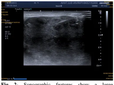
Fig 2- Sonographic features show a large
Aniruddha Mundhada et al JMSCR Volume 08 Issue 08 August 2020 Page 252
Fig 3- Gross appearance of specimen showing large
variegated mass
Fig 4 - Microscopic examination showing poorly differentiated carcinoma with sarcomatoid differentiation
Fig 5. Immunohistochemistry: (a)High Ki67
labelling index (b)Pan-Ck Positive
Discussion
According to epidemiologists from the American Cancer Society, 279100 new cases of breast cancer will occur in 2020 in the USA.[1] MBC is rare, accounting for 0.25%–1% of breast cancers diagnosed annually, or approximately 700–2790 new cases.[2,3] Meta-plastic breast cancer is
classified as a distinct pathologic entity by the WHO and characterized by mixed epithelial and sarcomatoid histology.[4]
In our institute, 356 cases of Invasive Mammary carcinomas have been reported in SRMC & RI, with only 2 cases of Metaplastic Carcinoma, in the last year. Carcinosarcoma / metaplastic breast carcinoma is a sarcomatoid metaplasia of malignant epithelial cells. It is a rare, locally aggressive disease comprising only 0.08-0.2% of all malignant breast lesions.[5]
The cell of origin is unknown, but the hypothesis is that myoepithelial cells originate from a single stem cell like spindle-cells. The carcinomas have been reported to develop from existing cystosarcoma phyllodes, fibroadenoma and cystic backgrounds. A rapidly growing breast mass is the most common manifestation. [2,14]
Accurate diagnosis can be clinched from the histopathology images. These entities are usually triple negative and lymph node involvement is less.[6,8,13] Histology demonstrates Sarcomatous component which resembles fibromatosis; the epithelial component can reveal osteoclast-like giant cells too. The tumors are often cystic structures with lining composed of squamous cell carcinoma.
Different imaging methods can be used to diagnose MBC. Magnetic resonance imaging (MRI) features show a T2 hyperintense, irregular mass that may show spiculated margins. The mass is irregular and/or spiculated margins on mammograms. Sonographically a heterogeneous mass with both solid and cystic components present with complex echogenicity is appreciated. [5,9]
Aniruddha Mundhada et al JMSCR Volume 08 Issue 08 August 2020 Page 253 conservation therapy shows no significant change in
disease free survival when compared with mastectomy.[10] The tumor has been shown to be resistant to conventional breast cancer therapy. The most common sites of metastatic disease are the lungs and pleura[15], whereas brain, hepatic and skeletal metastases are uncommon.[16] Due to the nature of MBCs, a multidisciplinary approach should always be considered. Participation of patients in clinical trials incorporating agents targeting PI3K and EMT pathways should be considered.[12,17] Intensive follow-up of patients with MBC is mandatory due to the aggressive clinical course of the tumor.
Conclusions
Because of the rarity of the tumor, there are not enough data and standard guidelines for the optimal treatment and information about management and prognosis is based on small retrospective studies rather than randomized trials. Surgery is the main curative approach. An intensive follow-up is required for the detection of an early recurrence of the disease.
Declaration of Conflicting interests- Authors
report no conflicting interests
Ethical Approval- Not required from the institution
Funding- None
Consent for Publication- All authors hereby
consent for publication of this case report.
References
1. Siegel, R.L., Miller, K.D. and Jemal, A., 2020. Cancer statistics, 2020. CA: A Cancer Journal for Clinicians, 70(1), pp.7-30. 2. Hayat, M.J., Howlader, N., Reichman, M.E.
and Edwards, B.K., 2007. Cancer statistics, trends, and multiple primary cancer analyses from the Surveillance, Epidemiology, and End Results (SEER) Program. Oncologist, 12(1).
3. McKinnon, E. and Xiao, P., 2015. Metaplastic carcinoma of the breast.
Archives of Pathology and Laboratory Medicine, 139(6), pp.819-822.
4. Tzanninis, I.G., Kotteas, E.A., Ntanasis-Stathopoulos, I., Kontogianni, P. and Fotopoulos, G., 2016. Management and outcomes in metaplastic breast cancer. Clinical breast cancer, 16(6), pp.437-443. 5. Schwartz, T.L., Mogal, H., Papageorgiou,
C., Veerapong, J. and Hsueh, E.C., 2013. Metaplastic breast cancer: histologic characteristics, prognostic factors and systemic treatment strategies. Experimental hematology & oncology, 2(1), p.31.
6. Pezzi, C.M., Patel-Parekh, L., Cole, K., Franko, J., Klimberg, V.S. and Bland, K., 2007. Characteristics and treatment of metaplastic breast cancer: analysis of 892 cases from the National Cancer Data Base. Annals of surgical oncology, 14(1), pp.166-173.
7. Ong, C.T., Campbell, B.M., Thomas, S.M., Greenup, R.A., Plichta, J.K., Rosenberger, L.H., et. al., 2018. Metaplastic breast cancer treatment and outcomes in 2500 patients: a retrospective analysis of a national oncology database. Annals of surgical oncology, 25(8), pp.2249-2260.
8. Lai, H.W., Tseng, L.M., Chang, T.W., Kuo, Y.L., Hsieh, C.M., Chen, S.T., et. al., 2013. The prognostic significance of metaplastic carcinoma of the breast (MCB)–a case controlled comparison study with infiltrating ductal carcinoma. The Breast, 22(5), pp.968-973.
9. Velasco M, Santamaría G, Ganau S, et al. MRI of metaplastic carcinoma of the breast. Am J Roentgenol. 2005;184:1274–78. 10.Shah DR, Tseng WH, Martinez SR.
Treatment options for metaplastic breast cancer. ISRN Oncol. 2012;2012:706162. 11.Luini, A., Aguilar, M., Gatti, G., Fasani, R.,
Aniruddha Mundhada et al JMSCR Volume 08 Issue 08 August 2020 Page 254 Oncology and review of the literature.
Breast cancer research and treatment, 101(3), pp.349-353.
12.Plichta, J.K., Ren, Y., Thomas, S.M., Greenup, R.A., Fayanju, O.M., Rosenberger, L.H., et. al., 2020. Implications for breast cancer restaging based on the 8th edition AJCC staging manual. Annals of surgery, 271(1), pp.169-176.
13.Shrestha, R., Neupane, P.R. and Satyal, B., 2019. Metaplastic Breast Carcinoma: A Rare Entity. Nepalese Journal of Cancer, 3(1), pp.57-59.
14.Ilhan, E., Vardar, E., Ozkok, G., Sezgin, A., Sahin, S., Teker, K., Postaci, H. and Yildirim, M., 2010. A rare tumour of the breast: carcinosarcoma. Journal of clinical medicine research, 2(2), pp.96-98.
15.Esbah, O., Turkoz, F.P., Turker, I., Durnali, A., Ekinci, A.S., Bal, O., Sonmez, O.U., Budakoglu, B., Arslan, U.Y. and Oksuzoglu, B., 2012. Metaplastic breast carcinoma: case series and review of the literature. Asian Pacific Journal of Cancer Prevention, 13(9), pp.4645-4649.
16.Liu, C.H., Chang, C., Sy, E., Lai, H.W. and Kuo, Y.L., 2015. Metaplastic breast carcinoma with multiple muscle metastasis: A case report. Medicine, 94(17).
17.Basho, R.K., Gilcrease, M., Murthy, R.K., Helgason, T., Karp, D.D., Meric-Bernstam, et. al., 2017. Targeting the PI3K/AKT/mTOR pathway for the treatment of mesenchymal triple-negative breast cancer: evidence from a phase 1 trial of mTOR inhibition in combination with liposomal doxorubicin and bevacizumab. JAMA oncology, 3(4), pp.509-515.
List of Abbreviations