The coordinated movement of the spine
and pelvis during running
Preece, SJ, Mason, D and Bramah, CA
http://dx.doi.org/10.1016/j.humov.2015.11.014
Title The coordinated movement of the spine and pelvis during running
Authors Preece, SJ, Mason, D and Bramah, CA
Type Article
URL This version is available at: http://usir.salford.ac.uk/id/eprint/37173/
Published Date 2016
USIR is a digital collection of the research output of the University of Salford. Where copyright permits, full text material held in the repository is made freely available online and can be read, downloaded and copied for noncommercial private study or research purposes. Please check the manuscript for any further copyright restrictions.
The coordinated movement of the spine and pelvis during running
12
Author names: Stephen J. Preece1, Duncan Mason1 and Christopher Bramah1
3
1
School of Health Sciences, University of Salford, Salford, Manchester, M6 6PU.
4
Email: Stephen J. Preece1 : s.preece@salford.ac.uk ***corresponding author**
5
Duncan Mason1: d.mason@salford.ac.uk
6
Christopher Bramah1 : chris.bramah1@btinternet.com 7
8
9
Funding: This project was internally funded by the University of Salford.
10
11
Conflict of interest Disclosure: None
12
13
Correspondence Address: Stephen Preece, Blatchford Building, University of Salford,
14
Manchester, M6 6PU.
15
16
17
Abstract 19
20
Previous research into running has demonstrated consistent patterns in pelvic, lumbar and
21
thoracic motions between different human runners. However, to date, there has been limited
22
attempt to explain why observed coordination patterns emerge and how they may relate to
23
centre of mass (CoM) motion. In this study, kinematic data were collected from the thorax,
24
lumbar spine, pelvis and lower limbs during over ground running in n=28 participants. These
25
data was subsequently used to develop a theoretical understanding of the coordination of the
26
spine and pelvis in all three body planes during the stance phase of running. In the sagittal
27
plane, there appeared to be an antiphase coordinate pattern which may function to increase
28
femoral inclination at toe off whilst minimising anterior-posterior accelerations of the CoM.
29
In the medio-lateral direction, CoM motion appears to facilitate transition to the contralateral
30
foot. However, an antiphase coordination pattern was also observed, most likely to minimise
31
unnecessary accelerations of the CoM. In the transverse plane, motion of the pelvis was
32
observed to lag slightly behind that of the thorax. However, it is possible that the close
33
coupling between these two segments facilitates the thoracic rotation required to passively
34
drive arm motion. This is the first study to provide a full biomechanical rationale for the
35
coordination of the spine and pelvis during human running. This insight should help
36
clinicians develop an improved understanding of how spinal and pelvic motions may
37
contribute to, or result from, common running injuries.
38
39
40
Keywords: 42
43
Running; Coordination; Pelvis; Thorax, Centre of mass
44
Introduction 46
Running is complex movement which requires precise inter-segmental coordination to
47
create forward momentum. Given the integrated nature of running, it is possible that poorly
48
coordinated movement of the pelvis and spine could result in abnormal tissue stress not just
49
in the low back (Seay, Van Emmerik, & Hamill, 2011b), but also within more distal
50
structures of the lower limbs (Leetun, Ireland, Willson, Ballantyne, & Davis, 2004).
51
However, before interventions can be developed to address abnormalities in pelvis and spinal
52
movement, it is important to develop a clear biomechanical understanding of the coordination
53
between the spine and pelvis during normal running.
54
We suggest that there are two constraints which will play a pivotal role in determining
55
coordination patterns between the pelvis and spinal segments. In the sagittal and frontal plane
56
we suggest that coordination patterns will develop which will minimise excessive changes of
57
momentum in the anterior-posterior (AP) and medio-lateral (ML) directions respectively. It is
58
likely that this strategy, suggested as a mechanism for minimising energy consumption,
59
(Heise & Martin, 2001; Williams & Cavanagh, 1987), will lead to anti-phase coordination
60
between the pelvis and thorax. This is because rotational movements of the pelvis in either
61
the sagittal or frontal planes during stance will require a rotation of the thorax in the opposite
62
direction to minimise displacement of the centre of mass (CoM).
63
Rotations of the pelvis or trunk in the transverse plane will not displace the CoM.
64
However, it has been shown that arm motion during running functions to counterbalance the
65
rotational angular momentum of the swinging legs (Arellano & Kram, 2014; Hamner, Seth,
66
& Delp, 2010). Thus a coordination pattern between the pelvis and spine must emerge which
67
facilitates the necessary arm movement for angular momentum balance. It been suggested
68
(Pontzer, Holloway, Raichlen, & Lieberman, 2009) that this coordination is achieved via a
mass-damped system in which motion of the arms is driven passively by the motion of the
70
torso. Pontzer et al. (2009) also suggest that thorax motion is driven passively by motion of
71
the pelvis. If this is the case, then a phase lagged coordination pattern would be observed in
72
which rotation of the pelvis precedes that of the thorax.
73
A number of previous studies have published kinematic data describing the motions of
74
the pelvis and lumbar spine during running (MacWilliams, et al., 2014; Saunders, Schache,
75
Rath, & Hodges, 2005; Schache, Blanch, Rath, Wrigley, & Bennell, 2002). However, these
76
studies either failed to include a thoracic segment or did not analyse coordination patterns in
77
detail and therefore provide limited insight into pelvis-spinal coordination during running.
78
Only two studies have investigated the coordination patterns between the pelvis and thorax
79
during running (Seay, Van Emmerik, & Hamill, 2011a; Seay, et al., 2011b). However, these
80
studies did not include a lumbar segment, nor did they present accompanying data on CoM
81
motion. Furthermore, it was not possible to infer, from the presented analysis, whether
82
transverse plane motion of the thorax was driven by the pelvis.
83
The primary objective of this paper was to explore specific ideas around the
84
coordination of the pelvis and spine during running and to interpret these ideas in the context
85
of CoM motion. In order to address this objective, experimental data describing the
three-86
dimensional kinetics of the thorax, lumbar spine, pelvis and lower-limbs were collected from
87
a cohort of human subjects during over ground running. These data was then used to test a
88
number of specific hypotheses relating to the coordination between the thorax and pelvis
89
during stance phase. We hypothesised that there would be an anti-phase relationship between
90
the pelvis and thorax in the sagittal and frontal plane during stance. In the transverse plane,
91
we hypothesised that motion of the pelvis would lead motion of the thorax demonstrating a
92
phase-lagged coordination pattern. These kinematic descriptions were then interpreted in the
93
context of previously observed trunk EMG patterns.
95
Methods 96
97
2.1 Subjects and experimental set up
98
A cohort of 28 subjects (16 male) participated in the study. The mean (SD) age of the
99
subjects was 28 (4) years, mean (SD) height 175 (9) cm and mean weight 63 (9) Kg. Ethical
100
approval was obtained from the Local Ethics Committee before data collection and all
101
subjects gave informed consent to participate in the study. For each subject, kinematic data
102
were collected for the pelvis, thoracic spine, lumbar spine, lower limbs and feet. Each subject
103
ran along a 32m running track at a target speed of 3.9 ms-1 whilst data were collect using a
104
12-camera Qualisys Pro-reflex system (240Hz). In order to obtain event information, kinetic
105
data were collected from 3 AMTI force plates (1200Hz) embedded in the track. Running
106
speed was measured using optical timing gates and 7-10 trials within ±2.5% of the target
107
speed were collected for each subject.
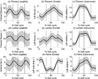
108
2.2 Protocol and kinematic calculations
109
A global optimisation algorithm (Mason, Preece, Bramah, & Herrington, 2014) was
110
used to obtain segmental kinematics. With this approach, joint constraints are applied to a
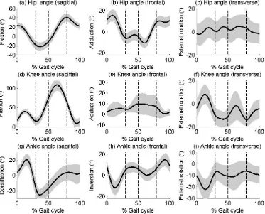
111
multi-link model in which segments could rotate with three degrees of freedom but not
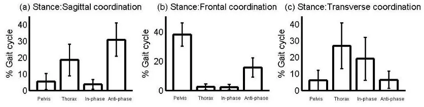
112
translate relative to adjacent segments. Within the nine-segment-model model, constraint
113
points were positioned at the origins of all segment coordinate frames distal to the pelvis and
114
expressed in the pelvis coordinate frame. In our previous analysis (Mason, et al., 2014) we
115
defined a pelvic segment which had an anterior-posterior axis pointing from the midpoint of
116
the posterior superior iliac spines (PSIS) to the midpoint of the anterior superior iliac spines
(ASIS). However, with this approach, between-subject differences in bony geometry of the
118
pelvis can lead to increased inter-subject variability in pelvic tilt (Preece, et al., 2008).
119
Therefore, for the present study, the Z (vertical) axis of the pelvic frame was aligned with the
120
laboratory in standing. The origin of this segment was modelled by a virtual marker that was
121
created midway between two iliac crest makers. These iliac crest markers were positioned at
122
the level of the iliac crests and above the hip centres (which were predicted from the ASIS
123
and PSIS locations (Bell, Brand, & Pedersen, 1989)). The X (ML axis) pointed from the
124
pelvic origin to the right ilicac crest marker and the Y (anterior-posterior) axis was the mutual
125
perpendicular. This pelvic segment was tracked using markers placed on the ASISs and
126
PSISs. The coordinate frames and corresponding tracking markers for the other eight
127
segments were the same as described in our previous repeatability paper analysis (Mason, et
128
al., 2014) and are therefore only reviewed briefly in the text below.
129
The anatomical coordinate system for the lumbar spine was aligned with the pelvic
130
frame with an origin that was positioned at the point 5% from the L5S1 marker to the
131
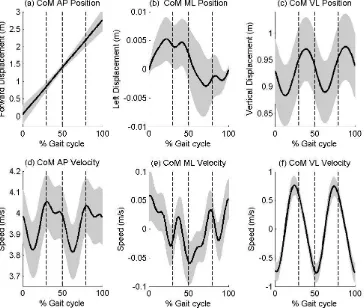
midpoint of the ASISs. This ensured a linked segment model for the global optimisation
132
calculations. The motion of this segment was tracked using a total of four markers placed on
133
the low back. This protocol was an adaptation of the method originally proposed by Seay et
134
al. (Seay, Selbie, & Hamill, 2008) and reported earlier in a repeatability study (Mason, et al.,
135
2014) in which 7 markers are used to track the lumbar spine. The decision to use only the
136
four markers placed lateral to the spine was based on a secondary analysis of data from n=15
137
participants which showed very little difference in lumbar orientation (average Standard
138
Error in the Measurement = 0.5˚-1.9˚) between the four-marker and the seven-marker
139
tracking approach.
140
Motion of the thoracic spine was tracked using three markers, mounted on a rigid
141
plate, which was attached to the sternum (van Andel, van Hutten, Eversdijk, Veeger, &
Harlaar, 2009). ISB recommendations (Wu, et al., 2005) were used to define a thoracic
143
reference frame, however, the origin of this frame was shifted to the point 5% along the line
144
from T12 to xiphiod process (XP). This ensured a linked segment model for the kinematic
145
calculations. Rigid plates, attached laterally, were used to track the motion of the thigh and
146
shank segments and the foot was tracked using markers positioned on the rear of the shoe and
147
over the first, second and fifth metatarsal heads. Anatomical coordinate systems for the thigh,
148
shank and foot were defined as reported earlier (Mason, et al., 2014).
149
Centre of mass position and velocity of the nine-segment model was calculated in
150
order to interpret the coordination analysis in the sagittal and frontal planes. Data from
151
Dempster (1955) were used for these calculations in which the pelvis was assumed to be an
152
elliptical cylinder which ran from the iliac crest markers to the hip centres, a diameter equal
153
to the distance between the greater trochanters and a depth equal to the distance from the
154
middle of the ASISs to L5S1 (Seay, et al., 2008). The lumbar segment was also assumed to
155
be an elliptical cylinder, which spanned the distance from the origin of the lumbar frame to
156
the level of T12. This segment had a diameter equal to the distance between the iliac crest
157
markers and a depth calculated as twice the distance from the XP to the midpoint of the line
158
from XP to T12. The geometry of the thoracic segment was again represented as an elliptical
159
cylinder using markers placed over the acromioclavicular joints and the iliac crests to define
160
distal and proximal diameters. This segment ran from the level of T12 up to C7 and had the
161
same depth as the lumbar segment. The contribution to the centre of mass position of the
162
lower extremities was calculated by assuming each segment to be a frustum of a cone with
163
proximal and distal diameters equal to the segment diameters.
164
Right and left contact phases were identified from the force platform data and a
165
second right initial contact (RIC) obtained using a pattern recognition algorithm (Stanhope,
166
Kepple, McGuire, & Roman, 1990). Using these events, each kinematic curve was
interpolated and then time normalised to produce 101 data point corresponding to 0-100%
168
gait cycle (RIC to RIC). All kinematic, and centre of mass, calculations were implemented in
169
Visual 3D (C-Motion) and then exported to Matlab for ensemble averaging and further
170
analysis.
171
2.2 Coordination analysis
172
In order to understand coordination patterns between the pelvis and thorax in the
173
sagittal and frontal planes, we used a technique based around angle-angle diagrams.
174
Following the vector coding method suggested by Chang et al. (2008), a coupling angle (γ)
175
was obtained for each of the 100 time points which described the change in direction of the
176
angle-angle plot between that time point and the next. Each of these changes were classified
177
as either in-phase (segments moving in the same direction), anti-phase (segments moving in
178
opposite directions), pelvis only or thorax only movement. These data were then used to
179
quantify the relative period spent in each different coordination phases when the foot was in
180
contact with the ground (stance phase).
181
182
In order to analyse the phase lag between thorax and pelvic motion in the transverse
183
plane we identified the timing of peak angular velocity for each of the two segments. This
184
corresponds to the time of zero angular acceleration and therefore when the net torque acting
185
on the segment changes direction. Peak positive angular velocity was easily identified during
186
right stance phase for every participant. Pontzer et al. (2009) suggest that motion of the pelvis
187
and thorax can be described as a mass damped system in which rotational torques from the
188
pelvis are transmitted through the trunk and drive thorax rotation. In this scenario, the inertia
189
of the torso tends to resist the rotational torque applied by the pelvis and this leads to a
phase-190
lagged coordination pattern. If this is the case then we would expect the peak angular velocity
191
of the pelvis to precede the peak angular velocity of the thorax. To test this idea we used a
one-sample t-test to establish if the time lag, between the peak angular velocity of the pelvis
193
and thorax, was significantly different from zero. In addition to analysing coordination
194
patterns, we also included data on lower limb motions to provide the reader with a complete
195
understanding of how transverse plane motion of the pelvis and thorax is coordinated with the
196
swinging legs.
197
Results 198
In the sagittal plane, all three segments displayed a biphasic pattern, in which there
199
was a peak in flexion/anterior tilt either during or immediately after stance phase (Figure 1).
200
Motion in this plane occurred about a position of relative flexion for the thoracic spine and
201
the lumbar spine in (Figures 1a & 1d) and a position of relative anterior tilt for the pelvis
202
(Figure 1g). Timing of peak thoracic forward flexion (Mean(SD) 15(4)% of gait cycle)
203
corresponded with the timing of peak posterior pelvic tilt (Mean(SD) 14(6)% of gait cycle)
204
(Figure 1a & 1g). Peak anterior tilt (Mean(SD) 28(12)% of gait cycle) occurred in early flight
205
phase at a similar time to peak hip extension (Mean(SD) 29(6)% of gait cycle) (Figure 2a).
206
Visual inspection of the kinematic trajectories for the pelvis and thorax suggested anti-phase
207
movement (Figures 1a & 1g). This was confirmed by the coordination analysis (Figure 4a)
208
which showed the anti-phase pattern to be the most common during stance. However, this
209
motion was sometimes classified as thorax-only movement due to the relatively smaller
210
amplitude of motion of the pelvis compared to the thorax.
211
FIGURE 1 ABOUT HERE
212
In the frontal plane, the pelvis was laterally tilted away from the stance limb (i.e.
213
lower on the contralateral side) at initial contact (Figure 1h). Following initial contact there
214
was a slight increase in this drop after which there was a rapid elevation of the contralateral
215
side of the pelvis which resulted in the pelvis being elevated relative to the stance limb at toe
off. During flight there was minimal frontal movement of the pelvis and then the cycle
217
repeated on the contralateral leg. Most of the movement of the thorax relative to the pelvis
218
(Figure 3b) occurred at the lumbo-pelvic junction (Figure 3h) with only minimal motion at
219
the thoraco-lumbar junction (Figure 3e).
220
FIGURE 2 ABOUT HERE
221
There appeared to be an anti-phase relationship in the frontal plane between thorax
222
and pelvic motion (Figures 1b & 1h) during stance. Specifically the thorax was laterally
223
flexed towards the stance limb during early stance and then moved towards a neural position
224
during the latter half of stance, as the pelvis became elevated on the contralateral side. The
225
coordination analysis classified the frontal plane pelvis-thorax motion as either anti-phase or
226
pelvis-only during stance (Figure 4b). This latter classification resulted from the increased
227
motion of the pelvis compared to the thorax which resulted in a more vertically aligned
228
coupling vector (Seay, et al., 2011b) and therefore a coupling angle which was classified as
229
pelvis-only motion.
230
FIGURE 3 ABOUT HERE
231
In the transverse plane, the thorax rotated towards the contralateral leg during stance.
232
Figure 2a shows the motion of the right hip which reaches maximal extension and begins to
233
flex in late swing phase. This flexion, which continues through stance, is accompanied by a
234
corresponding rotation of the thorax (Figure 1c) towards the swing limb. The kinematic
235
trajectory of the pelvis appears to follow a similar pattern to that of the thorax (Figure 1i).
236
However, during early stance the pelvis rotates towards the stance limb before starting to
237
rotate in the same direction as the thorax (away from the stance limb) for the remainder of
238
stance. Analysis of the relative segment motion showed that motion between the thorax and
239
pelvis occurred primarily at the thoraco-lumbar junction (Figure 3f). The transverse plane
coordination analysis identified a pattern in which motion of the thorax preceded motion of
241
the pelvis in 22 out of the 28 subjects. The mean (SD) time lag was 4(6)% of the gait cycle
242
and this time lag was significantly different from zero (p<0.05).
243
FIGURE 4 ABOUT HERE
244
The AP CoM velocity profile illustrated the characteristic braking and acceleration
245
phases of running during stance phase (Figure 5d). However, these changes were relatively
246
small (±0.1ms-1) compared to the target running speed of 3.9ms-1. In the ML direction the
247
CoM moved towards the contralateral limb during stance. However the ML changes in
248
velocity of the CoM velocity (±0.06ms-1) were smaller than those in the AP direction (Figure
249
5e). It is interesting to note that the point of zero ML velocity of the CoM occurred at 22% of
250
the gait cycle, coinciding with the point when the pelvis reaches its neutral position in the
251
frontal plane (Figure 1h).
252
FIGURE 5 ABOUT HERE
253
254
Discussion 255
256
This study was undertaken to understand the coordinated movement of the pelvis and
257
thorax during running in healthy individuals. We hypothesised that AP accelerations of the
258
CoM would be minimised and that this would lead to an anti-phase coordination pattern in
259
the sagittal plane during stance. This idea was supported by the observation of relatively
260
small changes in the CoM velocity (Figure 5d) and a predominantly anti-phase coordination
261
pattern (Figure 4a). Posterior pelvic tilt occurred during early stance and this was
262
accompanied by flexion of the thorax. During late stance the pelvis moved into anterior tilt
and there was a corresponding extension of the thorax. We suggest that this anterior tilting of
264
the pelvis during late stance is a mechanism for increasing femoral inclination at toe off and
265
thereby extending stride length. In addition to the anti-phase motions between the thorax and
266
the pelvis, we also observed the thorax and the pelvis to be in a position of flexion and
267
anterior tilt respectively, relative to standing. This segmental alignment will have the effect of
268
shifting the CoM anteriorly thus creating a more posteriorly directed ground reaction force
269
which will facilitate the generation of forward momentum (Novacheck, 1998).
270
Previous EMG studies of running have shown the lumbar extensor muscles to be
271
active at foot contact and during early stance (Thorstensson, Carlson, Zomlefer, & Nilsson,
272
1982). This early activation of the back extensors will act to limit forward flexion of the trunk
273
as energy is absorbed in the lower limbs and the CoM decelerates. During this deceleration
274
phase, the thorax moves into forward flexion and there is a small corresponding posterior tilt
275
of the pelvis as gluteus maximus acts to extend the hip. In the second half of stance, the lower
276
limbs act to accelerate the CoM and so active muscle control is required to decelerate the
277
extension of the thorax. This control is most likely provided by the oblique abdominal
278
muscles which have been shown to be active later in stance (Saunders, et al., 2005). As
279
suggested above, this extension of the thorax is coordinated with anterior tilting of the pelvis
280
in order to extend stride length whilst controlling the AP CoM velocity.
281
We hypothesised that changes in the ML CoM velocity are minimised and that this
282
leads to an anti-phase coordination pattern between the pelvis and thorax in the frontal plane
283
during stance. This idea was partially supported by the data which showed relatively small
284
changes in the ML velocity of the CoM (Figure 5b) and either an anti-phase or pelvis only
285
coordination pattern during stance phase (Figure 4b). We propose that frontal plane motion of
286
the pelvis has two primary functions. Firstly, during early stance, the pelvis is laterally tilted
287
(dropped) away from the stance limb. This results in a more medial position of the CoM,
which in turn creates a moment about the base of support, facilitating transition onto the
289
contralateral foot. From midstance onwards the pelvis lifts on the contralateral stride until it
290
reaches its maximum position at toe off (Figure 1h). This movement serves to elevate the
291
swing leg to ensure foot clearance and also to extend stride length. We further suggest that
292
thorax motion is precisely coordinated with this pelvic kinematic pattern in order to minimise
293
the ML acceleration of the CoM. This coordination requires a smaller range of movement of
294
the thorax and explains the pelvis only classification observed in our frontal plane
295
coordination analysis (Figure 4b).
296
Gluteus medius has been shown to be active prior to foot contact and for the most of
297
stance phase of running (Gazendam & Hof, 2007; Willson, Petrowitz, Butler, & Kernozek,
298
2012). We suggest that this muscle functions to control the downward acceleration of the
299
CoM following foot contact, then later to lift the pelvis on the contralateral side. This is
300
consistent with the observation of a large proportion of frontal plane movement occurring at
301
the lumbo-pelvic junction (Figure 3e & 3h). During the latter stages of stance, the lumbar
302
spine is laterally flexed towards the contralateral limb, relative to the pelvis (Figure 3h). It is
303
possible that this motion is assisted by the contralateral oblique abdominal muscles which
304
have been shown to be active during this period (Saunders, et al., 2005).
305
Previous modelling studies have clearly shown that motion of the arms effectively
306
counterbalances the angular momentum of the lower extremities during running (Hamner &
307
Delp, 2013; Hamner, et al., 2010). It has further been suggested that arm motion is driven
308
passively by rotation of the thorax (Pontzer, et al., 2009), an idea which is supported by
309
shoulder muscle EMG data, consistent with the shoulders as spring-like linkages (Ballesteros,
310
Buchthal, & Rosenfalck, 1965). Our data are consistent with this idea, showing motion of the
311
thorax to be in the opposite direction to that of the swinging leg. Pontzer et al. (2009) also
312
suggested that motion of the thorax is driven passively by motion of the pelvis. However, our
data shows that the thorax reaches its peak angular velocity earlier than the pelvis, indicating
314
that thorax motion is not completely passively driven by pelvic movements.
315
The pelvic rotation observed in our study matches the patterns observed in previous
316
studies (MacWilliams, et al., 2014; Schache, et al., 2002). Specifically, the pelvis rotates
317
slightly towards the stance limb during early stance after which it rotates away from the
318
stance limb. This initial rotation towards the stance limb has been suggested to function to
319
reduce horizontal braking (Novacheck, 1998; Schache, et al., 2002), however the subsequent
320
rotation away from the stance limb may decrease stride length. We suggest the pattern of
321
transverse plane pelvic motion during running is a secondary consequence of gluteus
322
maximus activity. This muscle is active for most of stance phase (Gazendam & Hof, 2007;
323
Willson, et al., 2012) and functions primarily to extend the hip. However, gluteus maximus
324
will also act to externally rotate the hip (Delp, Hess, Hungerford, & Jones, 1999) or,
325
equivalently, rotate the pelvis away from the stance limb.
326
Although the pattern of pelvic rotation in the transverse plane would appear to reduce
327
stride length, the effect is minimal. If we assume a rotation of 10˚ (Figure 1i) and a distance
328
between hip centres of 15-30cm, then stride length would be reduced by only 1-2%. It is
329
therefore unlikely that the muscle work required to oppose the action of gluteus maximums
330
and produce transverse rotation of the pelvis towards the stance limb would be worth the
331
metabolic cost. Instead, we suggest pelvic motion follows the motion of the thorax in order to
332
minimise the muscle work required to passively drive arm motion. This can be understood by
333
analysing the relative transverse plane motion between the thorax and the pelvis (Figure 3c).
334
This figure shows that, from midswing until early stance, the thorax moves from a rotated to
335
a neutral position relative to the pelvis. During this period the abdominal muscles are inactive
336
(Saunders, et al., 2005) and the relative motion between the thorax and pelvis most likely
337
results from stored elastic energy in connective tissues. Around midstance, the abdominal
muscles become active (Saunders, et al., 2005), working both to limit extension of the trunk
339
(in the sagittal plane) and also to actively rotate the thorax relative to the pelvis. This active
340
rotation results in a larger a movement of thorax compared to the pelvis.
341
It is interesting to compare the kinematic data described in this paper with the data
342
obtained from a study in which bone pins were inserted into the individual lumbar and sacral
343
vertebrae (MacWilliams, et al., 2014). Importantly, there is good agreement between the
344
pattern of pelvic movement in each body plane. However, the range of motion observed in
345
the bone pin data is slightly lower in both the frontal and transverse planes. This difference
346
may have resulted from skin movement artefact or from differences in running speed between
347
the two studies, which may affect pelvic range of movement. MacWilliams et al. (2014) also
348
reported on the relative motion of the individual lumbar spine segments with respect to the
349
pelvis. Again, our data matches these data closely with the same caveat of lower ranges of
350
movements in the bone pin data. Our data on the thorax also matches that reported by Seay et
351
al. (2008) who used skin mounted markers to characterise the rotation of the thorax relative to
352
the lumbar spine during the stance phase of over ground running. Seay et al. (2011b) later
353
investigated treadmill running and observed that the thorax rotated through approximately
354
25˚ of motion throughout the whole gait cycle, slightly higher than that shown in Figure 1c.
355
It is important to identify the limitations of the present study. Firstly, in order to
356
develop a practical skin mounted marker set, we chose to segment the spine into two rigid
357
segments. Although this represents a major simplification of the multi-articular structure of
358
the spine, our data compares well with the bone pin data presented by MacWilliams et al.
359
(2014). This suggests that that our relatively easy-to-implement laboratory protocol can be
360
used to extract the salient features of pelvic-spinal coordination during running. This protocol
361
therefore appears appropriate for future studies aimed at investigating the association
362
between musculoskeletal pain and abnormal motion of the spine and pelvis. Another
limitation of this study was that we investigated a single running speed. However, data on
364
multiple running speeds was deemed to be outside the scope of this paper and is therefore
365
presented in a subsequent publication.
366
Conclusion 367
This is the first study to provide an underlying biomechanical rationale for the
368
coordination pattern between the pelvis and thorax during running in all three body planes.
369
The data showed an anti-phase relationship between these two segments in the sagittal and
370
frontal planes and we suggest that this in a consequence of the requirement to minimise
371
accelerations of the CoM in the AP and ML directions. In the transverse plane, we observed a
372
phase lagged relationship in which motion of the pelvis lagged slightly behind that of the
373
thorax. This suggests that transverse plane thoracic motion is not completely passively driven
374
by pelvic motion. However, it is likely that the closely coupled movement of these two
375
segments facilitates the thoracic rotation required to passively drive arm motion.
376
378
References: 379
380
Arellano, C. J., & Kram, R. (2014). The metabolic cost of human running: is swinging the arms worth 381
it? Journal of Experimental Biology, 217, 2456-2461.
382
Ballesteros, M. L., Buchthal, F., & Rosenfalck, P. (1965). The Pattern of Muscular Activity during the 383
Arm Swing of Natural Walking. Acta Physiol Scand, 63, 296-310. 384
Bell, A. L., Brand, R. A., & Pedersen, D. R. (1989). Prediction of hip-joint center location from external 385
landmarks. Human Movement Science, 8, 3-16. 386
Chang, R., Van Emmerik, R., & Hamill, J. (2008). Quantifying rearfoot-forefoot coordination in human 387
walking. J Biomech, 41, 3101-3105. 388
Delp, S. L., Hess, W. E., Hungerford, D. S., & Jones, L. C. (1999). Variation of rotation moment arms 389
with hip flexion. J Biomech, 32, 493-501. 390
Dempster, W. T. (1955). Space requirements of the seated operator (Rep. No. Technical report 391
WADC-TR-55-159). Ohio: Wright-Patterson Air Force Base. 392
Gazendam, M. G. J., & Hof, A. L. (2007). Averaged EMG profiles in jogging and running at different 393
speeds. Gait & Posture, 25, 604-614. 394
Hamner, S. R., & Delp, S. L. (2013). Muscle contributions to fore-aft and vertical body mass center 395
accelerations over a range of running speeds. J Biomech, 46, 780-787. 396
Hamner, S. R., Seth, A., & Delp, S. L. (2010). Muscle contributions to propulsion and support during 397
running. J Biomech, 43, 2709-2716. 398
Heise, G. D., & Martin, P. E. (2001). Are variations in running economy in humans associated with 399
ground reaction force characteristics? European Journal of Applied Physiology, 84, 438-442. 400
Leetun, D. T., Ireland, M. L., Willson, J. D., Ballantyne, B. T., & Davis, I. M. (2004). Core stability 401
measures as risk factors for lower extremity injury in athletes. Medicine and Science in 402
Sports and Exercise, 36, 926-934.
403
MacWilliams, B. A., Rozumalski, A., Swanson, A. N., Wervey, R., Dykes, D. C., Novacheck, T. F., & 404
Schwartz, M. H. (2014). Three-Dimensional Lumbar Spine Vertebral Motion During Running 405
Using Indwelling Bone Pins. Spine, 39, E1560-E1565. 406
Mason, D. L., Preece, S. J., Bramah, C. A., & Herrington, L. C. (2014). Reproducibility of kinematic 407
measures of the thoracic spine, lumbar spine and pelvis during fast running. Gait & Posture 408
(http://dx.doi.org/doi:10.1016/j.gaitpost.2013.11.007).
409
Novacheck, T. F. (1998). The Biomechanics of Running. Gait and Posture, 7, 77-95. 410
Pontzer, H., Holloway, J. H., Raichlen, D. A., & Lieberman, D. E. (2009). Control and function of arm 411
swing in human walking and running (vol 212, pg 523, 2009). Journal of Experimental 412
Biology, 212, 894-894.
413
Preece, S. J., Willan, P., Nester, C. J., Graham-Smith, P., Herrington, L., & Bowker, P. (2008). Variation 414
in pelvic morphology may prevent the identification of anterior pelvic tilt. J Man Manip Ther, 415
16, 113-117. 416
Saunders, S. W., Schache, A., Rath, D., & Hodges, P. W. (2005). Changes in three dimensional lumbo-417
pelvic kinematics and trunk muscle activity with speed and mode of locomotion. Clin 418
Biomech (Bristol, Avon), 20, 784-793.
419
Schache, A. G., Blanch, P., Rath, D., Wrigley, T., & Bennell, K. (2002). Three-dimensional angular 420
kinematics of the lumbar spine and pelvis during running. Human Movement Science, 21, 421
273-293. 422
Seay, J., Selbie, W. S., & Hamill, J. (2008). In vivo lumbo-sacral forces and moments during constant 423
Seay, J., Van Emmerik, R. E., & Hamill, J. (2011a). Low back pain status affects pelvis-trunk 425
coordination and variability during walking and running. Clin Biomech (Bristol, Avon), 26, 426
572-578. 427
Seay, J., Van Emmerik, R. E. A., & Hamill, J. (2011b). Influence of Low Back Pain Status on Pelvis-428
Trunk Coordination During Walking and Running. Spine, 36, E1070-E1079. 429
Stanhope, S. J., Kepple, T. M., McGuire, D. A., & Roman, N. L. (1990). Kinematic-based technique for 430
event time determination during gait. Med Biol Eng Comput, 28, 355-360. 431
Thorstensson, A., Carlson, H., Zomlefer, M. R., & Nilsson, J. (1982). Lumbar back muscle activity in 432
relation to trunk movements during locomotion in man. Acta Physiol Scand, 116, 13-20. 433
van Andel, C., van Hutten, K., Eversdijk, M., Veeger, D., & Harlaar, J. (2009). Recording scapular 434
motion using an acromion marker cluster. Gait & Posture, 29, 123-128. 435
Williams, K. R., & Cavanagh, P. R. (1987). Relationship between distance running mechanics, running 436
economy and performance. J Appl Physiol (1985), 63, 1236-1245. 437
Willson, J. D., Petrowitz, I., Butler, R. J., & Kernozek, T. W. (2012). Male and female gluteal muscle 438
activity and lower extremity kinematics during running. Clin Biomech (Bristol, Avon), 27, 439
1052-1057. 440
Wu, G., van der Helm, F. C. T., Veeger, H. E. J., Makhsous, M., Van Roy, P., Anglin, C., Nagels, J., 441
Karduna, A. R., McQuade, K., Wang, X. G., Werner, F. W., & Buchholz, B. (2005). ISB 442
recommendation on definitions of joint coordinate systems of various joints for the 443
reporting of human joint motion - Part II: shoulder, elbow, wrist and hand. J Biomech, 38, 444
981-992. 445
446
Figures: 448
Figure 1: Ensemble average curves (across all n=28 subjects), with standard deviation
449
envelopes, for the thorax, lumbar spine and pelvis relative to the laboratory coordinate system
450
in each of the three body planes. Data is plotted from right initial contact (RIC) to the
451
following RIC with the three vertical lines showing the timing of right toe off, left initial
452
contact and left toe off respectively.
453
454
455
456
457
458
Figure 2: Ensemble average curves (across all n=28 subjects), with standard deviation
460
envelopes, for the right hip, knee and ankle in each of the three body planes. Data is plotted
461
from right initial contact (RIC) to the following RIC with the three vertical lines showing the
462
timing of right toe off, left initial contact and left toe off respectively.
463
464
Figure 3: Ensemble average curves (across all n=28 subjects), with standard deviation
466
envelopes, for relative motion between each of the pelvis and spinal segments in each of the
467
three body planes. Data is plotted from right initial contact (RIC) to the following RIC with
468
the three vertical lines showing the timing of right toe off, left initial contact and left toe off
469
respectively.
470
471
472
Figure 4: Average and SD pelvis-thorax coordination patterns across all n=28 subjects for
474
both stance and flight phase. Each plot illustrates the proportion of the gait cycle spent in
475
each of the four coordination phases: pelvis only, thorax only, in-phase or anti-phase.
476
477
478
Figure 5: Ensemble average centre of mass (CoM) displacements (a-c) and average CoM
480
velocities (d-f) across all n=28 subjects. Data is plotted from right initial contact (RIC) to the
481
following RIC with the three vertical lines showing the timing of right toe off, left initial
482
contact and left toe off respectively.
483