Journal of Chemical and Pharmaceutical Research, 2015, 7(10):745-754
Research Article
CODEN(USA) : JCPRC5
ISSN : 0975-7384
Transition metal (II) complexes of ligand with benzodiazole moiety:
Spectral characterization and biological evaluation
P. Latha, P. Kodisundaram, M. L. Sundararajan and T. Jeyakumar*
Chemistry Section, Faculty of Engineering and Technology, Annamalai University, Annamalainagar, Tamil Nadu, India
_____________________________________________________________________________________________
ABSTRACT
A bidentate ligand (NS type) with benzodiazole moiety has been synthesized by the condensation of 2-thiophenecarboxaldehyde with o-phenylenediamine. Complexes of type [ML2]⋅2H2O (M = Cu (II), Zn (II), Co (II),
Cd(II) and Ni(II)) have been derived from the ligand. The entire complexes have been characterized using UV-Vis, FT-IR, 1H NMR, Mass, TG-DTA and powder XRD techniques. The stoichiometry and geometry of the complexes were predicted based on the results obtained from the above analyses. Furthermore, all the metal complexes were screened for their in vitro biological activity against four bacterial strains viz., Salmonella typhi, Klebsiella pneumoniae, Pseudomonas aeruginosa and Escherichia coli and four fungal strains viz., Candida albicans, Rhizopus sp., Aspergillus fumigatus and Mucor sp. by disc diffusion method.
Keywords: 2-Thiophenecarboxaldehyde, o-Phenylenediamine, Transition metal complexes, Biological evaluation.
_____________________________________________________________________________________________
INTRODUCTION
Ligands derived using 1,2-phenylenediamine and their metal complexes have enormous applications including biological [1], clinical [2] and analytical [3]. In general, compounds with benzodiazole ring have been synthesized by condensation of 1,2-phenylenediamine with the corresponding aldehydes [4,5]. Schiff base ligands prepared using 2-thiophene-carboxaldehyde and their metal complexes having diverse biological activities [6–9]. Furthermore, compounds containing benzodiazole group exhibit antimicrobial [10–12], anticancer [13,14], antifungal [15,16], antiparasitic [17], antiviral [18] and anti-inflammatory [19] activities. In addition, the derivatives of benzimidazole behave as good ligands for transition metal ions [20,21]. Their metal complexes having potential medicinal applications including highly potent against human bladder, esophagus and cervical cancer [22] and high cytotoxicity against human lung cancer A549 and breast cancer MCF-7 cell lines in comparison with its ligand from which it was derived [23]. In view of these facts, we prepared transition metal (II) complexes of ligand, (thiophen-yl)-1-((thiophen-yl)methyl)-1H-1,3-benzodiazole (L) derived from 1,phenylenediamine and 2-thiophenecarboxaldehyde. The crystal structure and biological activity of this ligand was well documented in our previous work [24]. In this study we reported the spectral characterization, thermal analysis and biological evaluation of metal (II) complexes derived from ligand (L).
EXPERIMENTAL SECTION
______________________________________________________________________________
The percentage of C, H, N and S for all the complexes were determined by using Perkin Elmer 240 (USA) elemental analyzer and the percentage of metal content were determined by atomic absorption spectrophotometer (Perkin Elmer 5000). Infrared spectra of ligand and its metal complexes were recorded on Avatar 330 FT-IR, in the range of 4000– 400 cm–1 using KBr pellets. The UV-Vis spectra of the ligands and the complexes were recorded on a Shimadzu UV-1650PC spectrophotometer in the range of 200–800 nm. 1H NMR spectra (at room temperature) were recorded on a Bruker Magnet System (400 MHz/54 nm) ultra shield plus using DMSO as a solvent and TMS as internal standard. TG/DTA measurements were carried out under nitrogen atmosphere within the temperature range 25– 1200°C at a heating rate of 20°C/min using TA instrument, Model: Q6000SDT TG/DTA. The mass spectra were recorded by using JEOL GCMATE II GC-MS with data system is a high resolution, double focusing instrument with 6000 maximum resolution and 1500 maximum mass calibration. Melting points of the compounds were determined on a Gallenkamp melting point apparatus. Powder XRD was recorded using diffractometer system XPERT-PRO. Molar conductance of the Schiff base ligand and its transition metal complexes were determined in ethanol at room temperature using a CMD 750 WPA conductivity meter.
Antimicrobial activity
Antibacterial and antifungal activities of the standard antibiotics and the metal complexes were screened against four pathogenic bacteria viz., S. typhi, K. pneumoniae, P. aeruginosa and E. coli and four fungi viz., C. albicans, Mucor sp.,
Rhizopus sp. and A. fumigatus. Filter paper, disc agar diffusion method was used to detect the biological activities of
the complexes. The antimicrobial screening concentration of the complexes was estimated from minimum inhibitory concentrations (MIC). MIC is the lowest concentration of the compound that will inhibit the growth of microorganisms. Pure Ciprofloxacin and Amphotericin-B were separately used as standards for antibacterial and antifungal activity tests, respectively. Nutrient agar was the basal medium for bacterial culture. For the bacterial culture a series of different concentrations of the complexes in DMSO solvent were placed on the surface of the culture and incubated at 37°C for 24 h, whereas for fungal culture, potato agar medium was used as basal medium and the complexes in DMSO solvent were also incubated at 37°C for 24 h. The growth inhibition zone was measured in millimeter.
Preparation of complexes
The complex preparation was carried out by mixing methanolic (50 mL) solution of ligand (0.04 mmol) with metal chloride or nitrate or acetate in methanol solution (0.02 mmol). The 1:2 ratio metal-ligand mixtures were refluxed for 4 h. The volume of reaction mixture was reduced to one-half by evaporation of solvent. On cooling, solid product was precipitated. The filtered product was washed with hot methanol and dried in vacuum desiccators over CaCl2.
[CuL2]⋅2H2O
The crude complex was recrystallized with ethanol, yielding bluish green colour of 70%, m.p. 220°C. Anal. Calc. for C32H24CuN2S4 (%): C (55.51), H (4.08), N (8.09), S (18.52), M (9.18); found (%): C (55.48), H (4.03), N (8.11), S (18.50), and M (9.01). FT-IR (KBr, cm–1): 3413 ν(H2O), 2924, 2849 νasym, sym(C–H), 753, 720 νasym, sym(C–S), 561 ν(M– N), 463 ν(M–S).
[ZnL2]⋅2H2O
The complex was recrystallized with ethanol, yielding grey coloured solid of 70%, m.p. 248°C. Anal. Calc. for C32H28N4O2S4Zn(%): C (55.36), H (4.07), N (8.07), S (18.47), M (9.42); found (%): C (55.01), H (4.05), N (7.98), S (18.12), and M (9.39). FT-IR (KBr, cm–1): 3449 ν(H2O), 2967, 2761 νasym, sym(C–H), 1545 ν(C=N), 852 ν(C–S–C), 760, 721 νasym, sym(C–S), 560 ν(M–N), 444 ν(M–S).
[CoL2]⋅2H2O
The complex was recrystallized with ethanol, yielding blue coloured solid of 67%, m.p. 258°C. Anal. Calc. for C32H28CoN4O2S4 (%): C (53.10), H (2.33), N (7.54), S (17.97), M (8.53); found (%): C (55.88), H (4.10), N (8.15), S (18.65), and M (8.57). FT-IR (KBr, cm–1): 3440 ν(H2O), 2965, 2783 νasym, sym(C–H), 1540 ν(C=N), 860 ν(C–S–C), 755, 720 νasym, sym(C–S), 574 ν(M–N), 465 ν(M–S).
[CdL2]⋅2H2O
[NiL2]⋅2H2O
The complex was recrystallized with ethanol, yielding green solid of 71%, m.p. 220°C. Anal. Calc. for C32H28 -NiN4O2S4 (%): C (51.65), H (4.06), N (8.12), S (18.55), M (9.01); found (%): C (55.21), H (4.01), N (7.98), S (18.45), M (8.98). FT-IR (KBr, cm–1): 3445 ν(H2O), 2963, 2785 νasym, sym(C–H), 1538 ν(C=N), 865 ν(C–S–C), 760, 722
νasym, sym(C–S), 562 ν(M–N), 444 ν(M–S).
RESULTS AND DISCUSSION
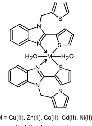
The ligand L derived by the condensation of 2-thiophenecarboxaldehyde and o-phenylenediamine in 2:1 molar ratio is converted to its corresponding metal complexes with metal-ligand ratio 1:2 (Fig. 1). The molar conductance (λm) of the metal complexes measured in ≈10
–3
[image:3.595.234.384.226.431.2]M solutions at room temperature (Table 1) is consistent with the non-electrolytic nature of the complexes. No counter ion in the proposed metal complex structure further confirms this nature.
[image:3.595.155.457.465.656.2]Fig. 1. Structure of complex
Table 1. Electronic spectral data (in DMSO) and molar conductivity of the ligand and its metal complexes
Ligand/complex λmaz (cm–1) Bond assignment Geometry λm (ohm–1 cm–1 mol–1)
L 38461 (π-π*) IMCT 4.58
31800 (n-π*) INCT
[CuL2]⋅2H2O 30800 (n-π*) LMCT Octahedral 6.17
15384 2
A2g(F) →2T1g(F)
10900 2A
2g(F) →2T2g(F)
10200 2T
1g(F) →2T2g(F)
[ZnL2]⋅2H2O 32800 LMCT (M←N) Octahedral 5.32
24100 LMCT (M←S)
[CoL2]⋅2H2O 29400 (n-π*) LMCT Octahedral 5.42
18200 4T
1g(F) →4T1g(P)
11200 4
T1g(F) →4A2g(F)
10100 4T
1g(F) →4T2g(F)
[CdL2]⋅2H2O 37000 LMCT (M←N) Octahedral 6.71
29100 LMCT (M←S)
[NiL2]⋅2H2O 30700 (n-π*) LMCT Octahedral 7.12
21929 3A
2g(F) →3T1g(P)
15151 3A
2g(F) →3T1g(F)
11284 3A
2g(F) →3T2g(F)
Table 2. Mass spectral data of metal complexes
Complex Calculated (m/z) Found (m/z) Peak assignment
[CuL2]⋅2H2O 691.04 690.63 [M]
[ZnL2]⋅2H2O 692.04 692.55 [M]
[CoL2]⋅2H2O 687.04 687.67 [M]
[CdL2]⋅2H2O 742.01 739.78 [M–2]
[image:3.595.193.421.681.744.2]______________________________________________________________________________
Table 3. Thermal analysis of metal complexes
Complex TG range (°C) Mass loss (%) found (calcd.) Assignment
[CuL2]⋅2H2O 200 5.20 (5.21) Loss of coordinated H2O molecule
200–1000 Decomposition of the ligand
[ZnL2]⋅2H2O 260 5.20 (5.20) Loss of coordinated H2O molecule
260–1000 Decomposition of the ligand
[CoL2]⋅2H2O 200 5.23 (5.24) Loss of coordinated H2O molecule
200–1000 Decomposition of the ligand
[CdL2]⋅2H2O 260 4.85 (4.85) Loss of coordinated H2O molecule
260–1000 Decomposition of the ligand
[NiL2]⋅2H2O 255 5.15 (5.21) Loss of coordinated H2O molecule
[image:4.595.169.442.226.334.2]255–1000 Decomposition of the ligand
Table 4. Minimum inhibitory concentration of ligand and its metal complexes
Ligand/complex
Minimum inhibitory concentration (µg/mL) Antibacterial activity Antifungal activity
a b c d e f g h
L 100 100 25 100 25 25 100 100
[CuL2]⋅2H2O 25 6.25 6.25 6.25 6.25 50 6.25 6.25
[ZnL2]⋅2H2O 200 200 25 6.25 200 200 50 6.25
[CoL2]⋅2H2O 100 100 12.5 25 6.25 100 200 6.25
[CdL2]⋅2H2O 25 – 200 6.25 6.25 200 200 6.25
[NiL2]⋅2H2O 100 100 25 25 6.25 100 100 6.25
Ciprofloxacin 5.25 4.50 6.25 5.50 – – – –
Amphotericin-B – – – – 6.25 6.25 4.50 6.25
a – S. typhi; b – K. pneumoniae; c – P. aeruginosa; d – E. coli; e – C. albicans; f – Mucor sp.; g – Rhizopus sp.; h – A. fumigatus.
Table 5. Antibacterial and antifungal activities of ligand and its metal complexes
Ligand/complex
Diameter zone of inhibition (mm) Antibacterial activity Antifungal activity
a b c d e f g h
L 8 8 10 8 10 8 11 10
[CuL2]⋅2H2O 8 12 12 14 15 8 12 14
[ZnL2]⋅2H2O 2 2 8 15 2 2 8 12
[CoL2]⋅2H2O 2 2 6 8 12 6 2 12
[CdL2]⋅2H2O 8 – 2 14 14 – 2 15
[NiL2]⋅2H2O 4 2 6 10 10 – 4 12
Ciprofloxacin 12 16 15 16 – – – –
Amphotericin-B – – – – 16 12 13 12
a – S. typhi; b – K. pneumoniae; c – P. aeruginosa; d – E. coli; e – C. albicans; f – Mucor sp.; g – Rhizopus sp.; h – A. fumigatus.
Electronic spectra
Electronic spectra provide the most detailed information regarding the electronic structure of ligand and complexes (Table 1). UV-Vis spectrum of the ligand (L) exhibits two charge transfer bands at 38500 and 31800 cm–1 assigned to π–π* and n–π* transition within the ligand molecule. A broad band observed at 31800 cm–1 in ligand is red shifted to 29400–28200 cm–1 in complexes is due to ligand to metal charge transfer (LMCT) transition. Likewise weak and undefined broad bands are found in spectra of complexes in the region 11800–10100 cm–1 are assigned to d-d transitions.
The electronic spectrum of Co(II) complex exhibit transition at 18200, 11200 and 10100 cm–1 suggesting an octahedral geometry [25]. The transition may be assignable to 4T1g(F) → 4T2g(F), 4T1g(F) → 4A2g(F) and 4T1g(F) → 4T2g(F), respectively. Three absorption bands observed at 21929, 15151 and 11248 cm–1 for Ni(II) complex assignable to 3
A2g(F) → 3
T1g(P), 3
A2g(F) → 3
T1g(F) and 3
A2g(F) → 3
T2g(F), respectively, suggesting octahedral geometry [26]. Cu(II) complex shows broad band at 10200, 10900 and 15384 cm–1 attributed to band assignment 2T1g(F) → 2T2g(F), 2A
2g(F) → 2T2g(F) and 2A2g(F) → 2T1g(F) respectively, also suggesting octahedral geometry. The octahedral geometry assigned for Cu(II), Ni(II) and Co(II) complexes are may be due to coordination of two water molecules with metal ion [27]. Due to diamagnetic nature of Zn(II) and Cd(II) complexes, no d-d transition was observed. However, according to empirical formula, octahedral geometry has been proposed for both Zn(II) and Cd(II) complexes.
FT-IR spectra
(ZnL2⋅2H2O), 3440 (CoL2⋅2H2O), 3450 (CdL2⋅2H2O) and 3445 cm –1
[image:5.595.165.450.168.622.2](NiL2⋅2H2O) may be due to O–H stretching of water molecule. A sharp band appeared at 848 cm–1 in ligand is assigned to ν(C–S–C) thiophene moiety [9], which on complexation shifted to higher frequency of range 4–17 cm–1, confirms the coordination of metal with thiophene sulfur [28]. Moreover, the asymmetric and symmetric stretching frequency of C–S in ligand appeared at 744 and 712 cm–1, respectively, shifted to higher frequency while complexation with metals. These higher frequency shifts further strengthen the involvement of thiophene sulfur in metal coordination. The new sharp bands appeared in the spectra of complexes in the range of 574–560 and 465–444 cm–1 are due to M–N and M–S bond, respectively [29].
Fig. 2. FT-IR spectra of (a) ligand, (b) Cu(II), (c) Zn(II), (d) Co(II), (e) Cd(II) and (f) Zn(II) complexes
1H NMR spectrum
______________________________________________________________________________
Fig. 3. 1H NMR spectrum of Zn(II) complex
Powder X-ray diffraction (XRD)
X-Ray diffraction spectra of ligand and its metal complexes are carried out with powdered compounds using XPERT-PRO diffractometer system at 25°C in Cu anode material [K-α1 = 1.54060 Å, K-α2 = 1.54443 Å, K-β = 1.39225 Å] and the generator settings as 30 mA, 40 kV from starting and end position (2θ) is equal to 10.02° to 79.92°, respectively. Among powder XRD of all the complexes (Fig. 4) only Cu(II) and Cd(II) complexes display defined sharp peaks indicating their crystalline nature, whereas, the broadening of peaks in all other complexes may reveals their amorphous nature. Crystalline nature of the complexes was indicated by comparing the XRD pattern of the ligand and complexes. The h2 + k2 + l2 values are determined for all the complexes by unit cell calculations. The result reveals that all the complexes are either tetrahedral or octahedral geometry due to the presence of forbidden number 7 [30]. The structure of complexes could not be determined due to amorphous nature of the complexes. Each complex has specific‘d’ values, which can be used in its characterization [31,32]. We, here calculate the crystallite size (dXRD) of the complexes using Scherrer’s formula [33,34].
dXRD = 0.9λ/β (Cosθ)
Where λ is the wavelength, β is the full width at half maxima, and θ is the diffraction angle. The calculated average crystallite sizes for the metal complexes Cu(II), Zn(II), Co(II), Cd(II) and Ni(II) are 36.01, 31.2, 35.03, 30.88 and 34.15 nm, respectively.
Mass spectra
Fig. 4. XRD patterns of (a) Cu(II), (b) Zn(II), (c) Co(II), (d) Cd(II) and (e) Zn(II) complexes
Thermogravimetric and differential thermal analysis
Thermogravimetric (TG) and differential thermal analysis (DTA) of the complexes are determined to confirm the thermal stability of the complexes as well as to confirm the presence of water molecule in coordination sphere. In this determination, heating rate was maintained at 20°C min–1, under nitrogen atmosphere. From the thermograms, it has been observed that the entire complexes show 4.85 to 5.23% weight loss in temperature range between 200 and 260°C, which confirms the coordinated water molecules in complexes. The decomposition ligand moiety occurs in the range of 400 to 700°C. In all cases the remaining residue are metal oxides. The results are in consistent with the composition of complexes (Table 3). A representative thermogram is given in Fig. 6.
Antimicrobial activity
The synthesized complexes were screened for in vitro antibacterial and antifungal activity against four bacterial (S.
typhi, K. pneumoniae, P. aeruginosa and E. coli) and four fungal (C. albicans, Rhizopus sp., A. fumigatus and Mucor
sp.) strains. MIC values of all the complexes are presented in Table 4. According to the antimicrobial activity results, Cu(II) complex exhibits good activity against K. pneumoniae, E. coli, P. aeruginosa, C. albicans, Rhizopus sp. and A. fumigatus at a MIC value of 6.25 µg/mL. Also complexes Zn(II) and Cd(II) show good activity with E. coli and A.
______________________________________________________________________________
[image:8.595.99.516.130.384.2]drugs. Moreover, the complexes [Cu(II), Zn(II), Co(II), Cd(II) and Ni(II)] exhibit more activity against the tested organism than the ligand [24]. The inhibition zone of all the compounds and standards against the bacterial and fungal strains have been summarized in Table 5. This result further strengthens the higher activity of complexes than ligand (Fig. 7).
Fig. 5. Mass spectrum of Zn(II) complex
[image:8.595.95.519.137.630.2]Fig. 7. Antibacterial and antifungal activities of ligand and its metal complexes: a – S. typhi; b – K. pneumoniae; c – P. aeruginosa; d – E. coli; e – C. albicans; f – Mucor sp.; g – Rhizopus sp.; h – A. fumigatus
CONCLUSION
Here, we report the synthesis of five coordinated Cu(II), Zn(II), Co(II), Cd(II) and Ni(II) complexes derived from ligand prepared by condensation of 2-thiophenecarboxaldehyde with o-phenylenediamine. The elemental analysis report suggests the stoichiometry 1:2 (metal:ligand). The stoichiometry of coordination complexes further strengthens by mass spectral results. The NS type bidentate nature of ligand was confirmed using FT-IR and 1H NMR results. The electronic spectral data suggest that all the complexes are in octahedral geometry. In TG-DTA analysis, the percentage decomposition confirms the presence of two coordinated water molecules in complexes. The biological evaluation, includes antibacterial and antifungal activity of the entire compounds were performed against four bacterial (S. typhi, K. pneumoniae, P. aeruginosa and E. coli) and four fungal (C. albicans, Mucor sp., Rhizopus sp. and A. fumigatus) strains. The results suggest that the complexes posses more activity than ligand, moderate or less activity than the standard used.
REFERENCES
[1] M Sugumaran; M Yokesh Kumar, Int. J. Pharm. Sci. Drug Res., 2012, 4, 80–83. [2] AM Mahindra; JM Fisher; M Rabinovitz, Nature, 1983, 303, 64–69. [3] PR Patel; BT Thaker; S Zele, Indian J. Chem., 1999, 38A, 563–567. [4] PL Beaulieu; B Hache; E von Moos, Synthesis, 2003, 1683–1692.
[5] RL Lombardy; FA Tanious; K Ramachandran; RR Tidwell; WD Wilson, J. Med. Chem., 1996, 39, 1452– 1462.
[6] P Sengupta; R Dinda; S Ghosh; WS Sheldrick, Polyhedron, 2003, 22, 447–453.
[7] AT Chaviara; PJ Cox; KH Repana; AA Pantazaki; KT Papazisis; AH Kortsaris; DA Kyriakidis; GS Nikolov; CA Bolos, J. Inorg. Biochem., 2005, 99, 467–476.
[8] GG Mohamed; MM Omar; AMM Hindy, Spectrochim. Acta A, 2005, 62, 1140–1150. [9] M Shakir; A Abbasi; M Azam; AU Khan, Spectrochim. Acta A, 2011, 79, 1866–1875. [10] KF Ansari; C Lal, Eur. J. Med. Chem., 2009, 44, 4028–4033.
[11] G Ayhan-Kilcigil; N Altanlar, IL Farmaco, 2003, 58, 1345–1350.
______________________________________________________________________________
[13] M Andrzejewska; LYepez-Mulia; R Cedillo-Rivera; A Tapia; L Vilpo, J Vilpo; Z Kazimierczuk, Eur. J. Med.
Chem., 2002, 37, 973–978.
[14] DV LaBarbera; EB Skibo, Bioorg. Med. Chem., 2005, 13, 387–395.
[15] H Kucukbay; R Durmaz; E Orhan; S Gunal, IL Farmaco, 2003, 58, 431–437. [16] NM Agh-Atabay; B Dulger; F Gucin, Eur. J. Med. Chem., 2003, 38, 875–881.
[17] G Navarrete-Vazquez; R Cedillo; A Hernandez-Campos; L Yepez; F Hernandez-Luis; J Valdez; R Morales; R Cortes; M Hernandez; R Castillo, Bioorg. Med. Chem. Lett., 2001, 11, 187–190.
[18] J Cheng; J Xie; X Luo, Bioorg. Med. Chem. Lett., 2005, 15, 267–269.
[19] SM Sondhi; R Rani; J Singh; P Roy; SK Agarwal; AK Saxena, Bioorg. Med. Chem. Lett., 2010, 20, 2306–2310. [20] M Gocke; S Utku; S Gur; A Rozkul; F Gumus, Eur. J. Med. Chem., 2005, 40, 135–141.
[21] SA Galal; KH Hegab; AM Hashem; NS Youssef, Eur. J. Med. Chem., 2010, 45, 5685–5691.
[22] F Saczewski; E Dziemidowicz-Borys; PJ Bednarski; R Grunert; M Gdaniec;P Tabin, J. Inorg. Biochem.,
2006, 100, 1389–1398.
[23] SA Galal; KH Hegab; AS Kassab; ML Rodriguez; SM Kerwin; AA El-Khamry; HI El Diwani, Eur. J. Med.
Chem., 2009, 44, 1500–1508.
[24] P Latha; P Kodisundaram; ML Sundararajan; T Jeyakumar, Spectrochim. Acta A, 2014, 129, 429–437.
[25] MA Neelakantan; F Rusalraj; J Dharmaraja; S Johnsonraja; T Jeyakumar; M Sankaranarayana Pillai,
Spectrochim. Acta A, 2008, 71, 1599–1609.
[26] MA Neelakantan; SS Marriappan; J Dharmaraja; T Jeyakumar; K Muthukumaran, Spectrochim. Acta A, 2008, 71, 628–635.
[27] CJ Ballhausen, Introduction to Ligand Field Theory, McGraw-Hill, New York, 1962. [28] M Hossain; SK Chattopadhyay; S Ghosh, Polyhedron 1997, 16, 1793–1802. [29] MM Moustafa, J. Therm. Anal., 1997, 50, 463–471.
[30] MR Karekal; MBH Mathada, Turk. J. Chem., 2013, 37, 775–795.
[31] AD Khalaji; SM Rad; G Grivani; D Das, J. Therm. Anal. Calorim., 2011, 103, 747–751. [32] RS Joseyphus; CJ Dhanaraj; MS Nair, Transit. Met. Chem., 2006, 31, 699–702.