PRIMER
Reactive oxygen species in plant development
Amna Mhamdi* and Frank Van Breusegem*ABSTRACT
Reactive oxygen species (ROS) are produced by metabolic pathways in almost all cells. As signaling components, ROS are best known for their roles in abiotic and biotic stress-related events. However, recent studies have revealed that they are also involved in numerous processes throughout the plant life cycle, from seed development and germination, through to root, shoot and flower development. Here, we provide an overview of ROS production and signaling in the context of plant growth and development, highlighting the key functions of ROS and their interactions with plant phytohormonal networks.
KEY WORDS: Plants, Hydrogen peroxide, Redox metabolism, Cell cycle, Division, Meristem, Root, Gametophyte, Senescence
Introduction
Plant development, growth and survival are continuously shaped and driven by genotypic and environmental cues. As plants are sessile, they have evolved mechanisms that allow them to take advantage of their metabolism and thus grow in highly variable environments, for instance by integrating primary metabolic products into vital processes. Reactive oxygen species (ROS) are one such example of metabolic products that regulate plant growth and development (Foyer and Noctor, 2009; Mittler, 2017; Noctor et al., 2017). ROS levels are determined by a tightly controlled balance between production and breakdown that is achieved via sophisticated and highly complex antioxidant systems (Mittler et al., 2011; Noctor et al., 2012). Together, these systems and the tight control of ROS-associated pathways determine plant plasticity and flexibility under fluctuating conditions and, thus, control plant growth and survival (Mittler, 2017; Waszczak et al., 2018).
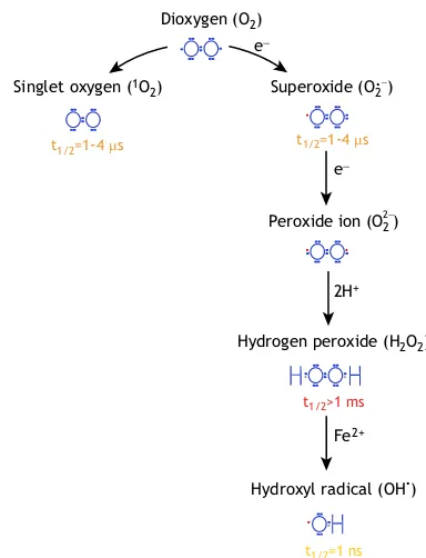
What are ROS? ROS refer to any oxygen derivative that is more reactive than an oxygen molecule (O2) itself (Foyer and Noctor, 2009; Mittler, 2017). Every type of ROS has unique and distinct chemical properties (Fig. 1). For example, singlet oxygen (1O
2) can oxidise lipids, proteins and guanidine residues of DNA; superoxide (O†2 ), like singlet oxygen, has a half-life time of 1-4 µs and reacts with Fe-S proteins; and hydroxyl (OH•) radicals are extremely reactive and unstable with a half-life time of 1 ns (Mittler, 2017; Waszczak et al., 2018). In contrast, hydrogen peroxide (H2O2) is fairly stable (more than 1 ms) and, therefore, is considered as the predominant ROS involved in cellular signaling. ROS can interact with various cellular components, including those that play a role in regulating ROS intracellular levels, hereafter referred to as‘ROS processing systems’(Fig. 2). Hydrogen peroxide, for instance, can be processed by several enzymes, including catalases (CATs) and ascorbate peroxidases (APXs), which are the main players involved
in H2O2metabolism. H2O2and other peroxides can also be processed by glutathione S-transferases (GSTs) (Dixon and Edwards, 2010) and peroxiredoxins (PRXs) (Dietz, 2011) including glutathione peroxidases (GPXs), which were misleadingly named because of their homology to animal GPX, but are now considered to act as thioredoxin (TRX)-dependent peroxiredoxins (Iqbal et al., 2006; Bela et al., 2015). These systems rely on the regeneration of reductants, such as ascorbate, glutathione and TRX, and ultimately depend on NADPH. Whereas the main superoxide-processing enzymes are superoxide dismutases (SODs), hydroxyl radicals and singlet oxygen are mainly metabolized by non-enzymatic reactions (Fig. 2) (Fridovich, 1997; Triantaphylidès and Havaux, 2009; Noctor et al., 2012; Richards et al., 2015).
In plants, ROS are produced during basal metabolism at various subcellular sites (Fig. 2), including during mitochondrial respiration, during photosynthesis in chloroplasts, in peroxisome-localized photorespiratory reactions, and by apoplastic NADPH oxidases [such as the respiratory burst oxidase homologs (RBOHs)] and other oxidases. This compartmentalization of ROS production and oxidation-reduction (redox)-associated reactions ensures the further control of ROS levels and allows redox signaling between organelles and the nucleus (Mignolet-Spruyt et al., 2016; Noctor and Foyer, 2017). ROS are also highly interconnected with other metabolites, including phytohormones such as salicylic acid (SA), jasmonic acid (JA), ethylene (ET), abscisic acid (ABA) and gibberellic acid (GA). Indeed, crosstalk between ROS and phytohormone-modulating stress response reactions, such as those involving SA and JA, is well documented (Noctor et al., 2015). In addition, interplay between ROS and development-associated hormones, such as auxin and cytokinin, has been reported, although specific insights are rather scarce and many questions remain outstanding (Considine and Foyer, 2014; Diaz-Vivancos et al., 2015; Tognetti et al., 2017).
ROS have long been recognized for their roles in mediating the response to abiotic and biotic stress conditions. However, in recent years, a number of studies have uncovered key roles for them during plant growth and development. Here, we discuss these emerging roles of ROS and redox-dependent mechanisms during plant development, highlighting their interactions with plant phytohormonal networks. First, we discuss how ROS can affect basic cellular processes, such as the cell cycle and division, and then review the roles of ROS at various stages of plant development, within seeds and meristems, and during organ and tissue development.
ROS-mediated control of the cell cycle, cell division, cell expansion and cell death
In plants, exposure to stress is often accompanied by decreased growth and cell cycle arrest, although the mechanisms underlying this response remain largely unexplored. In particular, the molecular factors of the cell cycle that are influenced by ROS or redox-dependent mechanisms are rather poorly studied in plants. It is known that redox cycles are conserved within the cell cycle and that reductive and oxidative signals are required for transitions within Department of Plant Biotechnology and Bioinformatics, Ghent University, 9052
Gent, Belgium, and Center for Plant Systems Biology, VIB, 9052 Gent, Belgium.
*Authors for correspondence (amna.mhamdi@psb.vib-ugent.be; frank.vanbreusegem@psb.vib-ugent.be)
A.M., 0000-0001-9959-1362; F.V.B., 0000-0002-3147-0860
DEVEL
O
the cell cycle phases (Menon and Goswami, 2007; Diaz-Vivancos et al., 2015; de Simone et al., 2017). These phase-to-phase progressions and transitions are mainly governed by a complex machinery of interacting cyclins (CYCs) and cyclin-dependent kinases (CDKs), and recent studies have begun to elucidate how ROS and changes in redox states can influence these factors.
Both the activities and transcript levels of CYCs and CDKs are affected by redox perturbations (Reichheld et al., 1999; Féher et al., 2008; Foyer et al., 2018). For instance, redox reactions directly affect cell cycle components via the TEOSINTE BRANCHED1-CYCLOIDEA-PROLIFERATING CELL FACTOR1 (TCP) transcription factors (Kadota et al., 2005). TCPs transcriptionally regulate CYCs levels, possibly through interactions with CYC promoters, and have a conserved redox-sensitive cysteine residue that is required for DNA binding. This suggests that, under oxidizing conditions, the interaction between a TCP transcription factor and its promoter might be inhibited as a result of disulfide bond formation (Viola et al., 2013, 2016).
CYKs and CDKs are functional in the S1-to-M phase transition of the cell cycle, and their differential expression has been associated with cell cycle arrest in the Arabidopsis glutathione-deficient ROOTMERISTEMLESS (rml1) mutant (Vernoux et al., 2000; Schnaubelt et al., 2015). Glutathione is the most important redox buffer in plants and, hence, the strong growth defect phenotype of rml1 mutants demonstrates the importance of glutathione-buffered redox homeostasis during cell division. Although glutathione is recruited into the nucleus during cell division, it has been reported that glutathione pools in the nuclei are in equilibrium with those in the cytosol but that glutathione is more
easily depleted from the cytosol than the nucleus after treatment with buthionine sulfoximine (García-Giménez et al., 2013; Pellny et al., 2009). Of note, a redox cycle within the cell cycle has been described in which ROS levels along with ascorbate and glutathione fluctuate, with the reduced versus oxidized pools of these metabolites regulating the transition through specific cell cycle checkpoints (Diaz-Vivancos et al., 2010; Schnaubelt et al., 2015; Diaz-Vivancos et al., 2015; Tognetti et al., 2017). In line with these reports, it has also been shown that ascorbate deficiency increases the oxidation degree of the nucleus and delays cell cycle progression (de Simone et al., 2017).
ROS and redox homeostasis are also required for cytokinesis. Pharmacological perturbation of ROS homeostasis in wheat (Triticum sp.) and Arabidopsis root tip cells induces mainly atypical tubulin polymer formation and affects efficient cell plate formation, ultimately resulting in perturbed cytokinesis (Livanos et al., 2012a,b). Similarly, the genetic disruption of NADPH oxidases (ROS generators) and mitogen-activated protein kinases involved in ROS signaling leads to tubulin disorganization and, hence, reinforces the necessity of a tightly controlled ROS balance during cytokinesis (Foreman et al., 2003; Takeda et al., 2008; Kosetsu et al., 2010; Yao et al., 2011).
ROS are also able to modulate cell expansion, via their effects on the cell wall. Apoplastic H2O2, hydroxyl radicals and superoxides, for example, influence cell wall stiffness and relaxation and hence affect cell expansion rates. Various oxidant sources are recognized, although their regulation remains poorly understood. In addition to NADPH oxidases, amine and oxalate oxidases, the peroxidative and hydroxylatic activities of apoplastic class III peroxidases have antagonistic effects on rigidity of cell walls (Passardi et al., 2004; Schmidt et al., 2016). In general, in a peroxidative modus, peroxidases regulate the levels of H2O2 by oxidizing various substrates. In this way, they contribute to the crosslinking of phenolics and extensins, which leads to increased stiffening, and hence reduced elongation capacity, of the cell walls. On the other hand, hydroxyl radical formation has been demonstrated to cleave xylogucans and pectins and thereby facilitate cell wall loosening (Fry, 1998; Passardi et al., 2004). This feature of peroxidases being associated with both cell elongation and growth-restricting processes is reflected by their contrasting effects on growth rates, as revealed by genetic perturbations of various class III peroxidases (Lu et al., 2014; Raggi et al., 2015; Schmidt et al., 2016). The concerted transcriptional repression of at least seven peroxidases by a MYB-like transcription factor, KUODA1, positively correlates with growth elongation capacities. Increased peroxidase activities lead to restricted leaf growth, without affecting cell division (Lu et al., 2014). The above concept of transcriptional repression of ROS production to favor organ growth can certainly not be generalized to different organs. For example, in Arabidopsis roots, the absence of the repressive transcription factor UPBEAT1 leads to an increased number of meristem cells and an increase in the length of cortical cells (Tsukagoshi et al., 2010).
Increased ROS production, in either a transient or a stable manner, also known as an oxidative burst, occurs in response to various stimuli, including development and bacterial challenges, and can initiate signaling towards cell death (Van Breusegem and Dat, 2006). Development-associated programmed cell death (PCD) occurs in various tissues and organs, such as the tapetum, seed coat, endosperm and lateral root cap (Daneva et al., 2016). For example, tapetal cells undergo PCD that is essential for
Dioxygen (O2)
Superoxide (O.2−) Singlet oxygen (1O
2)
e−
e−
2H+
Fe2+
Hydroxyl radical (OH ).
t1/2=1-4 µs
Peroxide ion (O2 )
Hydrogen peroxide (H2O2) 2−
t1/2=1-4 µs
t1/2>1ms
[image:2.612.74.266.54.305.2]t1/2=1ns
Fig. 1. Atmospheric oxygen-derived reactive oxygen species.A number of oxygen-derived reactive oxygen species (ROS) are known to exist in plants. The excitation of oxygen (O2) produces singlet oxygen (1O2), while
reduction produces superoxide radicals (O†2 ), hydrogen peroxide (H2O2) and
hydroxyl radicals (OH†). The Lewis structure of each of these ROS is presented in blue, with impaired electrons highlighted in red. The half life (t1/2)
is given for each type of ROS and is colour coded with highest value for H2O2(red) and lowest value for OH•(yellow).
DEVEL
O
microspore development. Interestingly, the rice (Oryza sativa) mutantdefective in tapetum cell death 1(dtc1) fails to accumulate ROS and shows delayed tapetum PCD resulting in male sterile plants (Yi et al., 2016). Despite the potential role for ROS here, the type of ROS and the mechanisms by which they trigger developmental cell death are unclear (Van Aken and Van Breusegem, 2015). In this context, the apoplastic RBOHs have been implicated in the control of development-related processes, such as proper growth of pollen tube and the self-incompatibility response. The altered expression of RBOH also drives altered PCD (Xie et al., 2014; Duan et al., 2014; Serrano et al., 2015; Jiménez-Quesada et al., 2016). Moreover, intracellular H2O2, produced via photorespiration, triggers lesion formation in leaves in a photoperiod-dependent manner (Queval et al., 2007). The factors necessary for the development of such lesions include salicylic acid and glutathione (Chaouch et al., 2010; Mhamdi et al., 2010b; Han et al., 2013a).
Overall, the effects of ROS on basic cellular processes–the cell cycle, cell division, cell expansion and cell death–are thought to contribute, acting in concert via interactions between plant phytohormonal pathways, to the multiple functions of ROS during the plant life cycle.
The role of ROS during germination
In dry and dormant seeds, plant embryos and the surrounding endosperm display very limited metabolic activities, and ROS production is thus speculated to be very low (Bailly et al., 2008). However, after seed imbibition and during germination, metabolism rapidly resumes (Rajjou et al., 2012) and such a swift metabolic start seems to be correlated with increased ROS production via various pathways and at various subcellular sites. This includes production via NADPH oxidases, lipid catabolism and lipidβ-oxidation in the glyoxysomes and mitochondrial respiration (Rajjou et al., 2012; Wojtyla et al., 2016; Ishibashi et al., 2017). The spatiotemporal correlation of increased ROS production and accumulation during the onset of germination has been corroborated with experiments in which exogenously applied oxidants, such as H2O2 (El-Maarouf-Bouteau et al., 2015), and a pharmacologically or genetically
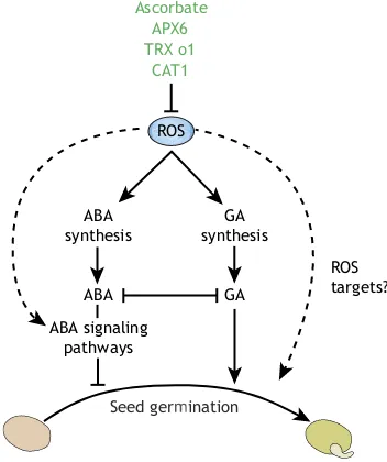
provoked decrease in catalase or in other antioxidant activities, were shown to positively influence the release of dormancy and the onset of germination (Fig. 3) (Leymarie et al., 2012; Cembrowska-Lech et al., 2015; Basbouss-Serhal et al., 2017). Reciprocally, overexpression of CAT in barley (Hordeum vulgare) seeds was shown to suppress precocious germination (Ishibashi et al., 2017). Therefore, increased ROS levels are key to proficient germination and are positive signals for the release of dormancy (Bailly et al., 2008; Singh et al., 2016).
ROS levels increase after seed imbibition and act as a positive signal for germination. However, above certain limits, ROS are either too low to allow germination or too high and affect embryo viability and therefore prevent or delay germination (Bailly et al., 2008). Thus, ROS homeostasis during germination needs to be tightly controlled and this creates an ‘oxidative window’ for germination that restricts proficient seedling development within certain borders of increased ROS levels (Stacey et al., 2006; Bailly et al., 2008). Consistently, several phenotypes are observed in mutants with perturbed antioxidant homeostasis. For instance, knocking out cytosolicAPX6, the transcript levels of which are usually high in dry seeds, leads to reduced germination rates owing to increased protein carbonylation (Chen et al., 2014). Theseapx6
mutants also exhibit increased sensitivity to stress and to ABA, triggered by disturbed ABA and auxin signaling. This suggests that these signaling pathways are interdependent, and that ABA and auxin accumulation and activation of ROS and redox signals are required. By contrast, mitochondrial thioredoxin O1 (trxo1) mutants exhibit accelerated germination together with increased H₂O₂levels (Ortiz-Espín et al., 2017). It was also recently shown that, in Arabidopsis thaliana, the transcription factor ABI5 regulates H2O2homeostasis in addition to its core role in ABA-dependent signaling; specifically, ABI5 assists the germination process by binding to the promoter of theCATALASE 1gene and regulating its expression and hence H2O2 levels (Skubacz et al., 2016; Bi et al., 2017).
ROS concentrations also increase during endosperm weakening, cell wall loosening and radicle elongation. Accordingly, the treatment of pea (Pisum sativum) seeds with H2O2facilitates seed germination and seedling growth (Barba-Espin et al., 2010). ROS-mediated effects on germination in Arabidopsis are inhibited by ABA, and this can be counteracted by the action of GA (Müller et al., 2009). In barley, H2O2is required for alleviating dormancy and this relies on GA accumulation and the expression of GA synthesis and signaling genes, rather than on the repression of ABA signaling (Bahin et al., 2011; Graeber et al., 2010). In the ascorbate-deficientArabidopsismutantvtc1, ABA levels are increased due to upregulation of synthesis genes (Pastori et al., 2003), and it has also been shown that ascorbate-defective vtc2 vtc5 mutants show seedling-lethal phenotypes that can be rescued by treating with ascorbate or its precursor galactose (Dowdle et al., 2007). In the same way, the apx6 mutants show moderate changes in the ascorbate pool (Chen et al., 2014).
Overall, these findings reinforce the notion that ROS action during seed germination relies heavily on interactions with ABA and GA, the two main phytohormones that antagonistically participate in regulation of the seed germination process (Fig. 3). Certainly, a better understanding of the molecular mechanisms that underlie ROS function in seed physiology (Oracz et al., 2007; Bazin et al., 2011; El-Maarouf-Bouteau et al., 2015; Wojtyla et al., 2016) will open up new routes for improving seed quality and tolerance to pathogen infection and provide new directions for engineering germination-recalcitrant species.
Apoplast Chloroplast Mitochondria Peroxisomes
Peroxisomes Chloroplast Mitochondria
Cytosol Apoplast
Carotenoids Tocopherols Ascorbate
Cell wall Membranes Chloroplast
Chloroplast
Flavonoids Ascorbate
SOD
Ascorbate Glutathione
CAT APX TRX/NTR GPX/PRX
Ascorbate Flavonoids Sugars
ROS generation sites
ROS processing pathways
H2O2 OH
.
1O
[image:3.612.57.293.55.247.2]2 O2 O.2−
Fig. 2. An overview of the major ROS production sites and processing pathways in plants.Oxygen and oxygen-derived ROS are aligned in the middle and highlighted in blue. Major subcellular sites involved in ROS production are listed below the ROS and the key ROS processing pathways are highlighted above.
DEVEL
O
The interplay between phytohormones and redox-linked reactions dictates meristem development
In both the shoot apical meristem (SAM) and the root apical meristem (RAM), stem cells are organized in a central zone (CZ) surrounding an organizing center, which is termed the organizing zone (OZ) or the quiescent center (QC) in shoots and roots, respectively. The maintenance of both meristems relies on signal exchange between the CZ and the OZ/QC but also on feedback from the already differentiated tissues. The major difference between both systems is the gene network that regulates their activity and their sensitivity to growth hormones, such as cytokinins and auxins. In short, while SAM activity is determined by WUSCHEL (WUS) and CLAVATA (CLV) peptides, QC establishment and stem cell maintenance in roots is ensured by SCARECROW (SCR), SHORT ROOT (SHR) and PLETHORA (PLT) (Stahl and Simon, 2010). Importantly, studies have revealed that the activities of both the SAM and the RAM are affected by interactions between ROS, redox components and phytohormones (Schippers et al., 2016).
RAM activity is highly sensitive to alterations in cellular redox status. For example, treatment with H2O2decreases the number of meristem cells (Tsukagoshi et al., 2010). DNA damage also promotes H2O2 accumulation, through FLAVIN-CONTAINING MONOOXYGENASE 1 (FMO1), and reduces root meristem size, hence indicating H2O2 as a negative regulator of the RAM (Tsukagoshi et al., 2010; Chen and Umeda, 2015). In addition, ROS gradients have been described in different zones of the root, with superoxide maxima correlating with cell division zones, and H2O2 maxima occurring in the elongation zone (Dunand et al., 2007; Tsukagoshi et al., 2010; Tsukagoshi, 2016), suggesting that superoxide and H2O2 act antagonistically. The molecular mechanism underlying the antagonistic actions of superoxide and H2O2has been elucidated in the context of the SAM (Zeng et al., 2017). This study showed that superoxide is associated with
transcriptional upregulation of the transcription factor WUS, whereas H2O2 displays an inhibitory action, accumulates in the peripheral zone and is associated with cell differentiation.
Within the RAM, QC cells are in a highly oxidized environment compared with their adjacent rapidly dividing cells. Both ascorbate and glutathione are mainly present as oxidized forms [dehydroascorbic acid (DHA) and glutathione disulfide (GSSG)] and NADPH is hardly detected, while higher antioxidant capacities and a more reducing environment is detected in the adjacent cells (Jiang et al., 2003). In line with this, cell type-specific transcriptomic analyses have revealed that ROS-associated genes are differentially expressed in specific SAM and RAM tissues (Tognetti et al., 2017 and references therein). In addition, specific glutathione- and thioredoxin-dependent reductive systems seem to be essential for appropriate meristem development. For instance, while the cytosolic form of glutathione reductase 1 (GR1) is not needed for development, loss of function of the chloroplast/mitochondrial form (GR2) is embryonic lethal, pointing to a key role for glutathione reduction in chloroplasts and mitochondria during early development (Chew et al., 2003; Tzafrir et al., 2004). In addition, a weak GR2 allele increases oxidized glutathione levels and provokes strong defects in the root meristem. This oxidizing environment (and the accumulation of GSSG) triggers decreased expression of the auxin efflux facilitator PIN-formed, PLT1 and PLT2 genes, clearly demonstrating that reduced glutathione is required for functional auxin signaling in the RAM (Yu et al., 2013).
Disrupted glutaredoxin (GRX) activity is also associated with meristem deficiencies. In Arabidopsis, GRXS17 regulates auxin sensitivity and transport (Cheng et al., 2011; Knuesting et al., 2015; Schippers et al., 2016) and in maize (Zea mays) GRX ABERRANT PHYLLOTAXY (ABPHYL2) influences shoot meristem size and phyllotaxy, probably through post-translational modification of the bZIP transcription factor FASCIATED EAR4 (Yang et al., 2015; Pautler et al., 2015). This was also demonstrated earlier for the
ArabidopsisGRXs ROXY1 and ROXY2, which reduce disulfide bonds in the heteromeric TGA9/TGA10 transcription factor complex, a reductive step that is necessary to activate gene expression during floral transition (Murmu et al., 2010). Intriguingly, the auxin-synthesizing flavin monooxygenase YUCCA6 also exhibits thiol reductase activity, thereby hinting towards an intimate link between redox and auxin pathways (Cha et al., 2015).
Besides affecting auxin signaling and transcription factors, the redox environment affects the cell-to-cell communication events and other hormonal pathways that are needed for SAM maintenance. The plastidial thioredoxin, TRXm3, regulates ROS homeostasis in the vicinity of plasmodesmata and is proposed to affect callose deposition and hence transport through plasmodesmata (Benitez-Alfonso et al., 2009). ROS also interact with the plant defense hormone SA. In both rice andArabidopsis, ABNORMAL INFLORESCENCE MERISTEM (AIM1), which is involved in SA biosynthesis, is needed for meristem development (Bussell et al., 2014; Xu et al., 2017). This interplay acts at the transcriptional level: SA downregulates a couple of plant-specific WRKY transcription factors and thereby alleviates their repressive effects on the expression of several antioxidative enzymes, such as CATs, GSTs and PRXs (Xu et al., 2017).
ROS homeostasis drives organ growth
The indeterminate growth characteristics of most plant roots not only entails continuous cell division and cell expansion of the primary root, but also the development of lateral roots (LRs) and
GA GA synthesis
Seed germination
Ascorbate APX6 TRX o1
CAT1
ROS targets? ABA
synthesis
ABA
ABA signaling pathways
[image:4.612.85.261.59.269.2]ROS
Fig. 3. ROS interactions with the ABA and GA pathways during seed germination.The accumulation of ROS (triggered by pharmacological or genetic approaches) positively influences the release of dormancy and favors the onset of germination. Metabolites and enzymes that have potentially important roles in keeping ROS levels under control in germinating seeds are shown at the top. In this context, ROS functions rely mainly on interactions with the ABA and GA signaling pathways, although some more direct effects (represented by dashed arrows) also occur.
DEVEL
O
root hairs. Studies have shown that altered ROS homeostasis affects all of these processes, restricting growth of the primary root, triggering LR emergence, and enhancing root hair growth (Table 1) (Foreman et al., 2003; Orman-Ligeza et al., 2016). An overview of ROS function in controlling root growth and development was recently provided by Tsukagoshi (2016) and highlights that interactions between ROS and auxin signaling, which play a crucial role in shaping root architecture (Du and Scheres, 2018), partially govern root growth and development.
In root hairs, for example, the auxin-controlled transcriptional regulation of NADPH oxidases and class III peroxidases promotes root hair elongation through at least two auxin-regulated transcriptional regulators: ROOT HAIR DEFECTIVE 6-LIKE 4 (RSL4) and MEDIATOR 25 (MED25) (Foreman et al., 2003;
Sundaravelpandian et al., 2013; Mangano et al., 2017). In addition, an analysis of ROS levels has suggested that a fine-tuned balance between H2O2and superoxide levels acts as a signal determining root hair cell differentiation (Sundaravelpandian et al., 2013).
[image:5.612.47.559.68.562.2]By contrast, the RBOH-peroxidase system, which also generates ROS, regulates LR emergence independently of auxin (Li et al., 2015; Manzano et al., 2014). DoublerbohD rbohFmutants exhibit early emerged LRs and enhanced density of LR primordia associated with increased levels of superoxides in the root tip (Li et al., 2015). Genetic manipulation of LR-specific peroxidases also abolishes LR emergence (Manzano et al., 2014). However, it is worth mentioning that all RBOH transcripts are auxin inducible and that H2O2generation mediated by RBOHD and RBOHE facilitates LR emergence by promoting cell wall remodeling in the overlying
Table 1. Overview of development and growth defects linked by perturbation of ROS and ROS-processing systems
Protein Gene locus Subcellular localization Mutant phenotypes References
Oxidases, superoxide dismutases and catalases
RBOHC AT5G51060 Plasma membrane rhd2, root hair defective Foreman et al. (2003)
RBOHD AT5G47910 Plasma membrane Atypical tubulin formation Yao et al. (2011)
RBOHD/RBOHF AT5G47910/AT1G64060 Plasma membrane Early emergence of LR and enhanced density
of LRs
Li et al. (2015)
RBOHE AT1G19230 Plasma membrane Aborted pollen and reduced fertility Xie et al. (2014)
RBOHH/RBOHJ AT5G60010/AT3G45810 Plasma membrane Root hair defective Mangano et al. (2017)
RBOHH/RBOHJ AT5G60010/AT3G45810 Plasma membrane Reduced fertility and impaired pollen tube growth Kaya et al. (2014)
MSD1 AT3G10920 Mitochondria Defect in embryo sac development Martin et al. (2013)
CAT2 AT4G23100 Peroxisomes Delayed growth and small hyponastic leaves Queval et al. (2007)
Ascorbate synthesis and dependent enzymes
VTC1 AT2G39770 Cytosol, nucleus Early flowering and senescence Barth et al. (2004)
VTC2 AT4G26850 Cytosol, nucleus Early flowering and senescence Kotchoni et al. (2009)
VTC3 VTC3 – Early flowering and senescence Kotchoni et al. (2009)
VTC4 AT3G02870 Cytosol Early flowering and senescence Kotchoni et al. (2009)
VTC1/VTC2 AT2G39770/AT4G26850 Cytosol, nucleus Seedling lethal Dowdle et al. (2007)
APX1 AT1G07890 Cytosol Reduced growth and embryo defects Pagnussat et al. (2005)
APX6 AT4G32320 Cytosol Reduced germination Chen et al. (2014)
Glutathione synthesis and reduction
GSH1 AT4G23100 Chloroplasts rml1, arrest of cell cycle on G1 Vernoux et al. (2000)
cad2,pad2,rax2, defect in LR development Marquez-Garcia et al. (2014)
GSH2 AT5G27380 Chloroplasts/cytosol Seedling lethal Pasternak et al. (2008)
GR2 AT3G54660 Chloroplasts/
mitochondria
Embryo lethal Tzafrir et al. (2004)
Defects in root growth and in RAM maintenance Yu et al. (2013)
Glutaredoxins and thioredoxins
GRXS17 AT4G04950 Nucleus, cytosol Compromised SAM, growth arrest and delayed
bolting
Knuesting et al. (2015)
GRXS13 AT1G03850 Nucleus, cytosol Reduced growth Laporte et al. (2012)
ROXY1 AT3G02000 Nucleus, cytosol Impaired petal development Xing et al. (2005)
ROXY2 AT5G14070 Nucleus, cytosol Defective anther development Xing and Zachgo (2008)
GRXC11/ROXY4 AT3G62950 Nucleus, cytosol Defective anther development Hou et al. (2008)
MIL1 OS07G05630 Nucleus, cytosol Defective anther development and impaired
meiosis
Hong et al. (2012)
MSCA1 CAX52135 Nucleus, cytosol Male sterile Chaubal et al. (2003)
NTRa/NTRb AT2G17420/AT4G35460 Cytosol/nucleus/
mitochondria
Growth defect and reduced fertility Reichheld et al. (2007)
NTRc AT2G41680 Chloroplasts Retarded growth of shoots and roots and
defective LR formation
Kirchsteiger et al. (2012)
TRXm3 AT2G15570 Chloroplasts Embryo lethal, impaired meristem development Benitez-Alfonso et al.
(2009)
TRX z AT3G06730 Chloroplasts Albino phenotype Arsova et al. (2010)
TRX o AT2G35010 Mitochondria Accelerated germination Ortiz-Espín et al. (2017)
TRX h9 AT3G08710 Cytosol Impaired growth of shoots and roots Meng et al. (2010)
NRX1 AT1G60420 Nucleus, cytosol Impaired fertility Marchal et al. (2014)
PDI1 AT2G47470 Cytosol Defect in embryo development Pagnussat et al. (2005)
Glutathione peroxidases and peroxiredoxins
GPX5 AT3G63080 Plasma membrane Defect in embryo development Pagnussat et al. (2005)
GPX1/GPX7 AT2G25080/AT4G31870 Chloroplasts Altered root architecture Passaia et al. (2014)
For genes that are described together, single mutants do not show phenotypes, and phenotypes are revealed only by additive mutations for the respective genes.
DEVEL
O
cell layers. RBOH loss-of-function mutants show delayed LR emergence, whereas targeted RBOHD expression in LR primordia promotes organ development (Orman-Ligeza et al., 2016). Interestingly, H2O2treatment restores LR formation in mutants in which auxin-mediated cell wall accommodation and remodeling are disrupted, such as the aux1 lax3 and pCASP1::shy2-2 mutants (Orman-Ligeza et al., 2016).
Consistent with the described roles for glutathione in cell cycle regulation and meristem development, glutathione-deficient mutants, such as pad2, cad2 and rax2, exhibit defects in LR formation (Table 1) (Marquez-Garcia et al., 2014; Schnaubelt et al., 2015). Furthermore, the pharmacological inhibition of glutathione synthesis affects root development and associated gene expression, similarly to phytohormone treatments and, in particular, exogenous auxin treatment (Koprivova et al., 2010). Unlike glutathione, ascorbate functions in root development are controversial and seem to be more subtle; ascorbate-deficient vtc mutants display only slightly altered root architecture and gravitropism (Olmos et al., 2006; Barth et al., 2010). The importance of redox control is further evidenced by altered root architecture phenotypes in individual mutants of allArabidopsisGPX genes, although the chloroplastic isoforms GPX1 and GPX7 were found to be the major players in this context (Passaia et al., 2014; Attacha et al., 2017).
A number of mutants exhibiting growth defects related to the misexpression of ROS processing system components have been reported, and these include a non-exhaustive list of mutants with leaf growth defects (Table 1). The detailed analysis of some of these mutants has, again, revealed interplay between ROS processing systems and hormone signaling pathways. In particular, new insights have been gained from the analysis of cat2 mutants, which are characterized by growth inhibition due to increased availability of photorespiratory H2O2, which triggers SA accumulation and activation of a pathogenesis-related pathway in a photoperiod-dependent manner (Queval et al., 2007; Mhamdi et al., 2010a). Furthermore, although some ROS components are dispensable for the normal growth and placement of leaves (i.e. into a‘rosette’formation) inArabidopsis, they have been shown to play specific functions in transmitting H2O2signals and in linking H2O2 to phytohormone pathways (Mhamdi et al., 2010b; Tognetti et al., 2010; Vanderauwera et al., 2011; Han et al., 2013a,b; Kerchev et al., 2015, 2016; Waszczak et al., 2016; Rahantaniaina et al., 2017).
Redox signaling in flower development
The crucial involvement of ROS during the development of plant reproductive organs and tissues has recently been reviewed (Jiménez-Quesada et al., 2016; Schippers et al., 2016). Briefly, and as we highlight below, ROS play key roles in petal development, pollen tube development and gametophyte development.
The functions of glutathione/GRX systems in flower development have been evidenced by the analysis of plant-specific class III CC-type GRXs, known as ROXYs (Fig. 4) (Gutsche et al., 2015). TheArabidopsis roxy1mutant was shown to exhibit an intriguing defect in petal development (Xing et al., 2005). Furthermore, it was shown that the phenotype of PETAL LOSS (ptl) mutants (Lampugnani et al., 2013) depends on ROXY1 function, and that PTL and ROXY1 interact to limit growth within and between sepals but to promote petal initiation (Quon et al., 2017). In this context, ROXY1 regulates petal development through TGA transcription factors, including PERIANTHIA and TGA2/ TGA3/TGA7 (Li et al., 2009).
Pharmacological approaches and ROS-staining experiments have also indicated that ROS accumulation at the tip of pollen
tubes is necessary for their efficient growth toward the female gametophyte (Potocký et al., 2012). Genetic evidence for the role for two RBOH genes (RBOHH and RBOHJ; Fig. 4) in pollen tube growth has been reported and has demonstrated the need for the activation of these NADPH oxidases by calcium and phosphorylation to allow proper growth (Duan et al., 2014; Kaya et al., 2014; Lassig et al., 2014). In particular, it was revealed that the growth rate oscillations of rbohH rbohJ pollen tubes show strong fluctuations in amplitude and frequency, ultimately leading to pollen tube collapse (Lassig et al., 2014). Interestingly, Rho-type GTPase (ROP1)-mediated spatial localization of these NADPH oxidases might steer ROS production and pollen tube growth (Kaya et al., 2014; Duan et al., 2014). Similar to the ROS-driven directional growth of root hairs, the presumed mode of action of ROS is to affect cell wall extensibility and strength. Within this specific context, cell wall loosening of the female tissues has been proposed to allow a more fluent pollen tube penetration (Smirnova et al., 2014; Wudick and Feijó, 2014). The spatiotemporal expression of RBOHE has also been reported, and mutation of
RBOHEorRBOHCwas reported to result in a significant proportion of aborted pollen grains, severely compromised pollen development and reduced fertility (Xie et al., 2014). In female gametophytes, by contrast, mitochondrial ROS sources rather than RBOHs seem to be required; in particular, the absence of the mitochondrial manganese SOD (MSD1) is associated with defective embryo sac development (Martin et al., 2013, 2014).
Glutathione, GRXs and TRXs have been shown to be required for proper gametophyte development (Fig. 4; Table 1). Arabidopsis ntra ntrbmutants, in which the genes encoding for two NADPH-dependent thioredoxin reductases are knocked out, show decreased fertility and slower growth (Reichheld et al., 2007). When glutathione deficiency (i.e. crossing withcad2andrml1mutants) is introduced in this background, meristem maintenance, growth
ROXY1 MSD1 ROXY1 ROXY2 Male gametophyte
development
Pistil
Anther RBOHC
RBOHE RBOHH RBOHJ NTRA NTRB GR1 PAD2
ROXY1 ROXY2 MSCA1 MIL1
Female gametophyte development
Petal and
sepal development
Microspore mother cell
Meiosis Pollen tube
Mitosis
Pollen
[image:6.612.314.562.56.285.2]Sepal
Fig. 4. ROS-associated genes involved in the control of flower and gametophyte development.Genes involved in ROS production and processing are presented. Genetic analyses have reported that loss of function of these candidates is associated with abnormalities during flower and gametophyte development and thus revealed their functions in petal development and in determining fertility and development of both gametophytes.
DEVEL
O
and flower development are severely inhibited (Reichheld et al., 2007; Bashandy et al., 2010). Thentra ntrbphenotypes can also be exacerbated, resulting in male sterility, if the gr1 mutation is introduced, whereas the lack of GR1 alone does not trigger developmental defects (Marty et al., 2009; Mhamdi et al., 2010b). Altogether, these results indicate the importance of cell thiol status and the interplay between TRX/NTR and glutathione systems during plant reproductive organ development.
ROXY1andROXY2are also expressed with overlapping patterns during anther development. TheArabidopsissingleroxy1androxy2
mutants produce normal anthers whereas roxy1 roxy2 double mutants are sterile (Xing and Zachgo, 2008). This effect is not only due to the function of ROXY1/ROXY2 in pollen production, but also to their function in female gametophyte development, with both functions being mediated via the regulation of gene expression. Consistently, the nuclear activity of ROXY genes and their interaction withTGA9/TGA10has been shown to be necessary for anther development (Murmu et al., 2010). The roles of GRX activity are conserved in rice and maize. Over-accumulation of ROS, lack ofMALE STERILE CONVERTED ANTHER1(MSCA1) or MICROSPORELESS1 (MIL1) trigger defects in anther development and are linked to male sterility in maize and rice (Chaubal et al., 2003; Kelliher and Walbot, 2012; Hong et al., 2012). Moreover, the rice genes OsROXY1 and OsROXY2 fully complement theArabidopsis roxy1mutant (Wang et al., 2009).
ROS metabolism and senescence
Plant senescence is a slow process and is accompanied by extensive reprogramming of gene expression (Breeze et al., 2011). A number of studies have revealed that developmentally regulated senescence is also associated with increased availability of ROS, which assist in degradation of cellular contents for recycling purposes but also play a role in initiating the senescence process (Guo and Gan, 2012; Munné-Bosch et al., 2013; Rogers and Munné-Bosch, 2016). Moreover, it is now known that ROS signaling impinges on the diverse hormone pathways that regulate senescence (Lim et al., 2007), including the auxin pathway, which is involved in regulating the timing of senescence (Mueller-Roeber and Balazadeh, 2014), and signaling via cytokinin, which is described as a senescence-delaying hormone (Swartzberg et al., 2011).
The expression of several ROS-induced transcription factors, including a significant proportion of genes encoding members of the NAC and WRKY gene families, is deregulated during senescence (Rosenwasser et al., 2011; Allu et al., 2014). Interestingly, NAC genes induced by H2O2 were found to determine senescence responses and stress tolerance. The effects on senescence gene expression driven byNAC3/ORS1, similarly to those driven by NAC2/ORE1, involve crosstalk with H2O2 -dependent signaling pathways (Balazadeh et al., 2010, 2011). Overexpression of the NAC factor JUNGBRUNNEN 1 also results in stress tolerance and is accompanied by enhanced expression of ROS-responsive genes (Wu et al., 2012). More recently, reports suggest that the molecular link between age-dependent increased ROS and SA require WRKY75 (Guo et al., 2017), which promotes SA synthesis by inducing SA INDUCTION-DEFICIENT2(SID2) and suppresses H2O2metabolism by inhibitingCAT2transcription (Guo et al., 2017).
Redox metabolism has also been directly implicated in the regulation of senescence. CAT2 levels drop in senescing leaves, allowing peroxisomal H2O2to increase (Zimmermann et al., 2006). This CAT2 downregulation at the transcriptional level appears to be the initial trigger of the H2O2peak during bolting time, whereas a
decrease in APX1 activity is thought to be a secondary and amplifying effect (Zimmermann et al., 2006). Ascorbate levels also decrease during senescence (Bartoli et al., 2000); accordingly, ascorbate deficiency (e.g. in vtc1mutants) enhances senescence and senescence-associated gene expression (Barth et al., 2004; Kotchoni et al., 2009). Senescence timing is also dependent on the regeneration of reduced glutathione by GR2. The GR2-RNAi lines exhibit early senescence phenotypes and increased levels of the senescence markers SENESCENCE-ASSOCIATED GENES SAG12 and SAG13 (Ding et al., 2016). In line with the above findings, the profiling of redox compounds during Arabidopsis
rosette development has revealed that ascorbate levels are higher during bolting and decrease significantly after flowering. By contrast, glutathione levels are maintained throughout development and tend to increase significantly with developmental age (Queval and Noctor, 2007). Changes in the redox states of ascorbate and glutathione do not occur, and both metabolites remain more than 80% reduced at all stages (Queval and Noctor, 2007). Of note, the least variable redox metabolite is NADPH, which is required for the regeneration of reduced glutathione.
Concluding remarks and perspectives
Over the last few decades, accumulating evidence has pointed to a crucial role for redox homeostasis in plant development. ROS production and ROS-related signaling has been implicated in almost all aspects of plant growth and development in a variety of organs and tissues (Table 1). A significant part of our current understanding of ROS functions has been gained through analyses of ROS-related components, the lack of function of which triggers aberrant developmental phenotypes. The analyses of development defective mutants clearly indicates that the spatial, temporal and compartment-specific distribution of ROS is governed by a complex network. However, currently, comprehensive insights into ROS production units, their interactions with the antagonistic ROS-processing pathways, and the precisein vivo modes of action of various ROS on both cellular building blocks and molecular processes are not available. This is, in part, due to the currently imperfect means to accurately monitor changes in ROS levels and associated redox perturbations in plant cells and tissues (Box 1). It should be noted that, although the studies cited in this Primer show how ROS distribution controls various developmental processes, we need to be cautious when interpreting data that are solely based on tissue staining methodologies. Current protocols that are used to visualize or quantify ROS signals are debatable and, in some cases, are not suitable or reliable for quantification (Box 1). This might be due to specificity issues and interference with other metabolites that might be present in the same tissue (Noctor et al., 2015, 2016; Ortega-Villasante et al., 2017). Quantitative information on ROS levels (steady state or inducible) in organelles, in specific cell types, or tissues is hence very scarce. New tools that facilitate ROS quantificationin vivowith standardized protocols that are specific for individual ROS will hopefully help us to better elucidate the causes and consequences of ROS in plant developmental processes (Waszczak et al., 2014). Certainly, the development of sensors and reporters forin vivoimaging is a fast growing area that will allow us to solve the difficulties surrounding ROS assays. However, most of the commonly used fluorescent probes are from prokaryotic origin and are not plant specific and thus require further development. The recent discoveries of ROS gene networks, mainly via analyses of transcriptomes and protein-protein interactions, offer the possibility to test new candidates for the development of novel tools for ROS imaging that are plant specific. The use of such new technologies
DEVEL
O
will be particularly relevant to addressing the key outstanding questions related to retrograde signaling, compartment-specific functions during development and the role of each ROS in
regulating signaling and communication (Noctor and Foyer, 2017). Similarly, the advent of more sensitive redox proteomics tools will start to allow thein vivodetection of proteins for which function is directly modulated by ROS. In this context, the identification of ROS sensitive targets within cell cycle regulators is likely to provide a significant leap forward in our understanding how ROS and redox perturbations affect the growth of organs and organisms (Foyer et al., 2018). Overall, the implementation of these new technologies will hopefully enable us to identify ROS targets at the organ, cellular and subcellular levels, and will help us to further elucidate the pathways that are modulated by ROS during growth and development (Waszczak et al., 2014). Ideally, these targets will be amenable (e.g. through genome-editing technologies) to genetic alterations and might allow redox-based strategies to improve the growth and reproductive features of both model plants and economically relevant crops.
Acknowledgements
We apologize if we inadvertently omitted citations of major contributions to this area. We thank Martine De Cock for help with editing the manuscript.
Competing interests
The authors declare no competing or financial interests.
Funding
The work in the Van Breusegem lab was supported by the Fonds Wetenschappelijk Onderzoek (G0D7914N); Universiteit Gent (Bijzonder Onderzoeks Fonds, 01J11311); and Fonds De La Recherche Scientifique - FNRS and the Fonds Wetenschappelijk Onderzoek-Vlaanderen under the EOS project (O018218F).
References
Allu, A. D., Soja, A. M., Wu, A., Szymanski, J. and Balazadeh, S.(2014). Salt stress and senescence: identification of cross-talk regulatory components.
J. Exp. Bot.65, 3993-4008.
Arsova, B., Hoja, U., Wimmelbacher, M., Greiner, E., Ustün, S., Melzer, M., Petersen, K., Lein, W. and Börnke, F.(2010). Plastidial thioredoxin z interacts with two fructokinase-like proteins in a thiol-dependent manner: evidence for an essential role in chloroplast development in Arabidopsis and Nicotiana benthamiana.Plant Cell22, 1498-1515.
Attacha, S., Solbach, D., Bela, K., Moseler, A., Wagner, S., Schwarzländer, M., Aller, I., Müller, S. J. and Meyer, A. J.(2017). Glutathione peroxidase-like enzymes cover five distinct cell compartments and membrane surfaces in
Arabidopsis thaliana.Plant Cell Environ.40, 1281-1295.
Bahin, E., Bailly, C., Sotta, B., Kranner, I., Corbineau, F. and Leymarie, J.(2011). Crosstalk between reactive oxygen species and hormonal signalling pathways regulates grain dormancy in barley.Plant Cell Environ.34, 980-993.
Bailly, C., El-Maarouf-Bouteau, H. and Corbineau, F.(2008). From intracellular signaling networks to cell death: the dual role of reactive oxygen species in seed physiology.C. R. Biol.331, 806-814.
Balazadeh, S., Siddiqui, H., Allu, A. D., Matallana-Ramirez, L. P., Caldana, C., Mehrnia, M., Zanor, M.-I., Köhler, B. and Mueller-Roeber, B.(2010). Gene regulatory network controlled by NAC transcription factor ANAC092/AtNAC2/ ORE1 during salt-promoted senescence.Plant J.62, 250-264.
Balazadeh, S., Kwasniewski, M., Caldana, C., Mehrnia, M., Zanor, M. I., Xue, G.-P. and Mueller-Roeber, B.(2011). ORS1, an H2O2-responsive NAC transcription
factor, controls senescence inArabidopsis thaliana.Mol. Plant4, 346-360.
Barba-Espin, G., Diaz-Vivancos, P., Clemente-Moreno, M. J., Albacete, A., Faize, L., Faize, M., Pérez-Alfocea, F. and Hernández, J. A.(2010). Interaction between hydrogen peroxide and plant hormones during germination and the early growth of pea seedlings.Plant Cell Environ.33, 981-994.
Basbouss-Serhal, I., Pateyron, S., Cochet, F., Leymarie, J. and Bailly, C.(2017). 5′to 3′mRNA decay contributes to the regulation ofArabidopsisseed germination by dormancy.Plant Physiol.173, 1709-1723.
Barth, C., Moeder, W., Klessig, D. F. and Conklin, P. L.(2004). The timing of senescence and response to pathogens is altered in the ascorbate-deficient
Arabidopsismutant vitamin c-1.Plant Physiol.134, 1784-1792.
Barth, C., Gouzd, Z. A., Steele, H. P. and Imperio, R. M.(2010). A mutation in GDP-mannose pyrophosphorylase causes conditional hypersensitivity to ammonium, resulting inArabidopsisroot growth inhibition, altered ammonium metabolism, and hormone homeostasis.J. Exp. Bot.61, 379-394.
Bartoli, C. G., Pastori, G. M. and Foyer, C. H.(2000). Ascorbate biosynthesis in mitochondria is linked to the electron transport chain between complexes III and IV.Plant Physiol.123, 335-344.
Box 1. ROS detection assays in plants: limitations and uncertainties
Various methods have been used for the detection and visualization of ROS in plant tissues and organs. However, several points need to be considered before making firm conclusions on ROS measurements in plants when using these approaches; detailed guidelines are presented in Noctor et al. (2016).
Biochemical assays
• As for other redox metabolites, ROS should not be extracted in water or neutral buffers due to the presence of contaminating antioxidant enzymes.
• Chemiluminescence probes have low selectivity and display high background levels (e.g. luminol); these issues should be considered when analyzing data.
Histochemical methods: diaminobenzidine (DAB) and nitro blue tetrazolium (NBT) staining
Both methods are used to visualize hydrogen peroxide and superoxide radicals, respectively. They gained their credibility mostly from the argument that they are widely used and hence accepted within the community. However, DAB and NBT are not specific or direct measurements of both ROS. Even if the difference in staining can be manipulated by treatment with antioxidants, this is not a direct proof of ROS generation.
• Color formation does not always reflect measurement of the desired ROS (H2O2for DAB and O†2 for NBT).
• NBT staining can reflect the presence of ascorbate or the activity of dehydrogenases; DAB brownish color accumulates in the presence of higher peroxidase activity.
• Differences in the uptake or the permeability of both dyes can lead to misinterpretation of the data.
Dichlorofluorescein (DCF)-derived fluorescent dyes
DCF is widely used for quantification for H2O2 in different systems.
However, this method is not reliable and is not specific. ROS imaging can be further complicated by the presence of endogenous autofluorescent compounds, in particular in leaf tissues (e.g. chlorophyll, flavonoids, anthocyanins). The permeability of the dye and its stability over time are also factors that might contribute to the difficulties associated with ROS imaging.
• Dichlorofluorescin (DCFH) does not react with H2O2 or other
ROS directly.
• DCF radicals can in fact produce O†2 or H2O2via reaction with
oxygen and therefore an artificial increase of ROS can be generated. • Other cell components (transition metals, cytochrome c and
peroxidases) can also enhance DCFH oxidation to DCF. • Glutathione and NADPH can interact with the photoexcited DCF.
Genetically encoded probes
Genetically encoded sensors are more suitable for ROS imaging in plant systems because they are non-invasive, flexible (stable or transient expression in target tissues or compartments) and more stable over time. Ratiometric sensors offer the potential to overcome problems related to photobleaching and the expression of the proteins in different conditions (Ortega-Villasante et al., 2017).
• Fluorescent protein-based sensors are pH sensitive, and this has an impact on accurate quantification in organelles with different pH; therefore, pH controls need to be measured simultaneously. • Silencing and problems with stable expression of sensors has been
reported in plant systems.
• The dynamics of the intracellular thiol systems (glutathione, GRX, TRX, PRX, etc.) in plants expressing genetic probes (sensors are often fused to redox proteins) might be worth considering. The fluorescence signal of the probe depends on the equilibrium with thiol systems, and at the same time their H2O2-driven oxidation
needs to be reversed by glutathione.
DEVEL
O
Bashandy, T., Guilleminot, J., Vernoux, T., Caparros-Ruiz, D., Ljung, K., Meyer, Y. and Reichheld, J.-P. (2010). Interplay between the NADP-linked thioredoxin and glutathione systems inArabidopsisauxin signaling.Plant Cell
22, 376-391.
Bazin, J., Langlade, N., Vincourt, P., Arribat, S., Balzergue, S., El-Maarouf-Bouteau, H. and Bailly, C. (2011). Targeted mRNA oxidation regulates sunflower seed dormancy alleviation during dry after-ripening.Plant Cell
23, 2196-2208.
Bela, K., Horváth, E., Gallé, Á., Szabados, L., Tari, I. and Csiszár, J.(2015). Plant glutathione peroxidases: emerging role of the antioxidant enzymes in plant development and stress responses.J. Plant Physiol.176, 192-201.
Benitez-Alfonso, Y., Cilia, M., San Roman, A., Thomas, C., Maule, A., Hearn, S. and Jackson, D. (2009). Control of Arabidopsis meristem development by thioredoxin-dependent regulation of intercellular transport.Proc. Natl. Acad. Sci. USA106, 3615-3620.
Bi, C., Ma, Y., Wu, Z., Yu, Y. T., Liang, S., Lu, K. and Wang, X. F.
(2017). Arabidopsis ABI5 plays a role in regulating ROS homeostasis by activating CATALASE 1 transcription in seed germination. Plant Mol. Biol.
94, 197-213.
Breeze, E., Harrison, E., McHattie, S., Hughes, L., Hickman, R., Hill, C., Kiddle, S., Kim, Y., Penfold, C. A., Jenkins, D. et al.(2011). High-resolution temporal profiling of transcripts duringArabidopsisleaf senescence reveals a distinct chronology of processes and regulation.Plant Cell23, 873-894.
Bussell, J. D., Reichelt, M., Wiszniewski, A. A., Gershenzon, J. and Smith, S. M.
(2014). Peroxisomal ATP-binding cassette transporter COMATOSE and the multifunctional protein abnormal INFLORESCENCE MERISTEM are required for the production of benzoylated metabolites in Arabidopsis seeds.Plant Physiol.
164, 48-54.
Cembrowska-Lech, D., Koprowski, M. and Kępczyński, J.(2015). Germination induction of dormantAvena fatuacaryopses by KAR1and GA3involving the
control of reactive oxygen species (H2O2and O2−) and enzymatic antioxidants
(superoxide dismutase and catalase) both in the embryo and the aleurone layers.
J. Plant Physiol.176, 169-179.
Cha, J.-Y., Kim, W.-Y., Kang, S. B., Kim, J. I., Baek, D., Jung, I. J., Kim, M. R., Li, N., Kim, H.-J. and Nakajima, M.(2015). A novel thiol-reductase activity of
Arabidopsis YUC6 confers drought tolerance independently of auxin
biosynthesis.Nat. Commun.6, 8041.
Chaouch, S., Queval, G., Vanderauwera, S., Mhamdi, A., Vandorpe, M., Langlois-Meurinne, M., Van Breusegem, F., Saindrenan, P. and Noctor, G.
(2010). Peroxisomal hydrogen peroxide is coupled to biotic defense responses by ISOCHORISMATE SYNTHASE1 in a daylength-related manner.Plant Physiol.
153, 1692-1705.
Chaubal, R., Anderson, J. R., Trimnell, M. R., Fox, T. W., Albertsen, M. C. and Bedinger, P.(2003). The transformation of anthers in themsca1mutant of maize.
Planta216, 778-788.
Chen, P. and Umeda, M.(2015). DNA double-strand breaks induce the expression of flavin-containing monooxygenase and reduce root meristem size in
Arabidopsis thaliana.Genes Cells20, 636-646.
Chen, C., Letnik, I., Hacham, Y., Dobrev, P., Ben-Daniel, B.-H., Vanková, R., Amir, R. and Miller, G. (2014). ASCORBATE PEROXIDASE6 protects
Arabidopsisdesiccating and germinating seeds from stress and mediates cross
talk between reactive oxygen species, abscisic acid, and auxin.Plant Physiol.
166, 370-383.
Cheng, N. H., Liu, J. Z., Liu, X., Wu, Q., Thompson, S. M., Lin, J., Chang, J., Whitham, S. A., Park, S., Cohen, J. D. et al.(2011).Arabidopsismonothiol glutaredoxin, AtGRXS17, is critical for temperature-dependent postembryonic growth and development via modulating auxin response. J. Biol. Chem.
286, 20398-20406.
Chew, O., Whelan, J. and Millar, A. H. (2003). Molecular definition of the ascorbate-glutathione cycle inArabidopsismitochondria reveals dual targeting of antioxidant defenses in plants.J. Biol. Chem.278, 46869-46877.
Considine, M. J. and Foyer, C. H.(2014). Redox regulation of plant development.
Antioxid. Redox Signal.21, 1305-1326.
Daneva, A., Gao, Z., Van Durme, M. and Nowack, M. K.(2016). Functions and regulation of programmed cell death in plant development.Annu. Rev. Cell Dev. Biol.32, 441-468.
de Simone, A., Hubbard, R., de la Torre, N. V., Velappan, Y., Wilson, M., Considine, M. J., Soppe, W. J. J. and Foyer, C. H.(2017). Redox changes during the cell cycle in the embryonic meristem ofArabidopsis thaliana.Antioxid.
Redox Signal.27, 1505-1519.
Diaz-Vivancos, P., Wolff, T., Markovic, J., Pallardó, F. V. and Foyer, C. H.(2010). A nuclear glutathione cycle within the cell cycle.Biochem. J.431, 169-178.
Diaz-Vivancos, P., De Simone, A., Kiddle, G. and Foyer, C. H. (2015). Glutathione- linking cell proliferation to oxidative stress.Free Radic. Biol. Med.
89, 1154-1104.
Dietz, K.-J.(2011). Peroxiredoxins in plants and cyanobacteria.Antioxid. Redox Signal.15, 1129-1159.
Ding, S., Wang, L., Yang, Z., Lu, Q., Wen, X. and Lu, C.(2016). Decreased glutathione reductase2 leads to early leaf senescence inArabidopsis.J. Integr. Plant Biol.58, 29-47.
Dixon, D. P. and Edwards, R.(2010). Glutathione S-transferases.Arabidopsis Book8, e0131.
Dowdle, J., Ishikawa, T., Gatzek, S., Rolinski, S. and Smirnoff, N.(2007). Two genes inArabidopsis thalianaencoding GDP-l-galactose phosphorylase are required for ascorbate biosynthesis and seedling viability.Plant J.52, 673-689.
Du, Y. and Scheres, B.(2018). Lateral root formation and the multiple roles of auxin.
J. Exp. Bot.69, 155-167.
Duan, Q., Kita, D., Johnson, E. A., Aggarwal, M., Gates, L., Wu, H.-M. and Cheung, A. Y.(2014). Reactive oxygen species mediate pollen tube rupture to release sperm for fertilization inArabidopsis.Nat. Commun.5, 3129.
Dunand, C., Crevecoeur, M. and Penel, C.(2007). Distribution of superoxide and hydrogen peroxide inArabidopsisroot and their influence on root development: possible interaction with peroxidases.New Phytol.174, 332-341.
El-Maarouf-Bouteau, H., Sajjad, Y., Bazin, J., Langlade, N., Cristescu, S. M., Balzergue, S., Baudouin, E. and Bailly, C.(2015). Reactive oxygen species, abscisic acid and ethylene interact to regulate sunflower seed germination.Plant Cell Environ.38, 364-374.
Féher, A., Ötvös, K., Pasternak, T. P. and Pettkó-Szandtner, A. (2008). The involvement of reactive oxygen species (ROS) in the cell cycle activation (G0-to-G1 transition) of plant cells.Plant Signal. Behav.3, 823-826.
Foreman, J., Demidchik, V., Bothwell, J. H. F., Mylona, P., Miedema, H., Torres, M. A., Linstead, P., Costa, S., Brownlee, C., Jones, J. D. G. et al.
(2003). Reactive oxygen species produced by NADPH oxidase regulate plant cell growth.Nature422, 442-446.
Foyer, C. H. and Noctor, G.(2009). Redox regulation in photosynthetic organisms: signaling, acclimation, and practical implications. Antioxid. Redox Signal.
11, 861-905.
Foyer, C. H. Wilson, M. H. and Wright, M. H.(2018). Redox regulation of cell proliferation: Bioinformatics and redox proteomics approaches to identify redox-sensitive cell cycle regulators.Free Radic. Biol. Med.S0891-S5849, 30150-3.
Fridovich, I.(1997). Superoxide anion radical (O·−2), superoxide dismuatases, and related matters.J. Biol. Chem.272, 18515-18517.
Fry, S. C.(1998). Oxidative scission of plant cell wall polysaccharides by ascorbate-induced hydroxyl radicals.Biochem. J.332, 507-515.
Garcı́a-Giménez, J. L., Markovic, J., Dası́, F., Queval, G., Schnaubelt, D., Foyer, C. H. and Pallardó, F. V.(2013). Nuclear glutathione.Biochem. Biophys. Acta1830, 3304-3316.
Graeber, K., Linkies, A., Müller, K., Kunchova, A., Rott, A. and Leubner-Metzger, G. (2010). Cross-species approaches to seed dormancy and germination: conservation and biodiversity of ABA-regulated mechanisms and the Brassicaceae DOG1 genes.Plant Mol. Biol.73, 67-87.
Guo, Y. and Gan, S. S.(2012). Convergence and divergence in gene expression profiles induced by leaf senescence and 27 senescence-promoting hormonal, pathological and environmental stress treatments. Plant Cell Environ. 35, 644-655.
Guo, P., Li, Z., Huang, P., Li, B., Fang, S., Chu, J. and Guo, H.(2017). A tripartite amplification loop involving the transcription factor WRKY75, salicylic acid, and reactive oxygen species accelerates leaf senescence.Plant Cell29, 2854-2870.
Gutsche, N., Thurow, C., Zachgo, S. and Gatz, C.(2015). Plant-specific CC-type glutaredoxins: functions in developmental processes and stress responses.Biol.
Chem.396, 495-509.
Han, Y., Chaouch, S., Mhamdi, A., Queval, G., Zechmann, B. and Noctor, G.
(2013a). Functional analysis ofArabidopsismutants points to novel roles for glutathione in coupling H2O2to activation of salicylic acid accumulation and
signaling.Antioxid. Redox Signal.18, 2106-2121.
Han, Y., Mhamdi, A., Chaouch, S. and Noctor, G.(2013b). Regulation of basal and oxidative stress-triggered jasmonic acid-related gene expression by glutathione.
Plant Cell Environ.36, 1135-1146.
Hong, L., Tang, D., Zhu, K., Wang, K., Li, M. and Cheng, Z.(2012). Somatic and reproductive cell development in rice anther is regulated by a putative glutaredoxin.Plant Cell24, 577-588.
Hou, X., Hu, W. W., Shen, L., Lee, L. Y., Tao, Z., Han, J. H. and Yu, H.(2008). Global identification of DELLA target genes during Arabidopsis flower development.Plant Physiol.147, 1126-1142.
Iqbal, A., Yabuta, Y., Takeda, T., Nakano, Y. and Shigeoka, S. (2006). Hydroperoxide reduction by thioredoxin-specific glutathione peroxidase isoenzymes ofArabidopsis thaliana.FEBS J.273, 5589-5597.
Ishibashi, Y., Aoki, N., Kasa, S., Sakamoto, M., Kai, K., Tomokiyo, R., Watabe, G., Yuasa, T. and Iwaya-Inoue, M. (2017). The interrelationship between abscisic acid and reactive oxygen species plays a key role in barley seed dormancy and germination.Front. Plant Sci.8, 275.
Jiang, K., Meng, Y. L. and Feldman, L. J.(2003). Quiescent center formation in maize roots is associated with an auxin-regulated oxidizing environment.
Development130, 1429-1438.
Jiménez-Quesada, M. J., Traverso, J. Á. and Alché, J. D.(2016). NADPH oxidase-dependent superoxide production in plant reproductive tissues.Front. Plant Sci.7, 359.
Kadota, Y., Furuichi, T., Sano, T., Kaya, H., Gunji, W., Murakami, Y., Muto, S., Hasezawa, S. and Kuchitsu, K.(2005). Cell-cycle-dependent regulation of
DEVEL
O
oxidative stress responses and Ca2+
permeable channels NtTPC1A/B in tobacco BY-2 cells.Biochem. Biophys. Res. Commun.336, 1259-1267.
Kaya, H., Nakajima, R., Iwano, M., Kanaoka, M. M., Kimura, S., Takeda, S., Kawarazaki, T., Senzaki, E., Hamamura, Y., Higashiyama, T. et al.(2014). Ca2 +
-activated reactive oxygen species production byArabidopsisRbohH and RbohJ is essential for proper pollen tube tip growth.Plant Cell26, 1069-1080.
Kelliher, T. and Walbot, V.(2012). Hypoxia triggers meiotic fate acquisition in maize.Science337, 345-348.
Kerchev, P., Mühlenbock, P., Denecker, J., Morreel, K., Hoeberichts, F. A., Van Der Kelen, K., Vandorpe, M., Nguyen, L., Audenaert, D. and Van Breusegem, F. (2015). Activation of auxin signalling counteracts photorespiratory H2O2-dependent cell death.Plant Cell Environ.38, 253-265.
Kerchev, P., Waszczak, C., Lewandowska, A., Willems, P., Shapiguzov, A., Li, Z., Alseekh, S., Mühlenbock, P., Hoeberichts, F. A., Huang, J. et al.(2016). Lack of GLYCOLATE OXIDASE1, but not GLYCOLATE OXIDASE2, attenuates the photorespiratory phenotype of CATALASE2-deficient Arabidopsis. Plant Physiol.171, 1704-1719.
Kirchsteiger, K., Ferrández, J., Pascual, M. B., González, M. and Cejudo, F. J.
(2012). NADPH thioredoxin reductase C is localized in plastids of photosynthetic and nonphotosynthetic tissues and is involved in lateral root formation in
Arabidopsis.Plant Cell24, 1534-1548.
Knuesting, J., Riondet, C., Maria, C., Kruse, I., Bécuwe, N., König, N., Berndt, C., Tourrette, S., Guilleminot-Montoya, J., Herrero, E. et al.(2015).
Arabidopsis glutaredoxin S17 and its partner, the nuclear factor Y subunit
C11/negative cofactor 2α, contribute to maintenance of the shoot apical meristem under long-day photoperiod. Plant Physiol.167, 1643-1658.
Koprivova, A., Mugford, S. T. and Kopriva, S.(2010).Arabidopsisroot growth dependence on glutathione is linked to auxin transport. Plant Cell Rep.
29, 1157-1167.
Kotchoni, S. O., Larrimore, K. E., Mukherjee, M., Kempinski, C. F. and Barth, C.
(2009). Alterations in the endogenous ascorbic acid content affect flowering time inArabidopsis.Plant Physiol.149, 803-815.
Kosetsu, K., Matsunaga, S., Nakagami, H., Colcombet, J., Sasabe, M., Soyano, T., Takahashi, Y., Hirt, H. and Machida, Y. (2010). The MAP kinase MPK4 is required for cytokinesis in Arabidopsis thaliana. Plant Cell
22, 3778-3790.
Lampugnani, E. R., Kilinc, A. and Smyth, D. R.(2013). Auxin controls petal initiation inArabidopsis.Development140, 185-194.
Laporte, D., Olate, E., Salinas, P., Salazar, M., Jordana, X. and Holuigue, L.
(2012). Glutaredoxin GRXS13 plays a key role in protection against photooxidative stress inArabidopsis.J. Exp. Bot.63, 503-515.
Lassig, R., Gutermuth, T., Bey, T. D., Konrad, K. R. and Romeis, T.(2014). Pollen tube NAD(P)H oxidases act as a speed control to dampen growth rate oscillations during polarized cell growth.Plant J.78, 94-106.
Leymarie, J., Vitkauskaité, G., Hoang, H. H., Gendreau, E., Chazoule, V., Meimoun, P., Corbineau, F., El-Maarouf-Bouteau, H. and Bailly, C.(2012). Role of reactive oxygen species in the regulation ofArabidopsisseed dormancy.
Plant Cell Physiol.53, 96-106.
Li, S., Lauri, A., Ziemann, M., Busch, A., Bhave, M. and Zachgo, S.(2009). Nuclear activity of ROXY1, a glutaredoxin interacting with TGA factors, is required for petal development inArabidopsis thaliana.Plant Cell21, 429-441.
Li, N., Sun, L., Zhang, L., Song, Y., Hu, P., Li, C. and Hao, F. S.(2015). AtrbohD and AtrbohF negatively regulate lateral root development by changing the localized accumulation of superoxide in primary roots ofArabidopsis.Planta
241, 591-602.
Lim, P. O., Kim, H. J. and Nam, H. G.(2007). Leaf senescence.Annu. Rev. Plant Biol.58, 115-136.
Livanos, P., Apostolakos, P. and Galatis, B.(2012a).Plant Celldivision ROS homeostasis is required.Plant Signal. Behav.7, 771-778.
Livanos, P., Galatis, B., Quader, H. and Apostolakos, P.(2012b). Disturbance of reactive oxygen species homeostasis induces atypical tubulin polymer formation and affects mitosis in root-tip cells ofTriticum turgidumandArabidopsis thaliana.
Cytoskeleton69, 1-21.
Lu, D., Wang, T., Persson, S., Mueller-Roeber, B. and Schippers, J. H. M.(2014). Transcriptional control of ROS homeostasis by KUODA1 regulates cell expansion during leaf development.Nat. Commun.5, 3767.
Manzano, C., Pallero-Baena, M., Casimiro, I., De Rybel, B., Orman-Ligeza, B., Van Isterdael, G., Beeckman, T., Draye, X., Casero, P. and del Pozo, J. C.
(2014). The emerging role of reactive oxygen species signaling during lateral root development.Plant Physiol.165, 1105-1119.
Mangano, S., Denita-Juarez, S. P., Choi, H.-S., Marzol, E., Hwang, Y., Ranocha, P., Velasquez, S. M., Borassi, C., Barberini, M. L., Aptekmann, A. A. et al.(2017). Molecular link between auxin and ROS-mediated polar growth.
Proc. Natl. Acad. Sci. USA114, 5289-5294.
Marchal, C., Delorme-Hinoux, V., Bariat, L., Siala, W., Belin, C., Saez-Vasquez, J., Riondet, C. and Reichheld, J. P.(2014). NTR/NRX define a new thioredoxin system in the nucleus ofArabidopsis thalianacells.Mol. Plant7, 30-44.
Marquez-Garcia, B., Njo, M., Beeckman, T., Goormachtig, S. and Foyer, C. H.
(2014). A new role for glutathione in the regulation of root architecture linked to strigolactones.Plant Cell Environ.37, 488-498.
Martin, M. V., Fiol, D. F., Sundaresan, V., Zabaleta, E. J. and Pagnussat, G. C.
(2013). oiwa, a female gametophytic mutant impaired in a mitochondrial manganese-superoxide dismutase, reveals crucial roles for reactive oxygen species during embryo sac development and fertilization inArabidopsis.Plant Cell
25, 1573-1591.
Martin, M. V., Distéfano, A. M., Bellido, A., Córdoba, J. P., Soto, D., Pagnussat, G. C. and Zabaleta, E.(2014). Role of mitochondria during female gametophyte development and fertilization in A. thaliana. Mitochondrion
19, 350-356.
Marty, L., Siala, W., Schwarzländer, M., Fricker, M. D., Wirtz, M., Sweetlove, L. J., Meyer, Y., Meyer, A. J., Reichheld, J.-P. and Hell, R.
(2009). The NADPH-dependent thioredoxin system constitutes a functional backup for cytosolic glutathione reductase inArabidopsis.Proc. Natl. Acad. Sci. USA106, 9109-9114.
Meng, L., Wong, J. H., Feldman, L. J., Lemaux, P. G. and Buchanan, B. B.(2010). A membrane associated thioredoxin required for plant growth moves from cell to cell, suggestive of a role in intercellular communication.Proc. Natl. Acad. Sci. USA
107, 3900-3905.
Menon, S. G. and Goswami, P. C.(2007). A redox cycle within the cell cycle: ring in the old with the new.Oncogene27, 1101-1109.
Mhamdi, A., Queval, G., Chaouch, S., Vanderauwera, S., Van Breusegem, F. and Noctor, G.(2010a). Catalase function in plants: a focus onArabidopsis
mutants as stress-mimic models.J. Exp. Bot.61, 4197-4220.
Mhamdi, A., Hager, J., Chaouch, S., Queval, G., Han, Y., Taconnat, L., Saindrenan, P., Gouia, H., Issakidis-Bourguet, E., Renou, J.-P. et al.
(2010b).ArabidopsisGLUTATHIONE REDUCTASE 1 plays a crucial role in leaf responses to intracellular hydrogen peroxide and in ensuring appropriate gene expression through both salicylic acid and jasmonic acid signaling pathways.
Plant Physiol.153, 1144-1160.
Mignolet-Spruyt, L., Xu, E. J., Idänheimo, N., Hoeberichts, F. A., Mühlenbock, P., Brosché, M., Van Breusegem, F. and Kangasjärvi, J.
(2016). Spreading the news: subcellular and organellar reactive oxygen species production and signalling.J. Exp. Bot.67, 3831-3844.
Mittler, R.(2017). ROS are good.Trends Plant Sci.22, 11-19.
Mittler, R., Vanderauwera, S., Suzuki, N., Miller, G., Tognetti, V. B., Vandepoele, K., Gollery, M., Shulaev, V. and Van Breusegem, F.(2011). ROS signaling: the new wave?Trends Plant Sci.16, 300-309.
Mueller-Roeber, B. and Balazadeh, S. (2014). Auxin and its role in plant senescence.J. Plant Growth Regul.33, 21-33.
Müller, K., Linkies, A., Vreeburg, R. A. M., Fry, S. C., Krieger-Liszkay, A. and Leubner-Metzger, G.(2009). In vivo cell wall loosening by hydroxyl radicals during cress seed germination and elongation growth.Plant Physiol.150, 1855-1865.
Munné-Bosch, S., Queval, G. and Foyer, C. H.(2013). The impact of global change factors on redox signaling underpinning stress tolerance.Plant Physiol.
161, 5-19.
Murmu, J., Bush, M. J., DeLong, C., Li, S., Xu, M., Khan, M., Malcolmson, C., Fobert, P. R., Zachgo, S. and Hepworth, S. R.(2010).Arabidopsisbasic leucine-zipper transcription factors TGA9 and TGA10 interact with floral glutaredoxins ROXY1 and ROXY2 and are redundantly required for anther development.Plant Physiol.154, 1492-1504.
Noctor, G. and Foyer, C. H.(2017). Update on redox compartmentation intracellular redox compartmentation and ROS-related communication in regulation and signaling.Plant Physiol.171, 1581-1592.
Noctor, G., Mhamdi, A., Chaouch, S., Han, Y., Neukermans, J., Marquez-Garcia, B., Queval, G. and Foyer, C. H.(2012). Glutathione in plants: an integrated overview.
Plant Cell Environ.35, 454-484.
Noctor, G., Lelarge-Trouverie, C. and Mhamdi, A.(2015). The metabolomics of oxidative stress.Phytochemistry112, 33-53.
Noctor, G., Mhamdi, A. and Foyer, C. H.(2016). Oxidative stress and antioxidative systems: recipes for successful data collection and interpretation.Plant Cell Environ.39, 1140-1160.
Noctor, G., Reichheld, J.-P. and Foyer, C. H.(2017). ROS-related redox regulation and signaling in plants.Semin. Cell Dev. Biol.80, 3-12.
Olmos, E., Kiddle, G., Pellny, T., Kumar, S. and Foyer, C. H.(2006). Modulation of plant morphology, root architecture, and cell structure by low vitamin C in
Arabidopsis thaliana.J. Exp. Bot.57, 1645-1655.
Oracz, K., El-Maarouf-Bouteau, H., Farrant, J. M., Cooper, K., Belghazi, M., Job, C., Job, D., Corbineau, F. and Bailly, C.(2007). ROS production and protein oxidation as a novel mechanism of seed dormancy alleviation.Plant J.50, 452-465.
Orman-Ligeza, B., Parizot, B., de Rycke, R., Fernandez, A., Himschoot, E., Van Breusegem, F., Bennett, M. J., Périlleux, C., Beeckman, T. and Draye, X.
(2016). RBOH-mediated ROS production facilitates lateral root emergence in
Arabidopsis.Development143, 3328-3339.
Ortega-Villasante, C., Burén, S., Blázquez-Castro, A., Barón-Sola, Á. and Hernández, L. E.(2017). Fluorescent in vivo imaging of reactive oxygen species and redox potential in plants.Free Radic. Biol. Med.S0891-5849, 30169-30162.
Ortiz-Espı́n, A., Iglesias-Fernández, R., Calderón, A., Carbonero, P., Sevilla, F. and Jiménez, A.(2017). Mitochondrial AtTrxo1 is transcriptionally regulated by