REVIEW
Plant regeneration: cellular origins and molecular mechanisms
Momoko Ikeuchi, Yoichi Ogawa, Akira Iwase and Keiko Sugimoto*ABSTRACT
Compared with animals, plants generally possess a high degree of developmental plasticity and display various types of tissue or organ regeneration. This regenerative capacity can be enhanced by exogenously supplied plant hormonesin vitro, wherein the balance between auxin and cytokinin determines the developmental fate of regenerating organs. Accumulating evidence suggests that some forms of plant regeneration involve reprogramming of differentiated somatic cells, whereas others are induced through the activation of relatively undifferentiated cells in somatic tissues. We summarize the current understanding of how plants control various types of regeneration and discuss how developmental and environmental constraints influence these regulatory mechanisms.
KEY WORDS: Cellular reprogramming,De novoorganogenesis, Somatic embryogenesis
Introduction
When living organisms injure or lose part of their bodies, many are able to regenerate new tissues or organs to minimize the impact of local damage. Regeneration is a widely conserved physiological response in both animals and plants (Pulianmackal et al., 2014). What is collectively referred to as regeneration can range from the repair of a small amputation to the formation of new organs or individuals, and the mode of regeneration varies markedly among taxa (Birnbaum and Sánchez Alvarado, 2008). Plants possess a high capacity to regenerate, which has long been utilized for clonal propagation in the form of cutting and grafting (Hartmann et al., 2010; Melnyk and Meyerowitz, 2015). The attempts to regenerate whole individual plants from small tissues or single cellsin vitro started in the early 20th century when Haberlandt (1902) proposed the concept of tissue culture. A landmark breakthrough in the history of tissue culture was the discovery that the balance of two exogenously applied plant hormones, namely auxin and cytokinin, could determine the fate of regenerating tissue: high ratios of auxin to cytokinin generally led to root regeneration and high ratios of cytokinin to auxin tended to promote shoot regeneration (Skoog and Miller, 1957). Steward et al. (1958) further demonstrated that even single cells from carrot vascular phloem retain totipotency – the capacity to regenerate whole plants – thus highlighting the astonishing regenerative potential of plant somatic cells.
A common mode of plant regeneration both in nature andin vitro isde novoorganogenesis, in which plant cuttings or explants first form ectopic apical meristems and subsequently develop shoots and roots. Meristems are specialized plant tissues where new cells, tissues and organs are generated through cell division and differentiation. Plants can also regenerate through somatic embryogenesisin vitro, whereby isolated protoplasts or cells first
develop cellular structures similar to zygotic embryos and subsequently generate whole plant bodies. Both of these regeneration processes occur either directly from parental tissues or indirectly via the formation of a callus. Over recent decades, various culture conditions have been established for plant regeneration and utilized for clonal propagation and genetic transformation in diverse plant species. In this Review, we first describe the diverse forms of regeneration that occur in various plant species and compare the underlying cellular basis. We then summarize our current understanding of how plants control regeneration at the molecular level and discuss how developmental and environmental contexts influence the efficiency of plant regeneration.
Regeneration in green algae, liverworts and mosses
Many unicellular green algae have macroscopic bodies, and when these cells are injured they need to repair the damage quickly for survival. Regenerating the whole body directly from damaged cells is unique to these unicellular organisms, as multicellular organisms usually abandon damaged cells and use the remaining intact cells as a source of regeneration. Although most green algae simply seal cut sites by reconnecting the plasma membrane, some algae display astonishing regenerative responses. In the unicellular multinucleated alga Bryopsis plumosa, for example, nuclei squeezed out of cut sites aggregate in the seawater and construct the primary membranede novo(Kim et al., 2001) (Fig. 1). These new protoplasts subsequently regenerate the second membrane and eventually rebuild the complete body.
Bryophytes, a group of basal land plants comprising liverworts, hornworts and mosses, have relatively simple body structures and they generally display high regenerative capacities (Necker et al., 1774). Explants of the liverwort Marchantia polymorpha, for example, regenerate new apical meristems within 60 h after wounding (Fig. 1) (Nishihama et al., 2015). Regenerating tissues often derive from the ventral midrib near cut ends (Vöchting, 1885), but other cell types also seem to contribute to regeneration (Nishihama et al., 2015). The moss Physcomitrella patens regenerates sporelings called protonema from leaf explants (Fig. 1). Protonema are thread-like chains of cells normally produced from spores, but can also be produced within the first 48 h after wounding when leaf cells facing the cut sites change their cell fate into protonema stem cells (Ishikawa et al., 2011). These stem cells subsequently start to elongate and proliferate, leading to the generation of new plant bodies.
Diverse forms of regeneration in seed plants
Compared with basal land plants, seed plants possess more complex body structures and display more diverse modes of regeneration depending on the developmental and/or environmental context (Fig. 2). Seed plants harbor apical meristems at both the top and bottom end of their bodies, known as the shoot and root apical meristem, respectively. The meristems are responsible for repair after injury: when the central zone of the shoot apical meristem is locally ablated, surrounding cells in the peripheral zone reconstruct RIKEN Center for Sustainable Resource Science, 1-7-22 Suehiro-cho, Tsurumi,
Yokohama, Kanagawa 230-0045, Japan.
*Author for correspondence (keiko.sugimoto@riken.jp)
DEVEL
O
the functional meristem (Reinhardt et al., 2003) (Fig. 2). Similarly, when part of the root meristems is removed, remaining cells in the meristem undergo additional division and rebuild a complete meristem (Sena et al., 2009) (Fig. 2). Plants adopt alternative strategies when entire meristems are excised by macroscopic injuries. In shoots, meristems in axillary buds are kept dormant by
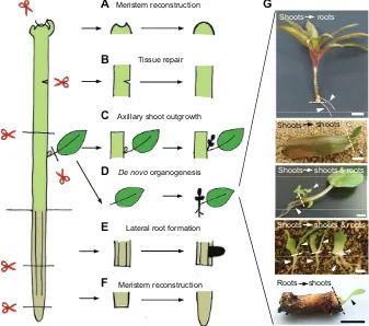
the presence of apical meristems. Upon loss of these apical meristems, apical dormancy is broken and axillary buds begin to grow (Shimizu-Sato and Mori, 2001) (Fig. 2). Similarly, when the whole root meristem is removed, new lateral and/or adventitious roots are formed from remaining roots and stems, respectively (Aloni et al., 2006; Bellini et al., 2014) (Fig. 2). In addition to these apical meristems, plant stems also display several types of tissue regeneration, including vascular reformation after debarking (Stobbe et al., 2002), tissue repair after partial incisions (Asahina et al., 2011) (Fig. 2) and vascular reconnection during grafting (Melnyk et al., 2015).
A unique feature of plant regeneration is the formation of new organs from cut sites (Hartmann et al., 2010) (Fig. 2), with the regeneration of roots from shoot cuttings seen in many different plant species. Some limited but phylogenetically diverse plant species, such as those in Crassulaceae (for exampleCrassulaspp., Echeveria spp., Kalanchoe spp. and Sedum spp.), Gesneriaceae (for example Saintpaulia ionantha, Sinningia speciosa and Streptocarpus xhybridus) and others (for exampleBegonia spp., Peperomiaspp. andSansevieria trifasciata), regenerate both shoots and roots from leaf cuttings (Fig. 2). In some bulbous plants, such as Hippeastrumspp.,Hyacinthusspp. andLiliumspp., detached bulb scales regenerate shoots and roots from cut sites (Fig. 2). Some plants, including the perennial herbaceous plantsPapaver orientale, Primula sieboldii and Taraxacum officinale, are capable of regenerating shoots from root cuttings (Fig. 2).
The regenerative capacity of plant cells can be enhancedin vitro when explants are cultured on nutrient media supplemented with plant hormones (Skoog and Miller, 1957; Murashige, 1974; George et al., 2008) (Fig. 3). Shoot explants of many ornamental plants are used for clonal propagation because multiple shoots can be formed from a shoot tip or stem node carrying a single bud (Fig. 3). Shoot or
Green algae
Liverworts
Mosses
Protoplast formation
Meristem formation
[image:2.612.54.297.56.233.2]Protonema formation
Fig. 1. Diverse forms of regeneration in green algae, liverworts and mosses.The unicellular green algaBryopsis plumosacan regenerate complete bodies through thede novoformation of protoplasts. The liverwort Marchantia polymorphacan regenerate new meristems (arrowhead) from the cut site. The mossPhyscomitrella patenscan regenerate new protonema cells (arrowheads) from leaf cuttings. Scissors indicate the cut site. Scale bars (from top to bottom): 10 µm, 1 mm, 500 µm. The top photograph is reprinted from Kim et al. (2001) with permission. The middle and bottom photographs were provided by Ryuichi Nishihama and Masaki Ishikawa, respectively.
Shoots
Meristem reconstruction
Tissue repair
Axillary shoot outgrowth
De novo organogenesis
Lateral root formation
Meristem reconstruction
roots
Shoots shoots
Shoots shoots & roots
Shoots shoots & roots
shoots Roots
A
E
B
C
D
F
G
Fig. 2. Diverse forms of regeneration in seed plants.Shoots and roots restore functional apical meristems when part of these meristems is removed (A,F). When they cannot repair existing meristems–for example when the whole meristems are excised–they develop new organs such as axillary shoots and lateral roots (C,E). Plant stems also repair tissues after partial incisions (B). Some plants undergode novoorganogenesis and develop new organs from cut sites (D,G). Photographs (G) show examples ofde novo organogenesis, from top to bottom: rootregeneration from a shoot cutting ofDracaenaspp.; shoot regeneration from a leaf cutting ofHaworthia spp.; shoot and root regeneration from a petiole of African violetSaintpaulia ionantha; shoot and root regeneration from detached bulb scales of lilyLilium longiflorum; and shoot regeneration from a root cutting of dandelionTaraxacum officinale. Scissors indicate the cut site and arrowheads mark regenerating organs. Scale bars: 5 mm, except 5 cm in the top photograph.
DEVEL
O
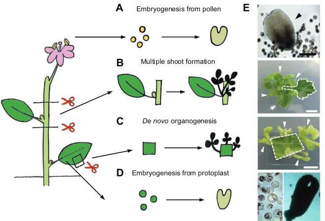
[image:2.612.48.385.438.736.2]root regeneration can also be inducedde novofrom various mature somatic tissues, and whole plants can be regenerated even from single protoplasts through de novo organogenesis or somatic embryogenesis (Takebe et al., 1971; Zhu et al., 1997; Chupeau et al., 2013) (Fig. 3). The regenerative capacity of pollen in some species, such asBrassica napus,Nicotiana tabacumandHordeum vulgare, is utilized to develop haploid plants via somatic embryogenesis (Maraschin et al., 2005) (Fig. 3), from which homozygous diploid plants can be chemically induced in a single generation. In many monocotyledonous cereals, including Oryza sativaandZea mays, mature somatic tissues are less regenerative, and thus zygotic embryos are commonly used forin vitroregeneration (Green and Phillips, 1975). For the remainder of this article we focus our attention onde novoorganogenesis and somatic embryogenesis in seed plants, from which we have gained substantial mechanistic insights into regeneration over the last few decades.
Cellular origins of regeneration
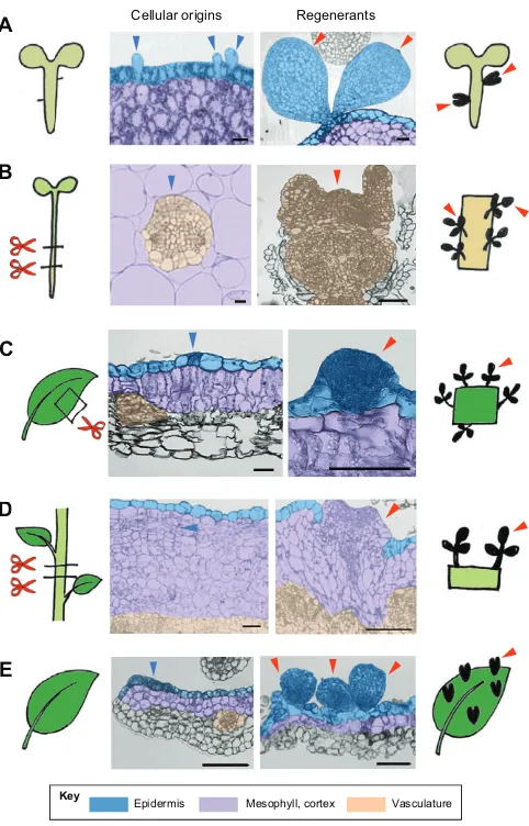
Plants possess at least two distinct cellular strategies to begin the process of regeneration. One is through the reactivation of relatively undifferentiated cells, and the other through the reprogramming of differentiated somatic cells. In both cases, regeneration relies on the phenomenon of cellular plasticity, which can be broadly defined as the ability to respecify cell fate. Plant cells in immature or juvenile bodies tend to have a high regenerative potential and, accordingly, those in zygotic embryos readily undergo somatic embryogenesis. A study by Kim et al. (2007) showed that even trichome initial cells on the embryonic hypocotyls of Tilia amurensis regenerate new somatic embryos (Fig. 4A), demonstrating that cellular fate can be redirected at an early stage of plant development.
During post-embryonic development, most somatic cells become differentiated and only a limited set of cell types remains competent for new tissue and organ formation. Plant roots, for example, have a cylinder of pericycle cells situated between the endodermis and stele, and they have the potential to produce new lateral roots (Beeckman and De Smet, 2014). These pericycle cells, together with neighboring vascular parenchyma and/or procambium cells, are often the source of root regeneration (Bellini et al., 2014) and in many cases they also serve as a primary source for shoot
regeneration and somatic embryogenesis in vitro (Atta et al., 2009; Che et al., 2007; Tarré et al., 2004) (Fig. 4B). On the other hand, plants can regenerate whole bodies from protoplasts or pollen (Fig. 3), and there are many other examples showing regeneration from differentiated cells. Shoot regeneration, for instance, initiates from mature leaf epidermal cells inChirita flavimaculata(Nakano et al., 2009) (Fig. 4C) or from stem cortex cells inChrysanthemum morifolium(Kaul et al., 1990) (Fig. 4D). Likewise, calli that give rise to somatic embryos have a cellular origin clearly distinct from pericycle or vascular cells in Medicago truncatula (Wang et al., 2011). It was reported that epidermal cell fate in developing leaves ofArabidopsis thaliana(Arabidopsis) can also be overwritten by overexpression of the RWP-RK protein RKD4 in order to initiate embryogenesis (Waki et al., 2011) (Fig. 4E). These observations indicate that‘youth’is not the prerequisite for plant regeneration and, at least underin vitroculture conditions, fully mature somatic cells can initiate regeneration.
Wound stress as a trigger for regeneration
Given that most naturally occurring regeneration starts at cut sites (Fig. 2), wound stimuli may in fact provide a primary inductive trigger for this phenomenon (Birnbaum and Sánchez Alvarado, 2008; Ikeuchi et al., 2013; Sugiyama, 2015). InArabidopsistissue culture where explants incubated on auxin-rich callus-inducing medium (CIM) were subsequently transferred on to cytokinin-rich shoot-inducing medium (SIM) (Valvekens et al., 1988), intact, uncut plants hardly regenerated shoots at all (Iwase et al., 2015), thus demonstrating a requirement for wound stimuli in initiating regeneration. Wounding induces numerous cellular responses, including the production of plant hormones (Ahkami et al., 2009), loss of cell-to-cell communication and disruption of long-distance signaling (Melnyk et al., 2015).
What exactly plants perceive as a wound signal and how they start regeneration are not well understood, but recent studies in Arabidopsisshowed that the AP2/ERF-type transcriptional regulator WOUND-INDUCED DEDIFFERENTIATION1 (WIND1) and its homologs WIND2, WIND3 and WIND4 are induced upon wounding and promote callus formation at cut sites (Iwase et al., 2011a,b). Importantly, callus induced by transient overexpression of De novo organogenesis
Embryogenesis from protoplast Embryogenesis from pollen
Multiple shoot formation
A E
B
C
[image:3.612.52.382.56.281.2]D
Fig. 3. Diverse forms of regeneration in thein vitro tissue culture environment.Regenerationin vitro can be induced from (A) pollen, (B) cut stems, (C) leaf cuttings and (D) protoplasts. (E) Photographs show, from top to bottom: regeneration of a somatic embryo (arrowhead) from isolated microspores of Chinese cabbageBrassica rapavar.pekinensis; regeneration of multiple shoots (arrowheads) from a node explant (dashed outline) carrying a single axillary bud of chrysanthemumChrysanthemum morifolium; regeneration of calli and shoots (arrowheads) from a leaf explant of tobaccoNicotiana tabacum(dashed outline); and regeneration of a somatic embryo from protoplasts isolated from calli of grapevineVitis vinifera. Scissors indicate the cut site. Scale bars: 5 mm, except 100 µm in the top photograph.
DEVEL
O
WIND1 regenerates shoots and roots when transferred to non-inducible media (Iwase et al., 2011a), suggesting that WIND1 can reprogram somatic cells to confer pluripotency. IntactArabidopsis plants ectopically expressing WIND1 regenerate shoots without wounding, and plants expressing the dominant-negative form of WIND1 display reduced efficiency of in vitroshoot regeneration (Iwase et al., 2015). These data support a role for WINDs as key
mediators of wound-induced cellular reprogramming (Fig. 5A). Consistent with this, sequential activation of WIND1 and an embryonic regulator, LEAFY COTYLEDON2 (LEC2), induces somatic embryogenesis at both cut and non-cut sites, whereas single activation ofLEC2permits embryogenesis only at cut sites (Iwase et al., 2015). Downstream genes regulated by WINDs are currently unknown, although WINDs have been implicated in the control of cytokinin signaling based on the observation that the repression of WIND activity abolishes the wound-induced cytokinin response (Iwase et al., 2011a,b). Further investigation of how wounding activates WIND gene expression and how, in turn, WINDs promote cellular reprogramming will be crucial to advance our molecular understanding of wound-induced regeneration. Precisely which cell types contribute to naturally occurring regeneration is also not fully established, and thus future studies should clarify the origin of cells in natural regeneration and determine whether it involves fate conversion of fully differentiated somatic cells and/or the activation of existing competent cells.
Molecular basis ofde novoshoot organogenesis
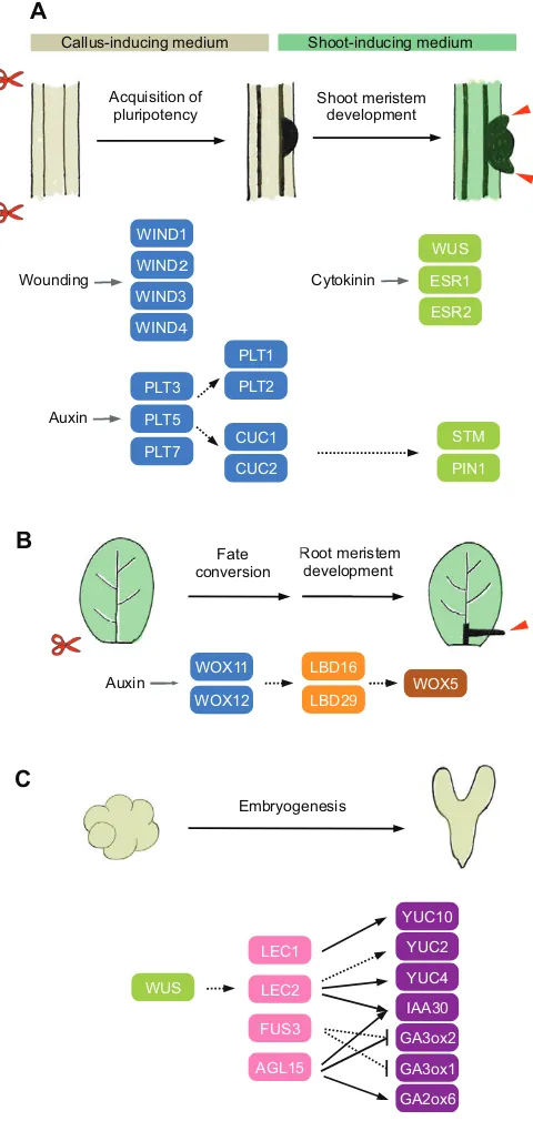
As in many other plant species,Arabidopsisexplants do not readily regenerate shoots, but incubation on CIM and SIM strongly enhances shoot regeneration from pericycle cells (Valvekens et al., 1988; Che et al., 2007; Atta et al., 2009). Recent histological and transcriptome analyses have revealed that CIM-induced callus resembles lateral root meristem, which is competent to regenerate shoots upon transfer to SIM (Che et al., 2007; Atta et al., 2009; Sugimoto et al., 2010; Duclercq et al., 2011). Induction of the AP2/ERF transcription factors PLETHORA3 (PLT3), PLT5 and PLT7 is among the earliest transcriptional responses induced by CIM, which in turn leads to the activation of the key root meristem regulatorsPLT1andPLT2to establish a pluripotent root meristem-like callus (Kareem et al., 2015). Expression of PLT3, PLT5 and PLT7 also induces the NAC family transcription factors CUPSHAPED COTYLEDON1 (CUC1) and CUC2, which are involved in shoot meristem initiation during zygotic embryogenesis (Kareem et al., 2015; Aida et al., 1997, 1999). Theplt3 plt5 plt7 triple mutants are defective in shoot regeneration, but dual overexpression of PLT1 and CUC2, but not their single expression, partially complements this phenotype, suggesting that PLT3, PLT5 and PLT7 promote both PLT1-mediated acquisition of pluripotency and CUC2-mediated initiation of shoot fate (Kareem et al., 2015) (Fig. 5A). Both CUC1 and CUC2 are uniformly expressed in CIM-induced callus (Gordon et al., 2007) and their presence is associated with cellular pluripotency in various experimental conditions (Cary et al., 2002; Daimon et al., 2003; Gordon et al., 2007; Motte et al., 2011).
Upon transfer to SIM, partitioning of the auxin and cytokinin responses in the pluripotent cell mass is thought to refine the shoot meristem fate.CUC2expression is restricted to the low cytokinin response domains, whereas the shoot meristem regulator WUSCHEL (WUS) is induced in the high cytokinin response domains (Gordon et al., 2007; Chatfield et al., 2013; Che et al., 2006) (Fig. 5A). TheCUC2-expressing cells continue to proliferate to form dome-like structures called promeristems, in which localized upregulation of other regulators such asPIN-FORMED1 (PIN1) andSHOOT MERISTEMLESS (STM) further defines the radial patterning of newly developing meristems and primordia initiation (Gordon et al., 2007). Similar to WUS, other AP2/ERF transcription factors, such as ENHANCER OF SHOOT REGENERATION1/DORNRÖSCHEN (ESR1/DRN) and ESR2/ DRN-LIKE (DRNL), which control embryonic patterning and E
Vasculature Mesophyll, cortex
Epidermis
A Cellular origins Regenerants
B
C
D D
[image:4.612.54.295.55.431.2]Key
Fig. 4. Cellular basis of plant regeneration.(A) Somatic embryogenesis from embryonic hypocotyls ofTilia amurensis. Transverse sections (middle panels) of hypocotyls show that epidermal trichome initial cells (blue arrowheads) give rise to new embryos (red arrowheads). (B)In vitroshoot regeneration from root or hypocotyl explants ofArabidopsis thaliana. Transverse sections of explants show that pericycle cells (blue arrowhead) give rise to regenerating shoots (red arrowheads). (C)In vitroshoot regeneration from leaf explants ofChirita flavimaculata. Transverse sections of leaf explants show that epidermal cells (blue arrowhead) give rise to regenerating shoots (red arrowhead). (D)In vitro shoot regeneration from stem explants ofChrysanthemum morifolium. Transverse sections of stem explants show that cortex cells (blue arrowhead) give rise to regenerating shoots (red arrowhead). (E) Somatic embryogenesis inArabidopsisleaves reprogrammed by overexpression of the embryonic regulator RKD4. Transverse sections of reprogrammed leaves show that epidermal cells (blue arrowhead) give rise to new embryos (red arrowheads). Scissors indicate the cut site. Scale bars: 100 µm. Photographs in A are modified from Kim et al. (2007) with permission from Oxford University Press; in B are modified from Atta et al. (2009) with permission from Blackwell Publishing; in C are modified from Nakano et al. (2009) with permission from Salvia Press; in D are modified from Kaul et al. (1990) with permission from Springer; in E are provided by Keiji Nakajima.
DEVEL
O
shoot formation (Kirch et al., 2003; Chandler et al., 2007), are also induced on SIM and stimulate shoot regeneration at least partially by enhancingCUC1 expression (Banno et al., 2001; Ikeda et al., 2006; Matsuo et al., 2009, 2011) (Fig. 5A).
Molecular basis ofde novoroot organogenesis
CulturingArabidopsisexplants on CIM and root-inducing medium (RIM) strongly enhances root regeneration from pericycle cells (Ozawa et al., 1998; Che et al., 2002). This is probably because CIM promotes the production of a root meristem-like pluripotent cell mass, which then becomes further specified by RIM to develop root meristems. Consistent with this, root explants, which already possess lateral root meristem primordia along their body axis,
regenerate roots from both cut and non-cut sites without pretreatment on CIM, whereas hypocotyl explants regenerate roots only from cut sites under these conditions (Ozawa et al., 1998). Pretreatment of hypocotyl explants on CIM allows root regeneration from non-cut sites (Ozawa et al., 1998), confirming the physiological role of CIM in endowing regenerative competence.
Some plant species naturally regenerate roots from cuttings and several plant hormones, including auxin and cytokinin, are known to control this process (da Costa et al., 2013; Bellini et al., 2014). A recent study by Liu et al. (2014) uncovered a novel molecular link connecting auxin accumulation at cut sites to the formation of new root meristems during regeneration. WhenArabidopsisleaves are detached, accumulation of auxin at cut sites induces the expression of two homeobox transcription factors, namely WUSCHEL RELATED HOMEOBOX11 (WOX11) and WOX12, in the procambium and surrounding parenchyma cells. Expression of these genes promotes the fate conversion from leaf procambium/ parenchyma cells to root founder cells (Fig. 5B). Both WOX11 and WOX12 subsequently participate in thede novoestablishment of root meristems, which further involves the expression ofLATERAL ORGAN BOUNDARIES DOMAIN16(LBD16),LBD29andWOX5 (Liu et al., 2014) (Fig. 5B). The LBDs and WOX5 are also involved in lateral root development, in which locally accumulated auxin promotes the formation of new root meristems from pericycle cells (Goh et al., 2012; Ditengou et al., 2008). These two pathways thus share some key regulators to facilitate the auxin-mediated establishment of root meristems. Of note, the induction of WOX11 in root regeneration requires auxin response elements (AuxREs) in its promoter, suggesting that some members of the AUXIN RESPONSE FACTOR (ARF) family directly activate WOX11expression in leaves (Liu et al., 2014).
Molecular basis of somatic embryogenesis
Some somatic cells in plants can restart embryogenesis in vitro when they are exposed to a wide range of severe abiotic stressors (Fehér, 2014). Somatic embryogenesis can be induced by salt, hypochlorite, osmotic pressure, heavy metal ions or high temperature in Daucus carota (Kiyosue et al., 1989, 1990; A
Callus-inducing medium Shoot-inducing medium
Shoot meristem development Acquisition of
pluripotency
STM
PIN1 WUS
ESR1
Cytokinin
WIND1
Wounding
PLT3
PLT5
PLT7
PLT1
PLT2
CUC1
CUC2 WIND
WIND3
WIND
LBD16
LBD29
WOX5 6
9
WOX5
Root meristem development
LBD16
LBD29
Root me develop
Auxin WOX11
WOX12
Fate conversion B
9 LBD29 WOX12
Embryogenesis C
LEC1
LEC2
FUS3
AGL15
IAA30 YUC10
YUC2
GA2ox6 GA3ox1 GA3ox2
WUS YUC4
ESR2
[image:5.612.53.293.59.564.2]Auxin
Fig. 5. A molecular framework for plant regeneration.(A) A schematic model showing howArabidopsisexplants regenerate shootsin vitro. Wounding inducesWIND1-4expression to promote the acquisition of pluripotency at cut sites. Culturing plant explants on auxin-rich callus-inducing medium upregulates the expression ofPLT3,PLT5andPLT7, which subsequently promotes the acquisition of pluripotency through the induction ofPLT1,PLT2, CUC1andCUC2. Upon transfer to cytokinin-rich shoot-inducing medium, the WUS,ESR1andESR2genes are induced, conferring cells with shoot fate. CUC2expression becomes spatially confined to promeristems, in which STM and PIN1 further regulate patterning and formation of the meristems (red arrowheads). (B) A schematic model showing how root regeneration is controlled inArabidopsisleaf explants. Accumulation of auxin at cut sites promotes the fate conversion from leaf procambium/parenchyma cells to root founder cells by activatingWOX11andWOX12expression. WOX11 and WOX12 subsequently induce the expression ofLBD16,LBD29, and then WOX5to initiate root meristem formation (red arrowhead). (C) A schematic model showing how indirect somatic embryogenesis is regulated in Arabidopsis. A gradient of auxin in the embryonic callus specifiesWUS expression to low auxin response domains. WUS subsequently induces expression ofLEC1,LEC2andFUS3, which together with AGL15 modulate the endogenous levels of auxin, GA and ABA to promote embryogenesis. Solid black lines indicate direct transcriptional regulation demonstrated by molecular evidence and dotted black lines indicate direct or indirect transcriptional regulation inferred from genetic evidence. Proteins that promote cellular competency are in blue; those that mediate shoot fate are in green, root fate in orange or brown, and embryonic fate in pink or purple.
DEVEL
O
Kamada et al., 1989, 1993, 1994), and similar stress-induced embryogenesis has also been reported inArabidopsis(Ikeda-Iwai et al., 2003). Many plant species also undergo somatic embryogenesis when they are cultured on auxin-containing medium and then transferred to auxin-free medium (Wernicke and Brettell, 1980; Lu et al., 1983; Ikeda-Iwai et al., 2002). Among several synthetic auxin-like substances, 2,4-dichlorophenoxyacetic acid (2,4-D) is the most effective inducer of somatic embryos in many plants, possibly because it triggers both auxin and stress responses simultaneously (Gliwicka et al., 2013). During indirect somatic embryogenesis, by which most somatic embryos are formed, high levels of auxin in the culture medium first promote cell proliferation and embryonic callus formation (Ikeda-Iwai et al., 2002). A key physiological event after the transfer to auxin-free medium is the de novo establishment of auxin gradients in the embryonic callus. This initiates a developmental program similar to zygotic embryogenesis, and is also guided by polarized auxin distribution (Liu et al., 1993; Su et al., 2009). These auxin gradients subsequently lead to the localization of WUS expression to low auxin response domains, marking the position of future shoot meristem formation (Su et al., 2009) (Fig. 5C).
Several key regulators of zygotic embryogenesis and seed development, including the CCAAT box-binding transcription factor LEAFY COTYLEDON1 (LEC1), the B3 domain transcription factors LEC2 and FUSCA3 (FUS3), and the MADS box transcription factor AGAMOUS-LIKE15 (AGL15), are subsequently induced during somatic embryogenesis and control several downstream physiological responses to promote embryonic development (Braybrook and Harada, 2008). A key consequence of this transcriptional reprogramming is the further refinement of auxin production and signaling. LEC1 induces theYUCCA10(YUC10) gene, which encodes an auxin biosynthesis enzyme, and LEC2 activates the YUC2and YUC4 genes (Junker et al., 2012; Stone et al., 2008). LEC2 and AGL15 promote the expression ofINDOLE ACETIC ACID INDUCIBLE30 (IAA30), a negative regulator of auxin signaling, to modulate the auxin-mediated signaling (Braybrook et al., 2006; Zheng et al., 2009). Previous studies also suggest that a low level of gibberellin (GA) relative to abscisic acid (ABA) favors embryogenesis. Consistent with this, AGL15 positively regulates GA2ox6, which encodes a GA degrading enzyme, and negatively regulates the GA biosynthesis gene GA3ox2, resulting in the reduced endogenous GA level (Wang et al., 2004; Zheng et al., 2009). In addition, FUS3 downregulates GA biosynthesis by repressingGA3ox1andGA3ox2, and induces ABA biosynthesis through as yet unknown mechanisms (Curaba et al., 2004; Gazzarrini et al., 2004; Kagaya et al., 2005).
Epigenetic control of regeneration
The regenerative capacity of plant cells is required only when they experience damage. Recent studies have shown that several epigenetic mechanisms actively suppress regenerative potential during normal development (Ikeuchi et al., 2015a). POLYCOMB REPRESSIVE COMPLEX2 (PRC2) is a chromatin modifier that maintains transcriptional repression through the deposition of histone H3 lysine 27 trimethylation (H3K27me3) (Holec and Berger, 2012). A recent study showed that PRC2 mutants initially develop wild-type-like roots with fully differentiated, endoreplicated root hair cells, but that they subsequently reprogram and develop callus and embryo-like structures (Ikeuchi et al., 2015b). The reprogramming regulator WIND3 and the embryonic regulatorLEC2are among the key targets repressed by PRC2 in this context and elevated expression of these genes
contributes to cellular reprogramming in PRC2 mutants. This study thus enforces the idea that highly differentiated cells still retain the capacity to undergo embryogenesis, and that this potential must be tightly regulated–in this case, epigenetically repressed by PRC2 to maintain the differentiated status. Intriguingly, many other key regulators of regeneration, such asWOX11,WOX5,WUSandSTM, are also under PRC2-mediated repression (Liu et al., 2014, 2011; He et al., 2012; Lafos et al., 2011). An important question is whether the cells carrying these repressive marks on regeneration regulators initiate regeneration in the wild-type context and, if so, how these repressions are relieved to allow regeneration to proceed in nature or in vitroconditions. PRC2 is also required for root regeneration from leaves (Liu et al., 2014), suggesting that repression of original cell fate might be another important aspect of regeneration.
Histone deacetylation, which is also implicated in transcriptional repression, might serve as another safeguard to prevent the untimely onset of somatic embryogenesis. Wild-typeArabidopsisplants treated with an inhibitor of histone deacetylases, trichostatin A (TSA), produce embryo-like structures from true leaves (Tanaka et al., 2008). Similarly, loss-of-function mutants of two histone deacetylases, HDA19 and HDA6, generate embryo-like structures in shoots (Tanaka et al., 2008). These phenotypes are associated with the ectopic expression of several key embryonic regulators, such asLEC1 andLEC2, and can be suppressed by introducing thelec1mutation (Tanaka et al., 2008). Interestingly, TSA in combination with heat treatment greatly enhances the efficiency of somatic embryogenesis fromBrassica napusmicrospores (Li et al., 2014). It is plausible, then, that heat stress and histone deacetylation converge on the upregulation of embryonic regulators to initiate the embryonic program.
Genetic studies inArabidopsisalso suggest the involvement of other epigenetic mechanisms in the control of organ regeneration. Mutations in DNA METHYLTRANSFERASE1 (MET1) enhance shoot regeneration on SIM and this phenotype is accompanied by the elevated expression of several MET1 targets, includingWUS(Li et al., 2011). As with many other regeneration regulators, theWUS locus is marked by several other epigenetic signatures and these marks are modified when WUSexpression is upregulated during shoot regeneration (Li et al., 2011). Uncovering the causal relationships between these epigenetic modifications and transcriptional changes will be an important task for future studies.
Natural variations that impact plant regeneration
Small genetic variations within the same species can cause dramatic differences in the regenerative response. Genetic variability has been utilized to identify novel factors that modulate the efficiency of regeneration in many plant species (Armstrong et al., 1992; Taguchi-Shiobara et al., 1997; Ben Amer et al., 1997; Flores Berrios et al., 2000; Mano and Komatsuda, 2002; Trujillo-Moya et al., 2011). A leucine-rich repeat receptor-like kinase, RECEPTOR-LIKE PROTEIN KINASE1(RPK1), for instance, was identified as a major quantitative trait locus (QTL) that affects shoot regeneration inArabidopsisaccessions (Motte et al., 2014). RPK1 is implicated in ABA signaling, and although this hormone has not been studied extensively in the context of regeneration it has been reported to influence shoot regeneration in several plant species (Ghasemi Bezdi et al., 2007; Hoang and Raldugina, 2012; Huang et al., 2012). The single-nucleotide polymorphism responsible for the genetic variation lies within a putative ligand-binding domain, and thus the identification of its ligands should help to reveal its molecular functions.
InOryza sativa, some varieties or even cultivars within the same variety exhibit markedly different shoot regeneration capabilities
DEVEL
O
(Nishimura et al., 2005). Map-based cloning using the low regeneration cultivar Koshihikari (Japonica) and high regeneration cultivar Kasalath (Indica) identified a ferredoxin-nitrite reductase as a major QTL causing variations in shoot regeneration. Further studies showed that the reductase activity positively correlated with regeneration capacity in several other Japonicacultivars as well (Nishimura et al., 2005). Furthermore, introduction of the ferredoxin-nitrite reductase gene from Kasalath improved shoot regeneration in Koshihikari (Nishimura et al., 2005). Ferredoxin-nitrite reductase is involved in the nitrogen assimilation pathway, and thus it might be that low reductase activity in Koshihikari results in an accumulation of nitrite, which might hinder shoot regeneration.
The Regeneration1 (Rg1) locus in tomato was originally identified as a natural variation responsible for highly efficient shoot regeneration in the wild relative Solanum peruvianum (Koornneef et al., 1993). It was later shown thatRg1increases the competency for both root and shoot regeneration, and that this response does not involve alterations in auxin sensitivity orCUC expression (Lombardi-Crestana et al., 2012). Interestingly, the tomatoDELLAmutantprocera(pro), which shows a constitutive response to GA, displays a low regeneration phenotype, andRg1 rescues these defects in an Rg1 pro double mutant. These data suggest an involvement of DELLA-mediated GA signaling in the control of shoot regeneration (Lombardi-Crestana et al., 2012).
Developmental constraints that impact plant regeneration The regenerative capacity of explants varies markedly with the condition of parental plants, generally declining as plants get older. Compromised root regeneration in aged trees is a serious problem in horticulture, limiting the clonal propagation of elite cultivars. Histological studies using woody species Castanea sativa and Quercus sp. showed that the exogenous application of auxin reactivates cell proliferation but not the formation of new root meristems in mature explants (Ballester et al., 1999; Vidal et al., 2003). A recent study using Pisum sativa suggested that the vegetative-to-reproductive transition is linked to the reduced root regenerative capacity and that this is caused by the loss of auxin responsiveness in reproductive shoots (Rasmussen et al., 2015). By contrast, application of auxin improves the root regeneration efficiency of Arabidopsis leaves from aged plants (Chen et al., 2014), suggesting that there are multiple physiological constraints imposed by aging.
Explants from juvenile plants regenerate shoots more effectively than those from mature plants (Dong and Jia, 1991; Baker and Bhatia, 1993; Becerra et al., 2004; Zhang et al., 2015). The decline in shoot regeneration capacity with aging is at least partly due to a reduced responsiveness to plant hormones. The microRNA miR156 has been shown to regulate the juvenile-to-adult phase transition in plants (Wu et al., 2009), and a recent study suggests that a decline of miR156 expression in old plants is responsible for reduced shoot regeneration (Zhang et al., 2015). This reduction in miR156 increases the level of its target SQUAMOSA PROMOTER BINDING-LIKE9 (SPL9). SPL9, in turn, inhibits the transcriptional activity of B-type ARABIDOPSIS RESPONSE REGULATOR proteins (ARRs), leading to the reduced responsiveness to cytokinin and hence compromised shoot regeneration (Zhang et al., 2015).
Environmental constraints that impact plant regeneration The regenerative capacity of plant explants is also influenced by various environmental conditions, such as nutrient composition,
gelling agents, pH, light and temperature (George et al., 2008). A well-documented environmental condition that influences plant regeneration is the exposure to light, but its impact on regeneration appears to be highly context dependent. Light is required for shoot regeneration in some plant species (Reuveni and Evenor, 2007) and can also trigger organ regeneration (Saitou et al., 1992; Sorin et al., 2006; Gutierrez et al., 2009). On the other hand, exposure to light can have an inhibitory effect on root or shoot regeneration in some contexts (Bellini et al., 2014; Nameth et al., 2013). A series of tissue culture experiments usingArabidopsiscotyledons showed that light exposure during the first hours after tissue excision is mostly deleterious to shoot regeneration, and keeping explants in darkness for as little as 2-6 h is sufficient to improve regeneration (Nameth et al., 2013).
Light exposure invokes several parallel signaling pathways, some of which cause oxidative damage due to the production of reactive oxygen species. At least two photoreceptors are implicated in the light response of shoot regeneration: the blue/UV-A light receptor CRYPTOCHROME1 (CRY1), which mediates the strong inhibition of shoot regeneration; and the far-red light receptor PHYTOCHROME A (PHYA), which protects explants against initial light inhibition (Nameth et al., 2013). A key regulator acting downstream of light signaling is the transcription factor ELONGATED HYPOCOTYL5 (HY5), which appears to protect explants against light exposure by inducing anthocyanin accumulation. Several accessions inArabidopsisdisplay different responses to light in shoot regeneration, and an interesting topic for future studies will be the cause of such genetic variations.
Conclusions and future perspectives
During regeneration, select intrinsic developmental programs are ectopically activated in response to external stimuli. These responses require context-dependent integration of developmental and environmental signals, leading to diverse strategies and efficiencies of regeneration. Given that regeneration originates from a relatively small population of cells in somatic tissues, it is important to identify these cell populations and to study how external stress can cause them to undergo changes in cell fate. During normal development, many central regulators of regeneration are epigenetically silenced to prevent inappropriate cellular reprogramming. A central challenge, therefore, is to understand how these repressions are overcome by external stimuli. Molecular genetic studies in Arabidopsis have provided substantial insight into how plants regenerate from relatively undifferentiated cells, but other plants that regenerate from differentiated cells may utilize distinct mechanisms. With rapid advances in next-generation sequencing and genome editing technologies, we should be able to investigate the molecular mechanisms of these currently underexplored forms of regeneration and carry out functional studies in non-model plants. The QTL analysis on accessions with differing regeneration efficiencies has proved an excellent complementary approach and the further identification of new QTLs should help us to uncover novel mechanisms of plant regeneration.
An important goal of plant regeneration research is to use our knowledge of basic biology to design new molecular tools to analyze and improve regeneration efficiencies in crops. Indeed, the ectopic expression of key regeneration regulators, such as WUS and WIND1, has already been shown to promote organ regeneration and/or somatic embryogenesis in various crops (Srinivasan et al., 2007; Arroyo-Herrera et al., 2008; Heidmann et al., 2011; Iwase et al., 2013, 2015; Florez et al., 2015). Expression profiles of key
DEVEL
O
regeneration regulators have also been used in crops to identify cultivars with high regeneration capacities (Malik et al., 2008). Further mechanistic understanding of plant regeneration should help us to advance the classic but not fully exploited field of tissue culture, with numerous downstream implications for both basic and applied biology.
Acknowledgements
We thank Masahiro Mii (Chiba University), Bart Rymen, Anna Franciosini and Hirofumi Harashima (RIKEN) for providing comments on the manuscript. We are grateful to Masaki Ishikawa (National Institute for Basic Biology), Ryuichi Nishihama (Kyoto University) and Keiji Nakajima (Nara Institute of Science and Technology) for providing photographs in Figs 1 and 3 and John L. Bowman (Monash University) for providing advice on liverwort regeneration.
Competing interests
The authors declare no competing or financial interests.
Funding
Research in the authors’laboratory on the topic of this Review is supported by a grant from the Scientific Technique Research Promotion Program for Agriculture, Forestry, Fisheries and Food Industry and grants from the Ministry of Education, Culture, Sports, Science, and Technology of Japan to M.I. [15K18564], A.I. [15K18565] and K.S. [26291064, 15H05961]. M.I. is a recipient of the RIKEN Special Postdoctoral Researcher Programme.
References
Ahkami, A. H., Lischewski, S., Haensch, K.-T., Porfirova, S., Hofmann, J., Rolletschek, H., Melzer, M., Franken, P., Hause, B., Druege, U. et al.(2009). Molecular physiology of adventitious root formation in Petunia hybrida cuttings: involvement of wound response and primary metabolism. New Phytol. 181, 613-625.
Aida, M., Ishida, T., Fukaki, H., Fujisawa, H. and Tasaka, M.(1997). Genes involved in organ separation in Arabidopsis: an analysis of the cup-shaped cotyledon mutant.Plant Cell9, 841-857.
Aida, M., Ishida, T. and Tasaka, M.(1999). Shoot apical meristem and cotyledon formation during Arabidopsis embryogenesis: interaction among the CUP-SHAPED COTYLEDON and SHOOT MERISTEMLESS genes. Development
126, 1563-1570.
Aloni, R., Aloni, E., Langhans, M. and Ullrich, C. I.(2006). Role of cytokinin and auxin in shaping root architecture: regulating vascular differentiation, lateral root initiation, root apical dominance and root gravitropism.Ann. Bot.97, 883-893.
Armstrong, C. L., Romero-Severson, J. and Hodges, T. K.(1992). Improved tissue culture response of an elite maize inbred through backcross breeding, and identification of chromosomal regions important for regeneration by RFLP analysis.Theor. Appl. Genet.84, 755-762.
Arroyo-Herrera, A., Gonzalez, A. K., Moo, R. C., Quiroz-Figueroa, F. R., Loyola-Vargas, V. M., Rodriguez-Zapata, L. C., Burgeff D’Hondt, C., Suárez-Solıs,́
V. M. and Castaño E.(2008). Expression of WUSCHEL in Coffea canephora causes ectopic morphogenesis and increases somatic embryogenesis.Plant Cell Tissue Org. Cult.94, 171-180.
Asahina, M., Azuma, K., Pitaksaringkarn, W., Yamazaki, T., Mitsuda, N., Ohme-Takagi, M., Yamaguchi, S., Kamiya, Y., Okada, K. et al. (2011). Spatially selective hormonal control of RAP2. 6L and ANAC071 transcription factors involved in tissue reunion in Arabidopsis. Proc. Natl. Acad. Sci. USA 108, 16128-16132.
Atta, R., Laurens, L., Boucheron-Dubuisson, E., Guivarc’h, A., Carnero, E., Giraudat-Pautot, V., Rech, P. and Chriqui, D. (2009). Pluripotency of Arabidopsis xylem pericycle underlies shoot regeneration from root and hypocotyl explants grown in vitro.Plant J.57, 626-644.
Baker, B. S. and Bhatia, S. K. (1993). Factors effecting adventitious shoot regeneration from leaf explants of quince (Cydonia oblonga).Plant Cell Tissue Org. Cult.35, 273-277.
Ballester, A., San-José, M. C., Vidal, N., Fernandez-Lorenzo, J. L. and Vieitez, A. M. (1999). Anatomical and biochemical events during in vitrorooting of microcuttings from juvenile and mature phases of chestnut.Ann. Bot.83, 619-629.
Banno, H., Ikeda, Y., Niu, Q. W. and Chua, N. H.(2001). Overexpression of Arabidopsis ESR1 induces initiation of shoot regeneration. Plant Cell 13, 2609-2618.
Becerra, D. C., Forero, A. P. and Góngora, G. A.(2004). Age and physiological condition of donor plants affect in vitro morphogenesis in leaf explants of Passiflora edulisf. flavicarpa.Plant Cell Tissue Org. Cult.79, 87-90.
Beeckman, T. and De Smet, I.(2014). Pericycle.Curr. Biol.24, R378-R379.
Bellini, C., Pacurar, D. I. and Perrone, I.(2014). Adventitious roots and lateral roots: similarities and differences.Annu. Rev. Plant Biol.65, 639-666.
Ben Amer, I. M., Worland, A. J., Korzun, V. and Börner, A.(1997). Genetic mapping of QTL controlling tissue-culture response on chromosome 2B of wheat (Triticum aestivum L.) in relation to major genes and RFLP markers.Theor. Appl.
Genet.94, 1047-1052.
Birnbaum, K. D. and Sánchez Alvarado, A.(2008). Slicing across kingdoms: regeneration in plants and animals.Cell132, 697-710.
Braybrook, S. A. and Harada, J. J.(2008). LECs go crazy in embryo development. Trends Plant Sci.13, 624-630.
Braybrook, S. A., Stone, S. L., Park, S., Bui, A. Q., Le, B. H., Fischer, R. L., Goldberg, R. B. and Harada, J. J.(2006). Genes directly regulated by LEAFY COTYLEDON2 provide insight into the control of embryo maturation and somatic embryogenesis.Proc. Natl. Acad. Sci. USA103, 3468-3473.
Cary, A. J., Che, P. and Howell, S. H.(2002). Developmental events and shoot apical meristem gene expression patterns during shoot development in Arabidopsis thaliana.Plant J.32, 867-877.
Chandler, J. W., Cole, M., Flier, A., Grewe, B. and Werr, W.(2007). The AP2 transcription factors DORNRÖSCHEN and DORNRÖSCHEN-LIKE redundantly control Arabidopsis embryo patterning via interaction with PHAVOLUTA.
Development134, 1653-1662.
Chatfield, S. P., Capron, R., Severino, A., Penttila, P.-A., Alfred, S., Nahal, H. and Provart, N. J.(2013). Incipient stem cell niche conversion in tissue culture: using a systems approach to probe early events in WUSCHEL-dependent conversion of lateral root primordia into shoot meristems.Plant J.73, 798-813.
Che, P., Gingerich, D. J., Lall, S. and Howell, S. H.(2002). Global and hormone-induced gene expression changes during shoot development in Arabidopsis. Plant Cell14, 2771-2785.
Che, P., Lall, S., Nettleton, D. and Howell, S. H.(2006). Gene expression programs during shoot, root, and callus development in Arabidopsis tissue culture.Plant Physiol.141, 620-637.
Che, P., Lall, S. and Howell, S. H.(2007). Developmental steps in acquiring competence for shoot development in Arabidopsis tissue culture.Planta226, 1183-1194.
Chen, X., Qu, Y., Sheng, L., Liu, J., Huang, H. and Xu, L.(2014). A simple method suitable to study de novo root organogenesis.Front. Plant Sci.5, 208.
Chupeau, M.-C., Granier, F., Pichon, O., Renou, J.-P., Gaudin, V. and Chupeau, Y.(2013). Characterization of the early events leading to totipotency in an Arabidopsis protoplast liquid culture by temporal transcript profiling.Plant Cell25, 2444-2463.
Curaba, J., Moritz, T., Blervaque, R., Parcy, F., Raz, V., Herzog, M. and Vachon, G. (2004). AtGA3ox2, a key gene responsible for bioactive gibberellin biosynthesis, is regulated during embryogenesis by LEAFY COTYLEDON2 and FUSCA3 in Arabidopsis.Plant Physiol.136, 3660-3669.
da Costa, C. T., de Almeida, M. R., Ruedell, C. M., Schwambach, J., Maraschin, F. S. and Fett-Neto, A. G.(2013). When stress and development go hand in hand: main hormonal controls of adventitious rooting in cuttings.Front. Plant Sci.4, 133.
Daimon, Y., Takabe, K. and Tasaka, M.(2003). The CUP-SHAPED COTYLEDON genes promote adventitious shoot formation on calli.Plant Cell Physiol. 44, 113-121.
Ditengou, F. A., Teale, W. D., Kochersperger, P., Flittner, K. A., Kneuper, I., van der Graaff, E., Nziengui, H., Pinosa, F., Li, X., Nitschke, R. et al.(2008). Mechanical induction of lateral root initiation inArabidopsis thaliana.Proc. Natl.
Acad. Sci. USA105, 18818-18823.
Dong, J.-Z. and Jia, S.-R. (1991). High efficiency plant regeneration from cotyledons of watermelon (Citrullus vulgarisSchrad).Plant Cell Rep.9, 559-562.
Duclercq, J., Sangwan-Norreel, B., Catterou, M. and Sangwan, R. S.(2011).De novoshoot organogenesis: from art to science.Trends Plant Sci.16, 597-606.
Fehér, A.(2014). Somatic embryogenesis–Stress-induced remodeling of plant cell fate.Biochem. Biophys. Acta1849, 385-402.
Flores Berrios, E., Gentzbittel, L., Kayyal, H., Alibert, G. and Sarrafi, A.(2000). AFLP mapping of QTLs for in vitro organogenesis traits using recombinant inbred lines in sunflower (Helianthus annuusL.).Theor. Appl. Genet.101, 1299-1306.
Florez, S. L., Erwin, R. L., Maximova, S. N., Guiltinan, M. J. and Curtis, W. R.
(2015). Enhanced somatic embryogenesis in Theobroma cacao using the homologous BABY BOOM transcription factor.BMC Plant Biol.15, 121.
Gazzarrini, S., Tsuchiya, Y., Lumba, S., Okamoto, M. and McCourt, P.(2004). The transcription factor FUSCA3 controls developmental timing in Arabidopsis through the hormones gibberellin and abscisic acid.Dev. Cell7, 373-385.
George, E. F., Hall, M. A. and de Klerk, G.-J.(2008).Plant Propagation by Tissue Culture, 3rd edn. Dordrecht: Springer.
Ghasemi Bezdi, K., Karlov, G. I. and Ahmadikhah, A.(2007). Effects of genotype, explant type and nutrient medium components on canola (Brassica napusL.) shoot in vitro organogenesis.Afr. J. Biotechnol.6, 861-867.
Gliwicka, M., Nowak, K., Balazadeh, S., Mueller-Roeber, B. and Gaj, M. D.
(2013). Extensive modulation of the transcription factor transcriptome during somatic embryogenesis inArabidopsis thaliana.PLoS ONE8, e69261.
Goh, T., Joi, S., Mimura, T. and Fukaki, H.(2012). The establishment of asymmetry in Arabidopsis lateral root founder cells is regulated by LBD16/ASL18 and related
LBD/ASL proteins.Development139, 883-893.
DEVEL
O
Gordon, S. P., Heisler, M. G., Reddy, G. V., Ohno, C., Das, P. and Meyerowitz, E. M.(2007). Pattern formation during de novo assembly of the Arabidopsis shoot meristem.Development134, 3539-3548.
Green, C. E. and Phillips, R. L.(1975). Plant regeneration from tissue cultures of maize.Crop Sci.15, 417-421.
Gutierrez, L., Bussell, J. D., Pacurar, D. I., Schwambach, J., Pacurar, M. and Bellini, C.(2009). Phenotypic plasticity of adventitious rooting in Arabidopsis is controlled by complex regulation of AUXIN RESPONSE FACTOR transcripts and microRNA abundance.Plant Cell21, 3119-3132.
Haberlandt, G.(1902). Culturversuche mit isolierten Pflanzenzellen.Sitzungsber Akad. Wiss. Wien. Math. Nat.111, 69-91.
Hartmann, H. T., Kester, D. E., Davies, F. T. and Geneve, R.(2010).Hartmann & Kester’s plant propagation: principles and practices, 8th edn. Englewood Cliffs, NJ, USA: Prentice-Hall.
He, C., Chen, X., Huang, H. and Xu, L.(2012). Reprogramming of H3K27me3 is critical for acquisition of pluripotency from cultured Arabidopsis tissues.PLoS
Genet.8, e1002911.
Heidmann, I., de Lange, B., Lambalk, J., Angenent, G. C. and Boutilier, K.
(2011). Efficient sweet pepper transformation mediated by the BABY BOOM transcription factor.Plant Cell Rep.30, 1107-1115.
Hoang, T. G. and Raldugina, G. N.(2012). Regeneration of transgenic plants expressing the GFP gene from rape cotyledonary and leaf explants: effects of the genotype and ABA.Russ. J. Plant Physiol.59, 406-412.
Holec, S. and Berger, F. (2012). Polycomb group complexes mediate developmental transitions in plants.Plant Physiol.158, 35-43.
Huang, W.-L., Lee, C.-H. and Chen, Y.-R.(2012). Levels of endogenous abscisic acid and indole-3-acetic acid influence shoot organogenesis in callus cultures of rice subjected to osmotic stress.Plant Cell Tissue Org. Cult.108, 257-263.
Ikeda, Y., Banno, H., Niu, Q.-W., Howell, S. H. and Chua, N.-H.(2006). The ENHANCER OF SHOOT REGENERATION 2 gene in Arabidopsis regulates CUP-SHAPED COTYLEDON 1 at the transcriptional level and controls cotyledon development.Plant Cell Physiol.47, 1443-1456.
Ikeda-Iwai, M., Satoh, S. and Kamada, H.(2002). Establishment of a reproducible tissue culture system for the induction of Arabidopsis somatic embryos.J. Exp. Bot.53, 1575-1580.
Ikeda-Iwai, M., Umehara, M., Satoh, S. and Kamada, H.(2003). Stress-induced somatic embryogenesis in vegetative tissues ofArabidopsis thaliana.Plant J.34, 107-114.
Ikeuchi, M., Sugimoto, K. and Iwase, A.(2013). Plant callus: mechanisms of induction and repression.Plant Cell25, 3159-3173.
Ikeuchi, M., Iwase, A. and Sugimoto, K.(2015a). Control of plant cell differentiation by histone modification and DNA methylation.Curr. Opin. Plant Biol.28, 60-67.
Ikeuchi, M., Iwase, A., Rymen, B., Harashima, H. Shibata, M., Ohnuma, M., Breuer, C., Morao, A. K., de Lucas, M., de Veylder, L. et al.(2015b). PRC2 represses dedifferentiation of mature somatic cells in Arabidopsis.Nat. Plants1, 15089.
Ishikawa, M., Murata, T., Sato, Y., Nishiyama, T., Hiwatashi, Y., Imai, A., Kimura, M., Sugimoto, N., Akita, A., Oguri, Y. et al. (2011). Physcomitrella cyclin-dependent kinase A links cell cycle reactivation to other cellular changes during reprogramming of leaf cells.Plant Cell23, 2924-2938.
Iwase, A., Mitsuda, N., Koyama, T., Hiratsu, K., Kojima, M., Arai, T., Inoue, Y., Seki, M., Sakakibara, H., Sugimoto, K. et al. (2011a). The AP2/ERF transcription factor WIND1 controls cell dedifferentiation in Arabidopsis.Curr. Biol.21, 508-514.
Iwase, A., Ohme-Takagi, M. and Sugimoto, K.(2011b). WIND1: a key molecular switch for plant cell dedifferentiation.Plant Signal. Behav.6, 1943-1945.
Iwase, A., Mitsuda, N., Ikeuchi, M., Ohnuma, M., Koizuka, C., Kawamoto, K., Imamura, J., Ezura, H. and Sugimoto, K.(2013). Arabidopsis WIND1 induces callus formation in rapeseed, tomato, and tobacco. Plant Signal. Behav.8, e27432.
Iwase, A., Mita, K., Nonaka, S., Ikeuchi, M., Koizuka, C., Ohnuma, M., Ezura, H., Imamura, J. and Sugimoto, K.(2015). WIND1-based acquisition of regeneration competency in Arabidopsis and rapeseed.J. Plant Res.128, 389-397.
Junker, A., Mönke, G., Rutten, T., Keilwagen, J., Seifert, M., Thi, T. M. N., Renou, J.-P., Balzergue, S., Viehöver, P., Hähnel, U. et al.(2012). Elongation-related functions of LEAFY COTYLEDON1 during the development of Arabidopsis thaliana.Plant J.71, 427-442.
Kagaya, Y., Okuda, R., Ban, A., Toyoshima, R., Tsutsumida, K., Usui, H., Yamamoto, A. and Hattori, T.(2005). Indirect ABA-dependent regulation of seed storage protein genes by FUSCA3 transcription factor in Arabidopsis.Plant Cell Physiol.46, 300-311.
Kamada, H., Kobayashi, K., Kiyosue, T. and Harada, H.(1989). Stress induced somatic embryogenesis in carrot and its application to synthetic seed production. In Vitro Cell. Dev. Biol.25, 1163-1166.
Kamada, H., Ishikawa, K., Saga, H. and Harada, H.(1993). Induction of somatic embryogenesis in carrot by osmotic stress.Plant Tiss. Cult. Lett.10, 38-44.
Kamada, H., Tachikawa, Y., Saitou, T. and Harada, H. (1994). Heat stress induction of carrot somatic embryogenesis.Plant Tiss. Cult. Lett.11, 229-232.
Kareem, A., Durgaprasad, K., Sugimoto, K., Du, Y., Pulianmackal, A. J., Trivedi, Z. B., Abhayadev, P. V., Pinon, V., Meyerowitz, E. M., Ben Scheres et al.
(2015). PLETHORA genes control regeneration by a two-step mechanism.Curr. Biol.25, 1017-1030.
Kaul, V., Miller, R. M., Hutchinson, J. F. and Richards, D. (1990). Shoot regeneration from stem and leaf explants of Dendranthema grandiflora Tzvelev (syn. Chrysanthemum morifolium Ramat.).Plant Cell Tissue Org. Cult.21, 21-30.
Kim, G. H., Klotchkova, T. A. and Kang, Y. M.(2001). Life without a cell membrane: regeneration of protoplasts from disintegrated cells of the marine green alga Bryopsis plumosa.J. Cell Sci.114, 2009-2014.
Kim, T. D., Lee, B. S., Kim, T. S. and Choi, Y. E.(2007). Developmental plasticity of glandular trichomes into somatic embryogenesis inTilia amurensis.Ann. Bot.
100, 177-183.
Kirch, T., Simon, R., Grünewald, M. and Werr, W.(2003). The DORNRÖSCHEN/ ENHANCER OF SHOOT REGENERATION1 gene of Arabidopsis acts in the control of meristem cell fate and lateral organ development. Plant Cell 15, 694-705.
Kiyosue, T., Kamada, H. and Harada, H. (1989). Induction of somatic embryogenesis by salt stress in carrot.Plant Tiss. Cult. Lett.6, 162-164.
Kiyosue, T., Takano, K., Kamada, H. and Harada, H.(1990). Induction of somatic embryogenesis in carrot by heavy metal ions.Can. J. Bot.68, 2301-2303.
Koornneef, M., Bade, J., Hanhart, C. Horsman, K. Schel, J., Soppe, W., Verkerk, R. and Zabel, P.(1993). Characterization and mapping of a gene controlling shoot regeneration in tomato.Plant J.3, 131-141.
Lafos, M., Kroll, P., Hohenstatt, M. L., Thorpe, F. L., Clarenz, O. and Schubert, D.
(2011). Dynamic regulation of H3K27 trimethylation during Arabidopsis differentiation.PLoS Genet.7, e1002040.
Li, W., Liu, H., Cheng, Z. J., Su, Y. H., Han, H. N., Zhang, Y. and Zhang, X. S.
(2011). DNA methylation and histone modifications regulate de novo shoot regeneration in Arabidopsis by modulating WUSCHEL expression and auxin signaling.PLoS Genet.7, e1002243.
Li, H., Soriano, M., Cordewener, J., Muiño, J. M., Riksen, T., Fukuoka, H., Angenent, G. C. and Boutilier, K.(2014). The histone deacetylase inhibitor trichostatin a promotes totipotency in the male gametophyte.Plant Cell 26, 195-209.
Liu, C., Xu, Z. and Chua, N. H.(1993). Auxin polar transport is essential for the establishment of bilateral symmetry during early plant embryogenesis.Plant Cell
5, 621-630.
Liu, X., Kim, Y. J., Muller, R., Yumul, R. E., Liu, C., Pan, Y., Cao, X., Goodrich, J. and Chen, X.(2011). AGAMOUS terminates floral stem cell maintenance in Arabidopsis by directly repressing WUSCHEL through recruitment of polycomb group proteins.Plant Cell23, 3654-3670.
Liu, J., Sheng, L., Xu, Y., Li, J., Yang, Z., Huang, H. and Xu, L.(2014). WOX11 and 12 are involved in the first-step cell fate transition during de novo root organogenesis in Arabidopsis.Plant Cell26, 1081-1093.
Lombardi-Crestana, S., da Silva Azevedo, M., Silva, G. F. F. E., Pino, L. E., Appezzato-da-Glória, B., Figueira, A., Nogueira, F. T. S. and Peres, L. E. P.
(2012). The tomato (Solanum lycopersicum cv. Micro-Tom) natural genetic variation Rg1 and the DELLA mutant procera control the competence necessary to form adventitious roots and shoots.J. Exp. Bot.63, 5689-5703.
Lu, C., Vasil, V. and Vasil, I. K. (1983). Improved efficiency of somatic embryogenesis and plant regeneration in tissue cultures of maize (Zea mays L.).Theor. Appl. Genet.66, 285-289.
Malik, M. R., Wang, F., Dirpaul, J. M., Zhou, N., Hammerlindl, J., Keller, W., Abrams, S. R., Ferrie, A. M. R. and Krochko, J. E.(2008). Isolation of an embryogenic line from non-embryogenicBrassica napuscv. Westar through microspore embryogenesis.J. Exp. Bot.59, 2857-2873.
Mano, Y. and Komatsuda, T.(2002). Identification of QTLs controlling tissue-culture traits in barley (Hordeum vulgare L.).Theor. Appl. Genet.105, 708-715.
Maraschin, S. F., de Priester, W., Spaink, H. P. and Wang, M.(2005). Androgenic switch: an example of plant embryogenesis from the male gametophyte perspective.J. Exp. Bot.56, 1711-1726.
Matsuo, N., Mase, H., Makino, M., Takahashi, H. and Banno, H. (2009). Identification of ENHANCER OF SHOOT REGENERATION 1-upregulated genes during in vitro shoot regeneration.Plant Biotechnol.26, 385-393.
Matsuo, N., Makino, M. and Banno, H.(2011). Arabidopsis ENHANCER OF SHOOT REGENERATION (ESR)1 and ESR2 regulate in vitro shoot regeneration and their expressions are differentially regulated.Plant Sci.181, 39-46.
Melnyk, C. W. and Meyerowitz, E. M.(2015). Plant grafting. Curr. Biol. 25, R183-R188.
Melnyk, C. W., Schuster, C., Leyser, O. and Meyerowitz, E. M.(2015). A developmental framework for graft formation and vascular reconnection in Arabidopsis thaliana.Curr. Biol.25, 1306-1318.
Motte, H., Verstraeten, I., Werbrouck, S. and Geelen, D.(2011). CUC2 as an early marker for regeneration competence in Arabidopsis root explants. J. Plant Physiol.168, 1598-1601.
Motte, H., Vercauteren, A., Depuydt, S., Landschoot, S., Geelen, D., Werbrouck, S., Goormachtig, S., Vuylsteke, M. and Vereecke, D.(2014). Combining linkage and association mapping identifies RECEPTOR-LIKE PROTEIN KINASE1 as an essential Arabidopsis shoot regeneration gene.Proc. Natl Acad. Sci. USA111,
8305-8310.
DEVEL
O
Murashige, T.(1974). Plant propagation through tissue cultures.Annu. Rev. Plant Physiol.25, 135-166.
Nakano, M., Takagi, H., Sugawara, S. and Saito, T.(2009). Adventitious shoot regeneration and micropropagation of Chirita flavimaculata W.T. Wang, C. eburnea Hance, and C. speciosa Kurz.Propag. Ornam. Plants.9, 216-222.
Nameth, B., Dinka, S. J., Chatfield, S. P., Morris, A., English, J., Lewis, D., Oro, R. and Raizada, M. N. (2013). The shoot regeneration capacity of excised Arabidopsis cotyledons is established during the initial hours after injury and is modulated by a complex genetic network of light signalling.Plant Cell Environ.36, 68-86.
Necker, J. D.(1774). Physiologia Muscorum. Mannheim: C. F. Schwan.
Nishihama, R., Ishizaki, K., Hosaka, M., Matsuda, Y., Kubota, A. and Kohchi, T.
(2015). Phytochrome-mediated regulation of cell division and growth during regeneration and sporeling development in the liverwort Marchantia polymorpha. J. Plant Res.128, 407-421.
Nishimura, A., Ashikari, M., Lin, S., Takashi, T., Angeles, E. R., Yamamoto, T. and Matsuoka, M.(2005). Isolation of a rice regeneration quantitative trait loci gene and its application to transformation systems.Proc. Natl Acad. Sci. USA102, 11940-11944.
Ozawa, S., Yasutani, I., Fukuda, H., Komamine, A. and Sugiyama, M.(1998). Organogenic responses in tissue culture of srd mutants of Arabidopsis thaliana.
Development125, 135-142.
Pulianmackal, A. J., Kareem, A. V. K., Durgaprasad, K., Trivedi, Z. B. and Prasad, K.(2014). Competence and regulatory interactions during regeneration in plants.Front. Plant Sci.5, 142.
Rasmussen, A., Hosseini, S. A., Hajirezaei, M.-R., Druege, U. and Geelen, D.
(2015). Adventitious rooting declines with the vegetative to reproductive switch and involves a changed auxin homeostasis.J. Exp. Bot.66, 1437-1452.
Reinhardt, D., Frenz, M., Mandel, T. and Kuhlemeier, C.(2003). Microsurgical and laser ablation analysis of interactions between the zones and layers of the tomato shoot apical meristem.Development130, 4073-4083.
Reuveni, M. and Evenor, D.(2007). On the effect of light on shoot regeneration in petunia.Plant Cell Tissue Org. Cult.89, 49-54.
Saitou, T., Kamada, H. and Harada, H. (1992). Light requirement for shoot regeneration in horseradish hairy roots.Plant Physiol.99, 1336-1341.
Sena, G., Wang, X., Liu, H.-Y., Hofhuis, H. and Birnbaum, K. D.(2009). Organ regeneration does not require a functional stem cell niche in plants.Nature457, 1150-1153.
Shimizu-Sato, S. and Mori, H.(2001). Control of outgrowth and dormancy in axillary buds.Plant Physiol.127, 1405-1413.
Skoog, F. and Miller, C. O. (1957). Chemical regulation of growth and organ formation in plant tissues cultured in vitro.Symp. Soc. Exp. Biol. 54, 118-130.
Sorin, C., Negroni, L., Balliau, T., Corti, H., Jacquemot, M.-P., Davanture, M., Sandberg, G., Zivy, M. and Bellini, C.(2006). Proteomic analysis of different mutant genotypes of Arabidopsis led to the identification of 11 proteins correlating with adventitious root development.Plant Physiol.140, 349-364.
Srinivasan, C., Liu, Z., Heidmann, I., Supena, E. D. J., Fukuoka, H., Joosen, R., Lambalk, J., Angenent, G., Scorza, R., Custers, J. B. M. et al. (2007). Heterologous expression of the BABY BOOM AP2/ERF transcription factor enhances the regeneration capacity of tobacco (Nicotiana tabacumL.).Planta
225, 341.
Steward, F. C., Mapes, M. O. and Mears, K. (1958). Growth and organized development of cultured cells. II. Organization in cultures grown from freely suspended cells.Am. J. Bot.45, 705-708.
Stobbe, H., Schmitt, U., Eckstein, D. and Dujesiefken, D.(2002). Developmental stages and fine structure of surface callus formed after debarking of living lime trees (Tilia sp.).Ann. Bot.89, 773-782.
Stone, S. L., Braybrook, S. A., Paula, S. L., Kwong, L. W., Meuser, J., Pelletier, J., Hsieh, T.-F., Fischer, R. L., Goldberg, R. B. and Harada, J. J.(2008).
Arabidopsis LEAFY COTYLEDON2 induces maturation traits and auxin activity: implications for somatic embryogenesis. Proc. Natl Acad. Sci. USA 105, 3151-3156.
Su, Y. H., Zhao, X. Y., Liu, Y. B., Zhang, C. L., O’Neill, S. D. and Zhang, X. S.
(2009). Auxin-induced WUS expression is essential for embryonic stem cell renewal during somatic embryogenesis in Arabidopsis.Plant J.59, 448-460.
Sugimoto, K., Jiao, Y. and Meyerowitz, E. M.(2010). Arabidopsis regeneration from multiple tissues occurs via a root development pathway.Dev. Cell 18, 463-471.
Sugiyama, M.(2015). Historical review of research on plant cell dedifferentiation. J. Plant Res.128, 349-359.
Taguchi-Shiobara, F., Lin, S. Y., Tanno, K., Komatsuda, T. Yano, M., Sasaki, T. and Oka, S. (1997). Mapping quantitative trait loci associated with regeneration ability of seed callus in rice,Oryza sativaL.Theor. Appl. Genet.
95, 828-833.
Takebe, I., Labib, G. and Melchers, G.(1971). Regeneration of whole plants from isolated mesophyll protoplasts of tobacco.Naturwissenschaften58, 318-320.
Tanaka, M., Kikuchi, A. and Kamada, H. (2008). The Arabidopsis histone deacetylases HDA6 and HDA19 contribute to the repression of embryonic properties after germination.Plant Physiol.146, 149-161.
Tarré, E., Magioli, C., Margis-Pinheiro, M., Sachetto-Martins, G., Mansur, E. and Santiago-Fernandes, L. D. R.(2004). In vitro somatic embryogenesis and adventitious root initiation have a common origin in eggplant (Solanum melongena L.).Braz. J. Bot.27, 79-84.
Trujillo-Moya, C., Gisbert, C., Vilanova, S. and Nuez, F.(2011). Localization of QTLs for in vitro plant regeneration in tomato.BMC Plant Biol.11, 140.
Valvekens, D., Montagu, M. V. and Van Lijsebettens, M.(1988). Agrobacterium tumefaciens-mediated transformation of Arabidopsis thaliana root explants by using kanamycin selection.Proc. Natl. Acad. Sci. USA85, 5536-5540.
Vidal, N., Arellano, G., San-José, M. C., Vieitez, A. M. and Ballester, A.(2003). Developmental stages during the rooting ofin-vitro-cultured Quercus robur shoots from material of juvenile and mature origin.Tree Physiol.23, 1247-1254.
Vöchting, H.(1885). Über die regeneration der Marchantieen.Jahrb. Wiss. Bot.16, 367-414; Taf. XII-XV.
Waki, T., Hiki, T., Watanabe, R., Hashimoto, T. and Nakajima, K.(2011). The Arabidopsis RWP-RK protein RKD4 triggers gene expression and pattern formation in early embryogenesis.Curr. Biol.21, 1277-1281.
Wang, H., Caruso, L. V., Downie, A. B. and Perry, S. E.(2004). The embryo MADS domain protein AGAMOUS-Like 15 directly regulates expression of a gene encoding an enzyme involved in gibberellin metabolism.Plant Cell16, 1206-1219.
Wang, X.-D., Nolan, K. E., Irwanto, R. R., Sheahan, M. B. and Rose, R. J.(2011). Ontogeny of embryogenic callus in Medicago truncatula: the fate of the pluripotent and totipotent stem cells.Ann. Bot.107, 599-609.
Wernicke, W. and Brettell, R.(1980). Somatic embryogenesis fromSorghum bicolorleaves.Nature287, 138-139.
Wu, G., Park, M. Y., Conway, S. R., Wang, J.-W., Weigel, D. and Poethig, R. S.
(2009). The sequential action of miR156 and miR172 regulates developmental timing in Arabidopsis.Cell138, 750-759.
Zhang, T.-Q., Lian, H., Tang, H., Dolezal, K., Zhou, C.-M., Yu, S., Chen, J.-H., Chen, Q., Liu, H., Ljung, K. et al.(2015). An intrinsic microRNA timer regulates progressive decline in shoot regenerative capacity in plants.Plant Cell27, 349-360.
Zheng, Y., Ren, N., Wang, H., Stromberg, A. J. and Perry, S. E.(2009). Global identification of targets of the Arabidopsis MADS domain protein AGAMOUS-Like15.Plant Cell21, 2563-2577.
Zhu, Y.-M., Hoshino, Y., Nakano, M., Takahashi, E. and Mii, M.(1997). Highly efficient system of plant regeneration from protoplasts of grapevine (Vitis vinifera L.) through somatic embryogenesis by using embryogenic callus culture and activated charcoal.Plant Sci.123, 151-157.