R E S E A R C H A R T I C L E
Open Access
Soluble biglycan: a potential mediator of
cartilage degradation in osteoarthritis
Goncalo Barreto
1*, Antti Soininen
3, Pekka Ylinen
3, Jerker Sandelin
3, Yrjö T. Konttinen
1,3ˆ
, Dan C. Nordström
1and Kari K. Eklund
2Abstract
Background:Soluble biglycan (sBGN) and soluble decorin (sDCN), are two closely related essential components of extracellular matrix which both have been shown to possess proinflammatory properties. We studied whether sBGN or sDCN were present in synovial fluid (SF) of osteoarthritis (OA) or rheumatoid arthritis (RA) patients and studied sBGN or sDCN potential role in the degradation of OA cartilage.
Methods:SF obtained from meniscus tear, OA, and RA patients were analysed for sBGN and sDCN using enzyme-linked immunosorbent assays. OA chondrocytes and cartilage explants were stimulated for 48 h with 5μg/ml sBGN or 1μg/ml lipopolysaccharide. Messenger RNA (mRNA) levels of Toll-like receptors (TLRs), proteinases and cartilage matrix molecules were determined using quantitative real-time polymerase chain reaction. Protein levels of matrix metalloproteinases (MMPs) and cytokines were measured using Luminex xMap technology. Production of nitric oxide (NO), release of proteoglycans and soluble collagen were measured from conditioned culture media using biochemical assays. OA cartilage explant proteoglycans were stained for Safranin O and quantified using image analysis. TLR4 activation by sBGN and sDCN was studied in engineered HEK-293 cells with TLR4 signalling genes inserted together with a reporter gene. Results:sBGN was found in meniscus tear SF (14 ± 2 ng/ml), OA SF (582 ± 307 ng/ml) and RA SF (1191 ± 482 ng/ml). Low levels of sDCN could also be detected in SF of meniscus tear (51 ± 4) ng/ml, OA (52 ± 3 ng/ml), and RA (49 ± 4 ng/ml). Stimulation of chondrocytes with sBGN increased significantly the mRNA and protein expression of catabolic MMPs, including MMP1, MMP9 and MMP13, and of inflammatory cytokines interleukin (IL)-6 and IL-8, whereas the expression of anabolic markers aggrecan and collagen type II was decreased. sBGN induced release of proteoglycans, collagen and NO from chondrocytes and cartilage explants. The catabolic response in explants was dependent of OA cartilage degradation stage. The mechanism of action of sBGN was mainly mediated through the TLR4-nuclear factor-κB pathway.
Conclusions:High levels of sBGN was found in advanced OA and RA SF. sBGN activates chondrocytes mainly via TLR4, which results in net loss of cartilage. Thus, sBGN can be a mediator of OA cartilage degradation and also a potential biomarker for arthritis.
Keywords:Biglycan, Decorin, Osteoarthritis, Toll-like receptors, Chondrocytes, Cartilage, Innate immune response
* Correspondence:goncalo.barreto@helsinki.fi
ˆDeceased
1Department of Internal Medicine and Rehabilitation, University of Helsinki and Helsinki University (Central) Hospital, Biomedicum 1, PO Box 63, FIN-00290 Helsinki, Finland
Full list of author information is available at the end of the article
Background
Osteoarthritis (OA) is among the top five leading causes of disability worldwide [1]. To date, no effective long-term disease-modifying treatment for OA is available. At the root of the OA burden lies poor comprehension of the molecular pathophysiology of pre-symptomatic and clinically symptomatic OA.
Cartilage degeneration, a hallmark of OA, has its onset in wear, tear and mechanical injuries of the joint cartil-age. Although trauma is perhaps the first causal event in OA, the host inflammatory response plays an important role in the pathogenesis OA and inflammation is be-lieved to be the major driver to symptomatic OA [2, 3].
Cells of the cartilage, known as chondrocytes, maintain cartilage tissue homeostasis. They control the structural assembly of extracellular matrix (ECM) and regulate de-structive, remodelling and reparative processes [4]. In OA, cartilage-resident chondrocytes produce proteinases such as aggrecanases (ADAMTS-4, ADAMTS-5), matrix me-talloproteinases (e.g., MMP-13 and MMP-9) and cathep-sin K, and the anabolic synthesis of structural molecular components aggrecan and collagen type II (Col-II) are compromised [5]. Such a metabolic imbalance results in a failure of cartilage homeostasis and pathological cartilage destruction as well as eventual loss of cartilage.
ECM molecules have been thought to function as purely structural matrix-bound components, but evi-dence is emerging that they can also function as soluble ligands for pattern-recognising danger signalling recep-tors, mainly for Toll-like receptors (TLRs) [6–8]. Several of the ECM molecules found in OA joint and synovial fluid (SF) have been shown to trigger catabolic responses in TLR-equipped OA chondrocytes [9–11]. Biglycan (BGN) and decorin (DCN) are two small, closely related structural proteoglycans with leucine-rich repeats (small leucine-rich proteoglycan, SLRP) [12, 13]. The soluble forms of BGN (sBGN) or (sDCN) released from the car-tilage matrix as a result of tissue injury could potentially function as an endogenous danger signal [14]. For ex-ample, sBGN, the size of which is approximately 95 kDa, has been shown trigger TLR4/TLR2 signalling in human aortic valve and in acute ischemic kidney, as well as to activate the NLRP3 inflammasome in macrophages [15–17]. sDCN has been shown to activate TLR4/ TLR2 signalling in macrophages [18].
We hypothesised that BGN is a prototypic example of a major essential cartilage ECM component. Upon re-lease from stressed cartilage, it might activate TLR-mediated catabolic signalling pathways [19, 20]. In fact, several studies have shown that de novo BGN and DCN synthesis is increased in OA, that BGN and DCN frag-ments are produced and released by ADAMTSs and MMPs, and that OA SF also contains BGN and DCN autoantibodies [21–25]. However, to our knowledge, no
studies have addressed the concentration of intact sBGN and sDCN in OA or rheumatoid arthritis (RA) SF or in-cluded examination of the eventual direct role of these proteoglycans in chondrocyte-mediated cartilage degrad-ation in OA.
Our aim was to study whether sBGN and sDCN can be found in SF from arthritic joints and whether sBGN and sDCN can contribute to cartilage degradation by ac-tivating the TLR-mediated catabolic signalling in primary OA chondrocytes and OA tissue explants.
Methods
Tissue acquisition, cartilage explant culture and primary chondrocyte culture
Patient recruitment and participation and sample collec-tion were approved by the Helsinki and Uusimaa Hospital District ethics committee (Dnr_59/13/03/02/2013) and hospital board of directors (9310/407), and signed in-formed consent was obtained from the patients. Samples were collected from patients with OA who underwent total knee arthroplasty (TKA) (N = 12). Half of the pa-tients were women. The mean age of the papa-tients was 63.5 years (range 53–73). Tibial plateau containing cartil-age and subchondral bone was cut off to provide space for the tibial component of the TKA implant. Tibial plateau samples were collected into sterile sample containers con-taining cold phosphate-buffered saline solution (PBS) and processed within 2 h.
Before tissue samples were harvested, the macroscopic appearance of the visible pathological changes in differ-ent sampling areas was recorded according to the arthroscopic grading scale of Société Française d’ Ar-throscopie [26]. Explant samples were collected from two different types of areas: grade I OA samples with no macroscopically visible lesions (which represents early OA cartilage degradation stages) or grade IV samples with full-thickness osteochondral lesions [27]. This grading of the samples makes possible a stratified analysis based on the degree of OA cartilage, from very mild to severe. Cylin-drical full-thickness cartilage explants were obtained from graded tibial plateau areas using a 4-mm circular punch biopsy blade (Kai Medical, Oyana, Japan). Explants were cultured in Gibco Dulbecco’s modified Eagle’s medium (DMEM)/F-12 (Life Technologies, Carlsbad, CA, USA) supplemented with 10 % foetal calf serum (FCS) for 24 h before stimulation.
in 2.5 mg/ml pronase and 250 mg/ml collagenase P (Roche, Basel, Switzerland), with a PBS wash in between, for 60 minutes and overnight, respectively, under slow agi-tation at 37 °C. The resulting cell suspension was filtered through a 70-μm nylon cell strainer, centrifuged, washed twice with PBS and seeded at 1.5 × 10⁵/cm2 in Gibco DMEM/F-12 supplemented with 10 % FCS, 100 U/ml penicillin, 100 U/ml streptomycin and 0.25μg/ml ampho-tericin B (Life Technologies). Cells were balanced for 3–4 days before stimulation. For cryopreservation, cells were suspended in FCS containing 10 % (vol/vol) dimethyl sulphoxide and frozen in an isopropanol container for 24 h at−80 °C before storage in liquid nitrogen.
Arthritis group definitions for synovial fluid collection
Early osteoarthritis
SF samples were collected from ten patients scheduled for arthroscopic surgery. The main indication for surgery was a suspicion of a meniscal tear. Patients with inflammatory arthritis, severe arthritis, corticosteroid injection within 6 weeks, blood dyscrasias or active malignancy were ex-cluded. The prior use of nonsteroidal anti-inflammatory drugs was not considered an exclusion criterion. The diag-nosis of early arthritis was made during arthroscopy and based on the presence of minimal visible chondral lesions.
Advanced osteoarthritis
SF samples were collected from ten patients scheduled for elective total knee replacement for management of primary idiopathic OA. The exclusion criteria were simi-lar to those for patients with early OA.
Rheumatoid arthritis
SF samples were collected from ten patients with RA diag-nosed fulfilling the American College of Rheumatology/ European League Against Rheumatism classification cri-teria for RA [28].
Synovial fluid sample collection
Patients with early OA, advanced OA and RA were, de-pending on the schedule of the surgeon, randomly se-lected for collection of SF samples via needle aspiration before opening of the joint. Blood-contaminated samples were excluded. SF maintained at 4 °C was aliquoted within 2 h into sterile Eppendorf tubes, centrifuged at 1200g for 5 minutes at room temperature to separate solid debris and cells from the fluid phase, snap-frozen in liquid nitrogen and stored at −80 °C. When first thawed, SF was treated with a protease inhibitor cocktail (Roche Diagnostics, Meylan, France).
Enzyme-linked immunosorbent assay
SF was measured for intact-only sBGN and DCN molecules using a specific sandwich enzyme-linked immunosorbent
assay (ELISA) (Uscn Life Science Inc., Hubei, China, and BioVendor Laboratorní medicína, Brno, Czech Republic, respectively) for detection of intact sBGN and sDCN mole-cules. BGN and DCN fragments are not detected by the immunoassays. Absorbance was measured at 450 nm, as well as 450 nm and 630 nm, for sBGN and sDCN immuno-assays, respectively. All measurements were performed in duplicates.
SEAP NF-κB activity assays
TLR4 activity was measured using a cell-based assay according to the manufacturer’s instructions (InvivoGen, San Diego, CA, USA). HEK-hTLR4 cells express TLR4
andMD-2/CD14co-receptor genes of human origin and
contain the secreted embryonic alkaline phosphatase (SEAP) reporter gene for monitoring nuclear factor (NF)-κB activation. Upon interaction with the appropri-ate ligand, TLR4 transduces a signal which results in NF-κB activation and the expression of secreted alkaline phosphatase (AP), which can be detected by using detec-tion medium (QUANTI-Blue, a medium used for the de-tection and quantification of secreted AP; InvivoGen) and measured with a spectrophotometer.
Briefly HEK-hTLR4 cells were cultured at a density of 2.5 × 10⁴cells in 96-well plates and maintained in complete DMEM with selective antibiotics, as described in the manu-facturer’s instructions (InvivoGen). Cells were then stimu-lated with 5 μg/ml BGN (Sigma-Aldrich, St. Louis, MO, USA), 8 μg/ml sDCN (R&D Systems, Minneapolis, MN, USA) or 1μg/ml lipopolysaccharide (LPS) (positive control) for 24 h, and activation of TLR4 and NF-κB signalling was analysed by measuring SEAP from conditioned culture medium samples according to the manufacturer’s instruc-tions (InvivoGen).
Stimulation of chondrocyte and cartilage explant cultures
to primary chondrocyte cultures 30 minutes before sBGN or LPS. CLI-095, also known as TAK-242, specifically sup-presses TLR4 signalling mediated by the intracellular do-main of the receptor. Further receptor-blocking studies were performed with the addition of 20 μg/ml TLR2 (Santa Cruz Biotechnology, Santa Cruz, CA, USA) or 20μg/ml TLR4 (Santa Cruz Biotechnology) for 1 h before the addition of sBGN at 5μg/ml.
The optimal concentration for sBGN stimulation was determined in 24- and 48-h pilot experiments using 0.05, 0.5 and 5 μg/ml of sBGN, followed by analysis of
MMP13, CTSK (cathepsin K, cat K), IL6 (interleukin-6,
or IL-6) andCOL2A1(collagen type IIαchain 1, or Col-IIA) gene messenger RNA (mRNA) copy numbers rela-tive to the TATA box-binding protein (TBP) house-keeper, by using quantitative real-time polymerase chain reaction (qRT-PCR). Cartilage explant cultures were stimulated using the same conditions.
Real-time polymerase chain reactions
Total RNA was isolated from cartilage explants by using the RNAqueous® kit (Thermo Scientific) and from primary chondrocytes by using the RNeasy® Mini Kit (Qiagen, Val-encia, CA, USA). Relative quantification of the gene levels was performed by comparing the cycle threshold (Ct)
values of the different genes, correcting for glyceraldehyde 3-phosphate dehydrogenase (GAPDH) and TBP content (ΔCt) and for non-stimulated conditions (ΔΔCt) and
fi-nally expressed as fold changes. Primer sequences are pro-vided in Table 1.
Protein measurements using Luminex xMAP® technology
Measurement of protein levels was done using xMAP® technology (Luminex, Austin, TX, USA). To determine protein levels of MMPs (MMP-1, MMP-3, MMP-9 and MMP-13) as well as cytokines and chemokines (6, IL-8) in chondrocyte culture supernatant, xMAP® technology on the Bio-Plex 200® system (Bio-Rad Laboratories, Hercules, CA, USA) was used in combination with multi-plex MMP/cytokine kits (ProcartaPlex; eBioscience; San Diego, CA, USA). Protein levels were measured in 25μl of culture medium (diluted 1:2).
Measurement of nitric oxide
Nitric oxide (NO) was measured from conditioned culture medium samples using a nitrate/nitrite colorimetric assay kit (Cayman Chemical, Ann Arbor, MI, USA). Nitrate was converted to nitrite by adding nitrate reductase and its co-factor, followed by the addition of Griess reagent to de-velop a deep purple colour. The absorbance was measured at 544 nm using a plate reader (Chameleon; Hidex, Turku, Finland).
Measurement of soluble sulphated glycosaminoglycans and soluble collagen type II
[image:4.595.67.540.464.733.2]Soluble sulphated glycosaminoglycan (sGAG) standards, blanks and conditioned culture medium samples were mixed with the sGAG-binding Blyscan Dye Reagent® for 30 minutes, followed by separation of the GAG–dye com-plex by centrifugation and dissociation of the dye from the pellet (Biocolor Ltd., Carrickfergus, UK). Soluble Col-II
Table 1Primer pairs used for real-time polymerase chain reactions
Gene Protein name Primer sequence
ADAMTS4 A disintegrin and metalloproteinase with thrombospondin motifs 4 Forward: 5′-AAT CCT GTC AGC TTG GTG GT-3′ Reverse: 3′-CTT GGA GTT GTC ATG GAG CA-5′
ADAMTS5 A disintegrin and metalloproteinase with thrombospondin motifs 5 Forward: 5′-CTT CAC TGT GGC TCA CGA AA-3′ Reverse: 3′-AAT GTC AGG TTG CAC TGC TG-5′
MMP13 Matrix metalloproteinase 13 (MMP-13) Forward: 5′-CTA TGG TCC AGG AGA TGA AG-3′ Reverse: 3′-AGA GTC TTG CCT GTA TCC TC-5′
CTSK Cathepsin K Forward: 5′-ACC CAA CAG GCA AGG CAG CTAA-3′
Reverse: 3′-GCA ATG CCA CAGG CGT TGT TCT-3′
MMP9 Matrix metalloproteinase 9 (MMP-9) Forward: 5′-TTC TCC AGA AGC AAC TGT CC-3′ Reverse: 3′-CGG CAA GTC TTC CGA GTA GT-5′
ACAN Aggrecan Forward: 5′-TGG TGA TGA TCT GGC ACGA-3′
Reverse: 3′-TCT GCG TTT GTA GGT GGTG-5′
COL2A1 Collagen, type II, alpha 1 Forward: 5′-GAG TCA AGG GTG ATC GTG GT-3′ Forward: 5′-AAG CAC CTT GGT CTC CAG AA-5′
TBP TATA box-binding protein Forward: 5′-GAA GAA CAA TCC AGA CTA GCA GCA-3′ Reverse: 3′-CCT TAT AGG GAA CTT CAC ATC ACAG-5′
was similarly mixed with the collagen-binding Sircol Dye Reagent® (Biocolor Ltd.) for 30 minutes, followed by trifugation, pelleting, washes with salt wash reagent, cen-trifugation and dissociation of the collagen–dye complex using an alkali reagent vortexing wash. Absorbance was measured at 595 and 540 nm, respectively, using a plate reader (Chameleon).
Safranin O staining
Control and sBGN-stimulated cartilage explants were fixed in neutral buffered 10 % formalin for 2 weeks be-fore dehydration in ethanol series, clearing in xylene and embedding in paraffin. Deparaffinised tissue sections were stained with Fast Green dye, rinsed in 1 % acetic acid and stained in 0.1 % Safranin O before evaluation using a Nikon LV-DIA Base microscope (Nikon Instru-ments, Tokyo, Japan) with a motorized XY staging sys-tem (OptiScan III; Prior Scientific, Rockland, MA, USA) connected to a DS-Fi1 digital camera (Nikon Instru-ments) using NIS-Elements Basic Research analysis (Nikon Instruments). Image analysis was done using ImageJ software (National Institutes of Health, Bethesda, MD, USA).
Statistical analysis
Differences between groups were tested using the Mann–Whitney U test, the Wilcoxon signed-rank test or Student’s t test, as appropriate. Effects of covariates were analysed by multiple linear regression. All statis-tical analysis were performed using IBM SPSS version 21 software (IBM, Armonk, NY, USA).pValues <0.05 were considered significant.
Results
sBGN and sDCN can be found in synovial fluid obtained from patients with OA or RA
BGN and DCN are key components of the cartilage matrix. We hypothesised that some intact BGN or DCN could be released from matrix into SF in OA or RA and that in soluble form they could act as a proinflammatory stimulus. Indeed, sandwich ELISA disclosed clearly higher
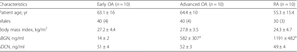
sBGN levels in advanced OA (582 ± 307 ng/ml) than the levels found in patients with meniscus tear who had very early OA (14 ± 2 ng/ml) (Table 2). The highest levels of sBGN were observed in SF obtained from patients with RA (1191 ± 482 ng/ml) (Table 2). In contrast to sBCN, the SF levels of sDCN were low, and no differences were ob-served between early OA (51 ± 4 ng/ml), OA (52 ± 3 ng/ml) and RA: (49 ± 4 ng/ml). Covariates (age, sex and body mass index) tested with multiple linear regression models were not associated with SF sBGN or SF sDCN levels.
sBGN upregulates catabolic factors in OA chondrocytes
As high concentrations of intact sBGN were found in the SF of OA and RA patients, we studied whether sBGN could have effects on the cartilage metabolism. First, we studied the effect of sBGN on the expression of catabolic cartilage factors. In primary monolayer chon-drocytes, sBGN significantly increased gene expression
of ADAMTS4, ADAMTS5, MMP13, CTSK and MMP9
almost as efficiently as LPS (Fig. 1a). Studies of cartilage explant tissues obtained from patients with varying de-grees of OA severity revealed that the response to sBGN was dependent on the grade of cartilage degradation. sBGN induced pronounced expression of catabolic factors in grade I OA cartilage, whereas a more modest increase of catabolic factors was observed in highly degenerated grade IV OA cartilage (Fig. 1b).
To further assess whether the studied gene mRNA transcripts were also translated into proteins, we used Luminex xMAP® technology. In cell culture superna-tants, the levels of MMP-1 and MMP-3 were substan-tially upregulated (1.6- and 3.6-fold, respectively) by sBGN compared with unstimulated controls (Fig. 2). Compared with positive control LPS, the effects of sBGN on protein levels for MMP-1, MMP-3, MMP-9 and MMP-13 were 3 %, 50 %, 82 % and 21 %, respect-ively, of that of LPS (LPS data not shown).
[image:5.595.58.539.622.707.2]Levels of proinflammatory cytokines IL-6 and IL-8 were also upregulated by sBGN compared with unstimu-lated control (1.4- and 2.2-fold, respectively) (Fig. 2). Compared with LPS, the effects of sBGN on cytokine
Table 2Intact sBGN and sDCN concentration levels in synovial fluid obtained from knee joints of early osteoarthritis, advanced osteoarthritis and rheumatoid arthritis patients
Characteristics Early OA (n= 10) Advanced OA (n= 10) RA (n= 10)
Patient age, yr 63.1 ± 16 64.4 ± 10 55.3 ± 15.4
Males 40 (4) 40 (4) 30 (3)
Body mass index, kg/m2 27.2 ± 4.4 27.8 ± 3.5 24.3 ± 4.7
sBGN, ng/ml 14 ± 2 582 ± 307a 1191 ± 482a
sDCN, ng/ml 51 ± 4 52 ± 3 49 ± 4
OAosteoarthritis,RArheumatoid arthritis,sBGNsoluble biglycan,sDCNsoluble decorin Data are presented as mean ± SD or count (%)
a
*
**
*
*
*
**
*
*
*
sBGN(5 µg/ml) LPS (1 µg/ml)
catabolic factors m
RNA
- primary chondrocytes
A
ADAMTS 4
ADAMTS 5
MMP 13
CTSK MMP
9
Folds of changes
0 2 4 6 8 10 12 14 16
Control
Grade I OA Grade IV OA
catabolic factors m
RNA
- explants
B
ADAMTS 4
ADAMTS 5
MMP 13
CTSK MMP
9
Folds of change
***
0 5 10 15 20 30 65 85 95
25 75
##
*
**
##
-5
Control
Fig. 1Effect of soluble biglycan (sBGN) on the expression of proteinases associated with matrix remodelling and cartilage degeneration in
osteoarthritis (OA).aIn primary chondrocytes, sBGN increased all proteinases examined. Lipopolysaccharide (LPS) was used as a positive control.
bIn cartilage explants, sBGN increasedADAMTS5,MMP13,CTSKandMMP9messenger RNA (mRNA) only in mild grade I OA and not in severe grade IV OA. The results are expressed as fold changes (mean ± SD) relative to TATA box-binding protein (TBP) housekeeper and compared with non-stimulated controls. Samples were run as technical duplicates, and each experiment was done using samples obtained from at least six different donors (biological replicates). *p< 0.05, **p< 0.01, ***p< 0.001 vs. non-stimulated controls;##p< 0.01 for pairwise comparisons between mild grade I OA and severe grade IV OA.ADAMTSa disintegrin and metalloproteinase with thrombospondin type 1 motif (aggrecanase),MMPmatrix metalloproteinase,
[image:6.595.61.538.84.625.2]levels for IL-6 and IL-8 were 18 % and 25 %, respectively (LPS data not shown).
sBGN impairs the synthesis of major cartilage matrix components in OA chondrocytes
To determine the effect of sBGN on overall cartilage turnover, we studied the effect of sBGN on the anabolic functions of OA chondrocytes. In primary OA chondro-cytes, sBGN significantly inhibited aggrecan and Col-II mRNA expression (Fig. 3a). The studies on cartilage ex-plants revealed that sBGN significantly decreased Col-II mRNA expression in highly degenerated grade IV OA but had no significant effect on the expression in grade I OA cartilage (Fig. 3b).
sBGN increases proteoglycan and collagen release from cartilage explants
Release of proteoglycans and soluble collagen from cartilage explants into cell culture media was determined to confirm that sBGN can also induce the degradation of OA cartilage by catabolic chondrocytes in situ. sBGN induced prominent release of sGAG in grade I OA explants, whereas sBGN-induced collagen release was observed only in grade IV OA cartilage (Fig. 4). Proteoglycan release was further con-firmed by Safranin O staining in sBGN-treated cartilage ex-plants. sBGN decreased the total quantity of proteoglycan in explants by approximately 50 % (Fig. 5).
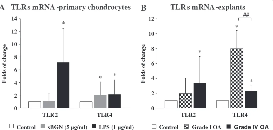
sBGN upregulates TLR4 expression in chondrocytes and in cartilage tissue explants
To study the mechanism of the effects of sBGN on cartilage metabolism, the effects of sBGN on TLR expression and function were first studied. Expression ofTLR2andTLR4 mRNA was observed in primary monolayer chondrocytes and cartilage explants of all patients with OA. In accord-ance with earlier studies, basal TLR2/TLR4 levels were slightly (1.3-fold) higher in grade IV OA than in grade I OA [30]. In primary monolayer chondrocyte cultures, sBGN significantly increased TLR4 expression but did not have any significant effect onTLR2mRNA expression (Fig. 6a).
Studies of cartilage explants revealed that the TLR re-sponse to sBGN is dependent on the grade of cartilage degradation. In grade I OA, the sBGN-induced increase of TLR4 mRNA expression was 352 % than that ob-served in grade IV OA cartilage (Fig. 6b). In contrast, there was no difference in sBGN-induced TLR2 mRNA expression between grade I and grade IV OA (Fig. 6b). Blocking TLR4 by using a low molecular weight inhibi-tor of TLR4 signalling (CLI095) abrogated the sBGN-induced increase ofTLR4mRNA expression (Fig. 7a).
Catabolic effect of sBGN in articular chondrocytes is mediated via TLR4
Inducible nitric oxide synthase (iNOS) is activated via TLR2/TLR4/NF-κB pathway activation. To further evaluate
0 100 200 300 400 500
MMP-3
0 100 200 300 400 500 600
IL-8 0
1 3 4 6 7
IL-6 0
3 6 9 12 15 18
MMP-1
19 20 21 22 23 24
MMP-9
0 750 1500 2250 3000 3750
MMP-13
ng/ml
pg/ml
ng/ml ng/ml
ng/ml ng/ml
MMPs and cytokines protein levels
in chondrocyte cell culture supernatant
CONTROL
BGN
Fig. 2Effect of soluble biglycan (sBGN) on the expression of matrix metalloproteinase (MMP)-1, MMP-3, MMP-9 and MMP-13 as well as interleukin
[image:7.595.56.540.88.341.2]Soluble collagen - explants
B
sBGN(5 µg/ml)
re
le
a
se
d
so
lubl
ec
ol
lage
n
(µ
g
/m
g
ti
ss
u
e)
Grade IV OA Grade I OA
0.0 2.0 4.0 6.0 8.0 10.0 12.0
Control
Sulphated glycosaminoglycans - explants
A
rel
ease
d
(µ
g
/mg
ti
ss
u
e)
GA
G
0.0 0.5 1.0 1.5 2.0 2.5 3.0 3.5
Grade IV OA Grade I OA
*
*
#
Fig. 4Effects of soluble biglycan (sBGN) on release of glycosaminoglycans (GAGs) and soluble collagen from cartilage explants compared with
non-stimulated controls.aIn grade I osteoarthritis (OA), sBGN increased GAG release.bIn grade IV OA, sBGN increased collagen release. The results are presented as mean ± SD. Samples were run as technical duplicates, and each experiment was done using samples from at least six different donors (biological replicates). *p< 0.05 vs. non-stimulated controls;##p< 0.01 for pairwise comparisons between mild grade I OA and
severe grade IV OA
matrix molecules m
RNA
-
primary
chondrocytes
A
-2.5 -2 -1.5 -1 -0.5
0 ACAN COL2A1
*
*
Fo
ld
s
o
f
chang
e
matrix molecules m
RNA
- explants
Fo
ld
s
o
f
chang
e
B
-35 -20 -5 10 25 40 55 70
CO L2A
1
AC AN
*
*
*
##
sBGN(5 µg/ml) LPS (1 µg/ml)
Control Control Grade I OA Grade IV OA
Fig. 3Effects of soluble biglycan (sBGN) on the expression of aggrecan and collagen type II (Col-II).aIn primary chondrocytes, sBGN decreased
aggrecan and Col-II. Lipopolysaccharide (LPS) was used as a positive control.bIn cartilage explants, sBGN did not affect aggrecan (ACAN) or
Col-II(COL2A1)in grade I osteoarthritis (OA) but decreased Col-II(COL2A1)in grade IV OA, with a different response in mild and severe OA. The results
are expressed as fold changes (mean ± SD) relative to the TATA box-binding protein (TBP) housekeeper and compared with non-stimulated con-trols. Samples were run as technical duplicates, and each experiment was done using samples obtained from at least six different donors (biological replicates). *p< 0.05 vs. non-stimulated controls;##p< 0.01 for pairwise comparisons between mild grade I OA and severe grade IV OA. mRNA
[image:8.595.65.539.90.324.2] [image:8.595.58.539.411.675.2]the role of TLRs in sBGN-induced signalling, we studied the effect of sBGN on NO release from primary OA chon-drocytes. In primary OA chondrocyte cultures, sBGN sig-nificantly increased NO production (Fig. 8a). In cartilage explant tissues, sBGN increased NO production slightly more in grade I OA than in grade IV OA (Fig. 8b). Blocking of TLR4 signalling with TLR4-neutralising antibody or syn-thetic inhibitor CLI-095 significantly reduced NO produc-tion to levels observed in non-stimulated controls (Fig. 8c), suggesting that the effect of sBGN is mediated via TLR4. To exclude the effect of contaminating LPS, polymyxin B was added, which had no effect on the sBGN-induced production of NO. In contrast, proteinase K reduced NO production to levels of non-stimulated controls, fur-ther supporting the finding that the observed effect of sBGN is not caused by LPS contamination. Moreover, blocking of TLR4 signalling abrogated the increase in
mRNA expression of the catabolic factors induced by both sBGN and LPS while also restoring to normal the levels of the mRNA expression of the key anabolic factors ACAN
andCOL2A1 (Fig. 7b and c). In contrast to TLR4,
TLR2-neutralising antibody had no significant effect on NO production. Together, these above suggest that the ef-fect of sBGN on cartilage metabolism is mediated via activation of TLR4 and that TLR2 has either no or only a minor role.
To confirm that sBGN signalling is mediated through TLR4/NF-κB pathway, we studied the activation of NF-κB after stimulation with sBGN. This was done using engineered HEK-hTLR4 cells, which can be stimulated only through activation of the TLR4 receptors. These cells have been stably transfected with SEAP plasmid containing NF-κB response elements, and the activation of NF-κB can be monitored by measuring activity of
Fig. 5Effects of soluble biglycan (sBGN) on proteoglycan content in osteoarthritis (OA) cartilage explants from five different patients.aSafranin O
[image:9.595.58.541.88.440.2]SEAP. Stimulation of HEK-hTLR4 cells with sBGN caused a substantial increase of SEAP, while inhibition of TLR4 signalling with TLR4 inhibitor CLI-095 pre-vented the increase of SEAP levels. sDCN and LPS caused a similar increase of SEAP levels. Proteinase K abrogated SEAP upregulation by sBGN and sDCN, while polymyxin B did not alter the response, thus ensuring that the studied effect was not caused by endotoxin con-tamination (Fig. 8d).
Discussion
BGN and DCN are two major non-collagenous SLRP products of chondrocytes which are deposited into ar-ticular cartilage matrix [13]. In this study, we show, for the first time to our knowledge, that intact sBGN is present in SF obtained from patients with meniscus tear lesions and early OA, advanced OA or RA, with the highest concentrations found in advanced OA and RA. DCN could also be found in SF, but, in contrast to BGN, only low levels were detected and no differences be-tween early OA, advanced OA and RA were observed. Thus, matrix-embedded chondrocytes as well as other cells in joints are exposed to fluid phase sBGN and sDCN. Intact sBGN and sDCN, but not their fragments, have been shown to engage proinflammatory responses [31, 32]. Therefore, while BGN fragments are known to be released into the SF of patients with OA, their
presence is merely a reflection of disease activity. Hence, they have been proposed to be solely a diagnostic bio-marker. The observed increased levels of intact sBGN are also in line with the observed upregulation of mRNA and protein levels of sBGN in human OA cartilage tissue and also in cartilage obtained using a sheep meniscec-tomy animal model of OA. Although DCN mRNA and protein levels have been shown to increase in OA, this was not reflected in the level of DCN in SF [22, 25, 33]. Interestingly, SF of OA and RA patients has been shown to contain immunoglobulin G autoantibodies against BGN, DCN and several other cartilage matrix molecules, suggesting their release from matrix and subsequent local loss of immunological tolerance [34]. Immune complex formation leading to complement and Fcγ re-ceptor activation plays a role in RA, as well as in OA ac-cording to recent studies [35–37]. The present finding of intact sBGN in SF, together with the recognition of sBGN as an endogenous damage-associated molecular pattern-type ligand for TLR2/TLR4 and for P2X7/4,
pro-vides a third mechanism of relevance for autoinflamma-tion, co-stimulation and autoimmunity.
Chondrocytes of OA cartilage are equipped with a full palette of TLRs, including TLR2 and TLR4, and the pro-portion of TLR+ chondrocytes increases with progres-sion of OA [38]. The results of the present study show that sBGN regulates TLR4 expression to an extent
A
TLR
s m
RNA
-primary chondrocytes
F
o
ld
s
o
f
chang
e
6
12
2 4 10
8
0
TLR
s m
RNA
-explants
F
o
ld
s
o
f
chang
e
B
*
*
0 2 4 6 8 10 12 14
*
*
*
*
##
TLR 2 TLR4
sBGN(5 µg/ml) LPS (1 µg/ml)
Control
TLR2 TLR4
Grade I OA Grade IV OA
Control
Fig. 6Effect of soluble biglycan (sBGN) on expression of Toll-like receptor 2 (TLR2) and Toll-like receptor 4 (TLR4) on primary chondrocytes.
aIn primary chondrocytes, sBGN increasedTLR4(p< 0.05) and did it as effectively as lipopolysaccharide (LPS), which was used as a positive control.
[image:10.595.66.538.90.315.2]0 5 10 15 20 25
)
M
(
e
di
x
O
ci
rti
N
µ
D
Nitric Oxide release - primary chondrocytes
-2 0 2 4 6 8 10 12 14 16
ADAMTS 5
CA T-K
MMP 9
MMP 13
ADAMTS 4
B
catabolic factor m
RNA
- primary chondrocytes
e
g
n
a
hc
f
o
s
dl
o
F
0 2 4 6 8 10 12 14
4 R L T 2
R L T
A
TLR
s m
RNA
- primary chondrocytes
e
g
n
a
hc
f
o
s
dl
o
F
-4 -3 -2 -1 0 1 2
C
catabolic factor m
RNA
- primary chondrocytes
e
g
n
a
hc
f
o
s
dl
o
F
1 A 2 L O C N
A C A
sBGN(5 µg/ml)
LPS (1 µg/ml) Control
sBGN+CLI-095
LPS+CLI-095 #
### #
### ### ###
###
#
#
## ##
#
#
###
[image:11.595.56.542.82.701.2]similar to that of LPS, which is a known TLR2/TLR4 lig-and. The effects of sBGN were not caused by LPS con-tamination, as the result of the endotoxin test was negative and polymyxin B did not have any effect on the results in HEK-hTLR4 cells or in primary chondrocytes.
Thus, sBGN can be conceived of as an endogenous damage-associated molecular pattern, a cartilage-derived LPS mimic. The mechanism of action of sBGN and DCN is mediated through activation of TLR4 and the NF-κB pathway. This is supported by several lines of (See figure on previous page.)
Fig. 7CLI-095 abrogates soluble biglycan (sBGN)–and lipopolysaccharide (LPS)–induced Toll-like receptor 4 (TLR4) signalling with CLI-095 in primary
chondrocytes.aPre-incubation with CLI-095 before sBGN and LPS stimulation abrogated the increase inTLR4messenger RNA (mRNA) expression.
bA minor increase in catabolic factor mRNA expression occurred when primary chondrocytes were treated with CLI-095 before sBGN and LPS stimulation.
cBlocking TLR4 signalling with CLI-095 inhibited the sBGN-induced upregulation of aggrecan (ACANorADAMTS) and collagen type II (COL2A1) before stimulation.dProduction of nitric oxide is reduced by CLI-095 incubation before sBGN or LPS stimulation. All in vitro experiments were technically repeated at least four times with primary chondrocytes (obtained from one biological sample). Differences between mean values were compared using Student’s
ttest, assuming normal distribution where four replicates were used.#p< 0.05,##p< 0.01,###p< 0.001 for pairwise comparisons between CLI-095 pre-incubated and non-CLI-095 pre-pre-incubated sBGN or LPS stimulation.CTSKcathepsin K,MMPmatrix metalloproteinase
0 5 10 15 20 25
sBGN(5 µg/ml) LPS (1 µg/ml)
)e us si t g m/ g µ( e di x O ci rti N 0.0 4.5 9.0 13.5 18.0 22.5 27.0 ) M µ( e di x O ci rti N
Grade IV OA Grade I OA
Control
Nitric Oxide release - primary chondrocytes
A B Nitric Oxide release - explants
*** ** *** 0 12.5 13.0 13.5 14.0 14.5 15.0 15.5 Contr ol sB GN+T LR2 sB GN+T LR4 sB GN+P L-B sBGN +P K roteinase sB GN+C LI0 95 sBGN *** *** *** ) M µ( e di x O ci rti N
C SEAP release - HEK-hTLR4 cells
*** *** *** *** *** *** 9 8 7 6 5 4 3 2 1 0 n oi tc u d ni f o s dl o F ) P A E S( yti vi tc a B K-f N Cont rol sB GN sB GN+P L-B sB GN +P roteinase K sD CN sD CN+P L-B sD CN+Pr oteinase K sD CN+L I09 5 sB GN+C LI0 95 LPS LP S+P L-B LP S+ Proteinase K LP S+L I095 D
Fig. 8Effects of soluble biglycan (sBGN) on HEK-hTLR4 secreted embryonic alkaline phosphatase (SEAP) production and chondrocyte production
[image:12.595.57.538.283.634.2]evidence. In HEK-hTLR4, which can be activated only through TLR4, sBGN and sDCN induced the secretion of AP, a reporter of NF-κB activation. This increase was significantly inhibited by CLI-095, an inhibitor of TLR4 signalling. Furthermore, in primary chondrocytes, sBGN induced production of NO, which was almost com-pletely impaired by CLI-095, an inhibitor of TLR4 sig-nalling, and also by TLR4-neutralising antibody. In contrast, addition of TLR2 antibody had no significant effect on sBGN-induced NO production (Fig. 8). The signalling of TLR4 has been shown to occur via NF-κB, which in turn is the principal inducer of iNOS [39, 40]. Furthermore, NO has been implicated as an important pro-inflammatory mediator of inflammation in OA [41, 42]. Thus, the sBGN induction of secreted AP in HEK-hTLR4 cells and the NO production in chondrocytes suggests that sBGN can induce a chondrocyte-mediated inflammatory response in cartilage.
sBGN has been shown in earlier in vitro experiments to exert dose-dependent effects at least up to 80 μg/ml concentration on peritoneal macrophages [43]. Although sBGN concentrations used in vitro are somewhat higher than those now measured in SF, they are probably pathophysiologically relevant. Given the fact that BGN is a pericellular matrix proteoglycan, local concentrations in the vicinity of chondrocytes can be expected to be higher than in SF [44]. Moreover, lack of biomechanic-ally cyclic compression of the cartilage explants causes a drop in interstitial fluid pressure, and therefore higher doses of sBGN are needed to ensure outside-in access. Nevertheless, current in vitro techniques cannot mimic in vivo conditions, and therefore the next step would be to replicate the findings of the present study in an OA animal model.
Interaction of sBGN with TLR4 (and possibly to a lesser extent the other sBGN receptor, P2X7/4) led to
in-creased production of aggrecanases and collagenolytic enzymes at the mRNA and protein levels [45]. After cleavage across the collagen triple helix, fragments undergo spontaneous helix-to-coil transition to gelatin at body temperature. Thus, the sBGN-induced proteo-glycanases (ADAMTS-4/5), collagenases (MMP-13, cat K) and gelatinases (MMP-9) are able to degrade all the major components of the hyaline articular cartilage. The ob-served elevated production of IL-6 and IL-8 levels induced by sBGN in chondrocytes may also lead to increase re-cruitment and influx of inflammatory cells such as neutro-phils and macrophages [46, 47]. Together the observed levels of MMPs and cytokines add further molecular stress to the OA joint and might create positive feedback loop of inflammation and cartilage degradation.
Soluble BGN induced expression of proteinases clearly more effectively in grade I than in grade IV OA. Grade IV OA represents an advanced stage of the disease and
the low response of grade IV OA to sBGN could
repre-sent functio laesa (i.e., function disturbed as a result of
inflammation and loss of cartilage). Interestingly BGN mRNA is more upregulated in advanced OA compared with early stages suggesting in fact that desensitisation to BGN in grade IV OA explants may have occurred due to long term exposure at the pericellular lacunae or by an attempt of the cartilage to compensate the proteogly-can loss [22]. In addition,TLR4 upregulation by sBGN was substantially higher in grade I OA vs grade IV OA. Thus as expected TLR4-mediated NF-κB pathway prod-ucts (i.e. proteinases and aggrecanases) were also pro-portionally upregulated. Likewise NO, a product of iNOS which is regulated by NF-κB pathway, was also in-creased in grade I OA vs. grade IV OA.
In primary chondrocytes sBGN inhibited synthesis of the collagen (fibre network) and aggrecans (ground substance). In relatively well preserved grade I OA explants sBGN had a slight stimulatory effects on matrix synthesis, whereas in more advanced grade IV OA Col-II synthesis decreased sig-nificantly. Such an imbalance between degradation and syn-thesis results in a net loss of cartilage [48, 49].
Due to their potential destructive properties, proteolytic enzymes are tightly regulated. ADAMTS-4/ADAMTS-5 and MMPs are regulated on the transcriptional level (e.g., by sBGN), as shown in the present study. They are synthe-sised as latent pro-enzymes, which for activation require proteolytic removal of the activation (pro)peptide [50, 51]. The MMPs are regulated by tissue inhibitors of metallopro-teinases. Therefore, the most direct way to demonstrate sBGN-induced proteolysis of cartilage is to demonstrate a release of proteoglycan and collagen degradation products. This was done by using GAG and collagen-binding dyes and light absorption. Proteoglycan release was statistically significant only in grade I OA, as expected given the sub-stantially higher catabolic response by grade I OA chondro-cytes. In contrast, collagen release was significant only in type IV OA, perhaps because collagen becomes accessible for collagenolytic enzymes only after proteoglycan deple-tion [52]. Release of proteoglycans was confirmed by stain-ing, which revealed their depletion from cartilage matrix in partly overlapping samples.
Conclusions
We demonstrate that intact sBGN is present in knee SF of patients with advanced knee OA or RA, whereas only low amounts of sDCN could be detected. sBGN upregulates
therapeutic target in inflammation- and catabolism-mediated cartilage degenerative disorders. Importantly, our findings strongly support the role of sBGN, a major ECM protein, as a potential biomarker, therapeutic target and contributor in the catabolic events that occur during progression of OA.
Abbreviations
ACAN:aggrecan; ADAMTS: aggrecanase; AP: alkaline phosphatase; Col-II: collagen type II; CTSK: cathepsin K; DMEM: Dulbecco’s modified Eagle’s medium; ECM: extracellular matrix; ELISA: enzyme-linked immunosorbent assay; FCS: foetal calf serum; GAPDH: glyceraldehyde 3-phosphate dehydrogenase; IL: interleukin; iNOS: inducible nitric oxide synthase; LPS: lipopolysaccharide; MMP: matrix metalloproteinase; mRNA: messenger RNA; NF-κB: nuclear factor-κB; NO: nitric oxide; OA: osteoarthritis; PBS: phosphate-buffered saline; RA: rheumatoid arthritis; sBGN: soluble biglycan; sDCN: soluble decorin; SEAP: secreted embryonic alkaline phosphatase; SF: synovial fluid; sGAG: sulphated glycosaminoglycan; SLRP: small leucine-rich proteoglycan; TBP: TATA box-binding protein; TKA: total knee arthroplasty; TLR: Toll-like receptor.
Competing interests
The authors declare that they have no competing interests.
Authors’contributions
BG participated in the conception and design of the study, cell culture functional studies and data analysis, interpretation of data and manuscript writing. SA performed chondrocyte isolation and participated in sGAG and collagen biochemical assay methods and analysis as well as manuscript writing. YP acquired and interpreted data and participated in manuscript. SJ performed surgical removal of cartilage and collection of synovial fluid, data interpretation and manuscript writing. KY participated in the conception and design of the study, interpretation of data and manuscript writing. ND participated in the conception and design of the study, interpretation of data and manuscript writing. EK participated in the conception and design of the study, interpretation of data and manuscript writing. All authors read and approved the final manuscript.
Acknowledgements
We acknowledge JPV Sainio for his valuable contribution in helping with cell culture experiments and RT-PCR analysis. This work was supported by ORTON Orthopaedic Hospital of the Invalid Foundation, the Finnish Medical Society, Maire Lisko Foundation, the Finnish Society for Rheumatology, the Medcare Foundation and Helsinki University Central Hospital funds.
Author details
1
Department of Internal Medicine and Rehabilitation, University of Helsinki and Helsinki University (Central) Hospital, Biomedicum 1, PO Box 63, FIN-00290 Helsinki, Finland.2Department of Rheumatology, University of Helsinki and Helsinki University (Central) Hospital, Helsinki, Finland.3ORTON Orthopaedic Hospital, Helsinki, Finland.
Received: 17 June 2015 Accepted: 14 December 2015
References
1. Woolf AD. The bone and joint decade: strategies to reduce the burden of disease: the Bone and Joint Monitor Project. J Rheumatol Suppl. 2003;67:6–9. 2. Felson DT. Osteoarthritis: priorities for osteoarthritis research: much to be
done. Nat Rev Rheumatol. 2014;10:447–8.
3. Berenbaum F. Osteoarthritis as an inflammatory disease (osteoarthritis is not osteoarthrosis!). Osteoarthritis Cartilage. 2013;21:16–21.
4. Gao Y, Liu S, Huang J, Guo W, Chen J, Zhang L, et al. The ECM-cell interaction of cartilage extracellular matrix on chondrocytes. Biomed Res Int. 2014;2014:648459.
5. Troeberg L, Nagase H. Proteases involved in cartilage matrix degradation in osteoarthritis. Biochim Biophys Acta. 2012;1824:133–45.
6. Kim HA, Cho ML, Choi HY, Yoon CS, Jhun JY, Oh HJ, et al. The catabolic pathway mediated by Toll-like receptors in human osteoarthritic chondrocytes. Arthritis Rheum. 2006;54:2152–63.
7. Johnson GB, Brunn GJ, Kodaira Y, Platt JL. Receptor-mediated monitoring of tissue well-being via detection of soluble heparan sulfate by Toll-like receptor 4. J Immunol. 2002;168:5233–9.
8. Chang MY, Tanino Y, Vidova V, Kinsella MG, Chan CK, Johnson PY, et al. A rapid increase in macrophage-derived versican and hyaluronan in infectious lung disease. Matrix Biol. 2014;34:1–12.
9. Okamura Y, Watari M, Jerud ES, Young DW, Ishizaka ST, Rose J, et al. The extra domain A of fibronectin activates Toll-like receptor 4. J Biol Chem. 2001;276:10229–33.
10. Campo GM, Avenoso A, Campo S, D’Ascola A, Nastasi G, Calatroni A. Molecular size hyaluronan differently modulates Toll-like receptor-4 in LPS-induced inflammation in mouse chondrocytes. Biochimie. 2010;92:204–15. 11. Schelbergen RFP, Blom AB, van den Bosch MHJ, Slöetjes A,
Abdollahi-Roodsaz S, Schreurs BW, et al. Alarmins S100A8 and S100A9 elicit a catabolic effect in human osteoarthritic chondrocytes that is dependent on Toll-like receptor 4. Arthritis Rheum. 2012;64:1477–87.
12. Gay NJ, Gangloff M, Weber AN. Toll-like receptors as molecular switches. Nat Rev Immunol. 2006;6:693–8.
13. Roughley PJ. The structure and function of cartilage proteoglycans. Eur Cell Mater. 2006;12:92–101.
14. Anders HJ, Schaefer L. Beyond tissue injury—damage-associated molecular patterns, Toll-like receptors, and inflammasomes also drive regeneration and fibrosis. J Am Soc Nephrol. 2014;25:1387–400.
15. Song R, Zeng Q, Ao L, Yu JA, Cleveland JC, Zhao KS, et al. Biglycan induces the expression of osteogenic factors in human aortic valve interstitial cells via Toll-like receptor-2. Arterioscler Thromb Vasc Biol. 2012;32:2711–20. 16. Moreth K, Frey H, Hubo M, Zeng-Brouwers J, Nastase MV, Hsieh LT, et al.
Biglycan-triggered TLR-2- and TLR-4-signaling exacerbates the
pathophysiology of ischemic acute kidney injury. Matrix Biol. 2014;35:143–51. 17. Moreth K, Brodbeck R, Babelova A, Gretz N, Spieker T, Zeng-Brouwers J, et al. The proteoglycan biglycan regulates expression of the B cell chemoattractant CXCL13 and aggravates murine lupus nephritis. J Clin Invest. 2010:120;4251–72. 18. Merline R, Moreth K, Beckmann J, Nastase MV, Zeng-Brouwers J, Tralhão JG,
et al. Signaling by the matrix proteoglycan decorin controls inflammation and cancer through PDCD4 and microRNA-21. Sci Signal. 2011;4:ra75. 19. Blom AB, van den Berg WB. The synovium and its role in osteoarthritis. In:
Bronner F, Farach-Carson MC, editors. Bone and osteoarthritis. London: Springer; 2007. p. 65–79.
20. Ni GX, Li Z, Zhou YZ. The role of small leucine-rich proteoglycans in osteoarthritis pathogenesis. Osteoarthritis Cartilage. 2014;22:896–903. 21. Polgar A, Falus A, Koo E, Ujfalussy I, Sesztak M, Szuts I, et al. Elevated levels
of synovial fluid antibodies reactive with the small proteoglycans biglycan and decorin in patients with rheumatoid arthritis or other joint diseases. Rheumatology (Oxford). 2003;42:522–7.
22. Bock HC, Michaeli P, Bode C, Schultz W, Kresse H, Herken R, et al. The small proteoglycans decorin and biglycan in human articular cartilage of late-stage osteoarthritis. Osteoarthritis Cartilage. 2001;9:654–63. 23. Monfort J, Tardif G, Reboul P, Mineau F, Roughley P, Pelletier JP, et al.
Degradation of small leucine-rich repeat proteoglycans by matrix metalloprotease-13: identification of a new biglycan cleavage site. Arthritis Res Ther. 2006;8:R26.
24. Melching LI, Fisher WD, Lee ER, Mort JS, Roughley PJ. The cleavage of biglycan by aggrecanases. Osteoarthritis Cartilage. 2006;14:1147–54. 25. Melrose J, Fuller ES, Roughley PJ, Smith MM, Kerr B, Hughes CE, et al.
Fragmentation of decorin, biglycan, lumican and keratocan is elevated in degenerate human meniscus, knee and hip articular cartilages compared with age-matched macroscopically normal and control tissues. Arthritis Res Ther. 2008;10:R79.
26. Dougados M, Ayral X, Listrat V, Gueguen A, Bahuaud J, Beaufils P, et al. The SFA system for assessing articular cartilage lesions at arthroscopy of the knee. Arthroscopy. 1994;10:69–77.
27. Heinola T, Kouri VP, Clarijs P, Ciferska H, Sukura A, Salo J, et al. High mobility group box-1 (HMGB-1) in osteoarthritic cartilage. Clin Exp Rheumatol. 2010;28:511–8.
28. Kay J, Upchurch KS. ACR/EULAR 2010 rheumatoid arthritis classification criteria. Rheumatology (Oxford). 2012;51 Suppl 6:vi5–9.
29. Vlachakis D, Tsaniras SC, Feidakis C, Kossida S. Molecular modelling study of the 3D structure of the biglycan core protein, using homology modelling techniques. J Mol Biochem. 2013;2:85–93.
cartilage matrix synthesis is dependent on the presence of Toll-like receptor 4 and antagonized by bone morphogenetic protein 7. Arthritis Rheum. 2007;56:1880–93.
31. Nastase MV, Iozzo RV, Schaefer L. Key roles for the small leucine-rich proteoglycans in renal and pulmonary pathophysiology. Biochim Biophys Acta. 2014;1840:2460–70.
32. Frey H, Schroeder N, Manon-Jensen T, Iozzo RV, Schaefer L. Biological interplay between proteoglycans and their innate immune receptors in inflammation. FEBS J. 2013;280:2165–79.
33. Young AA, Smith MM, Smith SM, Cake MA, Ghosh P, Read RA, et al. Regional assessment of articular cartilage gene expression and small proteoglycan metabolism in an animal model of osteoarthritis. Arthritis Res Ther. 2005;7:R852–61.
34. Monach PA, Hueber W, Kessler B, Tomooka BH, BenBarak M, Simmons BP, et al. A broad screen for targets of immune complexes decorating arthritic joints highlights deposition of nucleosomes in rheumatoid arthritis. Proc Natl Acad Sci U S A. 2009;106:15867–72.
35. Zvaifler NJ. Rheumatoid synovitis: an extravascular immune complex disease. Arthritis Rheum. 1974;17:297–305.
36. Nandakumar KS, Holmdahl R. Antibody-induced arthritis: disease mechanisms and genes involved at the effector phase of arthritis. Arthritis Res Ther. 2006;8:223.
37. Wang Q, Rozelle AL, Lepus CM, Scanzello CR, Song JJ, Larsen DM, et al. Identification of a central role for complement in osteoarthritis. Nat Med. 2011;17:1674–9.
38. Barreto G, Sillat T, Soininen A, Ylinen P, Salem A, Konttinen YT, et al. Do changing Toll-like receptor profiles in different layers and grades of osteoarthritis cartilage reflect disease severity? J Rheumatol. 2013;40:695–702. 39. Akira S, Takeda K. Toll-like receptor signalling. Nat Rev Immunol. 2004;4:499–511. 40. Feng X, Guo Z, Nourbakhsh M, Hauser H, Ganster R, Shao L, et al. Identification
of a negative response element in the human inducible nitric-oxide synthase (hiNOS) promoter: The role of NF-κB-repressing factor (NRF) in basal repression of the hiNOS gene. Proc Natl Acad Sci U S A. 2002;99:14212–7.
41. Boileau C, Martel-Pelletier J, Moldovan F, Jouzeau JY, Netter P, Manning PT, et al. The in situ up-regulation of chondrocyte interleukin-1-converting enzyme and interleukin-18 levels in experimental osteoarthritis is mediated by nitric oxide. Arthritis Rheum. 2002;46:2637–47.
42. Pelletier JP, Martel-Pelletier J, Abramson SB. Osteoarthritis, an inflammatory disease: potential implication for the selection of new therapeutic targets. Arthritis Rheum. 2001;44:1237–47.
43. Babelova A, Moreth K, Tsalastra-Greul W, Zeng-Brouwers J, Eickelberg O, Young MF, et al. Biglycan, a danger signal that activates the NLRP3
inflammasome via Toll-like and P2X receptors. J Biol Chem. 2009;284:24035–48. 44. Miosge N, Flachsbart K, Goetz W, Schultz W, Kresse H, Herken R. Light and
electron microscopical immunohistochemical localization of the small proteoglycan core proteins decorin and biglycan in human knee joint cartilage. Histochem J. 1994;26:939–45.
45. Knight MM, McGlashan SR, Garcia M, Jensen CG, Poole CA. Articular chondrocytes express connexin 43 hemichannels and P2 receptors–a putative mechanoreceptor complex involving the primary cilium? J Anat. 2009;214:275–83.
46. Blom AB, van Lent PL, Holthuysen AE, van der Kraan PM, Roth J, van Rooijen N, et al. Synovial lining macrophages mediate osteophyte formation during experimental osteoarthritis. Osteoarthritis Cartilage. 2004;12:627–35. 47. Blom AB, van Lent PL, Libregts S, Holthuysen AE, van der Kraan PM, van
Rooijen N, et al. Crucial role of macrophages in matrix metalloproteinase-mediated cartilage destruction during experimental osteoarthritis: involvement of matrix metalloproteinase 3. Arthritis Rheum. 2007;56:147–57. 48. Mueller MB, Tuan RS. Anabolic/catabolic balance in pathogenesis of
osteoarthritis: identifying molecular targets. PM R. 2011;3(6 Suppl 1):S3–11. 49. Yang S, Kim J, Ryu JH, Oh H, Chun CH, Kim BJ, et al. Hypoxia-inducible
factor-2αis a catabolic regulator of osteoarthritic cartilage destruction. Nat Med. 2010;16:687–93.
50. Malfait AM, Arner EC, Song RH, Alston JT, Markosyan S, Staten N, et al. Proprotein convertase activation of aggrecanases in cartilage in situ. Arch Biochem Biophys. 2008;478:43–51.
51. Springman EB, Angleton EL, Birkedal-Hansen H, Van Wart HE. Multiple modes of activation of latent human fibroblast collagenase: evidence for the role of a Cys73 active-site zinc complex in latency and a“cysteine switch”mechanism for activation. Proc Natl Acad Sci U S A. 1990;87:364–8. 52. Pratta MA, Yao W, Decicco C, Tortorella MD, Liu RQ, Copeland RA, et al.
Aggrecan protects cartilage collagen from proteolytic cleavage. J Biol Chem. 2003;278:45539–45.
• We accept pre-submission inquiries
• Our selector tool helps you to find the most relevant journal
• We provide round the clock customer support
• Convenient online submission
• Thorough peer review
• Inclusion in PubMed and all major indexing services
• Maximum visibility for your research
Submit your manuscript at www.biomedcentral.com/submit